Stress distribution of Class V composite resin restorations: A three-dimensional finite element study
- Affiliations
-
- 1Department of Conservative Dentistry, School of Dentistry, Pusan National University, Busan, Korea.
- 2Department of Conservative Dentistry, School of Dentistry, Kyungpook National University, Daegu, Korea. skykim@knu.ac.kr
- KMID: 1986747
- DOI: http://doi.org/10.5395/JKACD.2008.33.1.028
Abstract
- This study was to investigate the influence of composite resins with different elastic modulus, cavity modification and occlusal loading condition on the stress distribution of restored notch-shaped noncarious cervical lesion using 3-dimensional (3D) finite element (FE) analysis. The extracted maxillary second premolar was scanned serially with Micro-CT. The 3D images were processed by 3D-DOCTOR. ANSYS was used to mesh and analyze 3D FE model. A notch-shaped cavity and a modified cavity with a rounded apex were modeled. Unmodified and modified cavities were filled with hybrid or flowable resin. After restoration, a static load of 500N was applied in a point-load condition at buccal cusp and palatal cusp. The stress data were analyzed using analysis of principal stress. The results were as follows: 1. In the unrestored cavity, the stresses were highly concentrated at mesial CEJ and lesion apex and the peak stress was observed at the mesial point angle under both loading conditions. 2. After restoration of the cavity, stresses were significantly reduced at the lesion apex, however cervical cavosurface margin, stresses were more increased than before restoration under both loading conditions. 3. When restoring the notch-shaped lesion, material with high elastic modulus worked well at the lesion apex and material with low elastic modulus worked well at the cervical cavosurface margin. 4. Cavity modification the rounding apex did not reduce compressive stress, but tensile stress was reduced.
Keyword
MeSH Terms
Figure
-
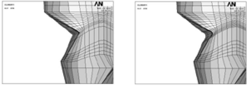
Figure 1 Schematic diagram represents unmodified and modified cavity configuration (Left; Unmodified cavity, Right; Modified cavity).

Figure 2 Schematic diagram of loading points (A; Perpendicular load on the upper third of the palatal slope of the buccal cusp, B; perpendicular load on the upper third of the buccal slope of the palatal cusp).

Figure 3 The principal stress distribution of notch-shaped cavity before restoration (Left; Minimum principal stress-Compressive stress, Right; Maximum principal stress-Tensile stress. Upper and lower view's scales were different each other).

Figure 4 Before restoration, the principal stress distribution on the lesion apex and CEJ (Apex & CEJ), cervical cavosurface margin (Cervical) under Load A and B (MP: Mesial point angle, DP: Distal point angle, Mes: Mesial node, Mid: Middle node, Dist: Distal node).

Figure 5 After restoration, the principal stress distribution in cervical cavity wall under Load A and B.

Figure 6 The principal stress distribution on lesion apex and CEJ after restoration under Load A and B (MP: Mesial point angle, DP: Distal point angle, Mes: Mesial node, Mid: Middle node, Dist: Distal node, M1: more mesial proximal point than MP).

Figure 7 The principal stress distribution on cervical cavosurface margin after restoration under Load A and B (MP: Mesial point angle, DP: Distal point angle, Mes: Mesial node, Mid: Middle node, Dist: Distal node).
Cited by 4 articles
-
The influence of combining composite resins with different elastic modulus on the stress distribution of Class V restoration: a three-dimensional finite element study
Jeong-Kil Park, Bock Hur, Sung-Kyo Kim
J Korean Acad Conserv Dent. 2008;33(3):184-197. doi: 10.5395/JKACD.2008.33.3.184.Stress analysis of maxillary premolars with composite resin restoration of notch-shaped class V cavity and access cavity; Three-dimensional finite element study
Seon-Hwa Lee, Hyeon-Cheol Kim, Bock Hur, Kwang-Hoon Kim, Kwon Son, Jeong-Kil Park
J Korean Acad Conserv Dent. 2008;33(6):570-579. doi: 10.5395/JKACD.2008.33.6.570.Finite element analysis of maxillary central incisors restored with various post-and-core applications
MinSeock Seo, WonJun Shon, WooCheol Lee, Hyun-Mi Yoo, Byeong-Hoon Cho, Seung-Ho Baek
J Korean Acad Conserv Dent. 2009;34(4):324-332. doi: 10.5395/JKACD.2009.34.4.324.Effect of restoration type on the stress distribution of endodontically treated maxillary premolars; Three-dimensional finite element study
Heun-Sook Jung, Hyeon-Cheol Kim, Bock Hur, Kwang-Hoon Kim, Kwon Son, Jeong-Kil Park
J Korean Acad Conserv Dent. 2009;34(1):8-19. doi: 10.5395/JKACD.2009.34.1.008.
Reference
-
1. Levitch LC, Bader JD, Shugars DA, Heymann HO. Non-carious cervical lesion. J Dent. 1994. 22:195–207.2. Litonjua LA, Bush PJ, Andreana S, Tobias TS. Effects of occlusal load on cervical lesion. J Oral Rehabil. 2004. 31:225–232.3. Telles D, Pegoraro LF, Pereira JC. Prevalence of non-carions cervical lesions and their relation to occlusal aspects: a clinical study. J Esthet Dent. 2000. 12:10–15.
Article4. Palamara D, Palamara JE, Tyas MJ, Messer HH. Strain patterns in cervical enamel of teeth subjected to occlusal loading. Dent Mater. 2000. 16:412–419.
Article5. Lee WC, Eakle WS. Possible role of tensile stress in etiology of cervical erosive lesions of teeth. J Prosthet Dent. 1984. 52:374–380.
Article6. Grippo JO. Abfractions: A new classification of hard tissue lesions of tooth. J Esthet Dent. 1991. 3:14–19.7. Rees JS. An investigation into the importance of the periodontal ligament and alveolar bone as supporting structures in finite element studies. J Oral Rehabil. 2001. 28:425–432.
Article8. Rees JS. The effect of variation in occlusal loading on the development of abfraction lesions: a finite element study. J Oral Rehabil. 2002. 29:188–193.
Article9. King PA. Adhesive techniques. Br Dent J. 1999. 186:321–326.
Article10. Osborne-Smith KL, Burke FJ, Wilson NH. The aetiology of the non-carious cervical lesion. Int Dent J. 1999. 49:139–143.
Article11. Vandewalle KS, Vigil G. Guidelines for the restoration of class V lesions. Gen Dent. 1997. 45:254–260.12. Marzouk MA, Bhaiji AH. Influence of enamel cavosurface configuration on marginal leakage in class V composite resin restorations. Am J Dent. 1989. 2:165–169.13. Saunders WP, Muirhead JM. Microleakage of composite restorations with Syntac Bond and Denthesive. Am J Dent. 1992. 5:255–257.14. Lee WC, Eakle WS. Stress-induced cervical lesions: review of advances in the past 10 years. J Prosthet Dent. 1996. 75:487–494.
Article15. Leinfelder KF. Restoration of abfracted lesions. Compendium. 1994. 15:1396–1400.16. Hakimeh S, Vaidyanathan J, Houpt ML, Vaidyanathan TK, Von Hagen S. Microleakage of compomer class V restorations: effect of load cycling, thermal cycling, and cavity shape differences. J Prosthet Dent. 2000. 83:194–203.
Article17. Katona TR, Winkler MM. Stress analysis of a bulk-filled class V light-cured composite restoration. J Dent Res. 1994. 73:1470–1477.
Article18. Geramy A, Sharafoddin F. Abfraction: 3D analysis by means of the finite element method. Quintessence Int. 2003. 34:526–533.19. Le SY, Chiang HC, Huang HM, Shih YH, Chen HC, Dong DR, Lin CT. Thermo-debonding mechanisms in dentin bonding systems using finite element analysis. Biomaterials. 2001. 22:113–123.
Article20. Litonjua LA, Andreana S, Patra AK, Cohen RE. An assessment of stress analyses in the theory of abfraction. Biomed Mater Eng. 2004. 14:311–321.21. Kuroe T, Itoh H, Caputo AA, Konuma M. Biomechanics of cervical tooth structure lesions and their restoration. Quintessence Int. 2000. 31:267–274.22. Kemp-Scholte CM, Davidson CL. Marginal integrity related to bond strength and strain capacity of composite resin restorative systems. J Prosthet Dent. 1990. 64:658–664.
Article23. Yaman SD, Sahin M, Aydin C. Finite element analysis of strength characteristics of various resin based restorative materials in class V cavities. J Oral Rehabil. 2003. 30:630–641.
Article24. Nakayama WT, Hall DR, Grenoble DE, Katz JL. Elastic properties of dental resin restorative materials. J Dent Res. 1974. 53:1121–1126.
Article25. Willems G, Lambrechts P, Braem M, Celis JP, Vanherle G. A classification of dental composites according to their morphological and mechanical characteristics. Dent Mater. 1992. 8:310–319.
Article26. Heymann HO, Sturdevant JR, Bayne S, Wilder AD, Sluder TB, Brunson WD. Examining tooth flexure effects on cervical restorations: a two-year clinical study. J Am Dent Assoc. 1991. 122:41–47.
Article27. Browning WD, Brackett WW, Gilpatrick RO. Retention of microfilled and hybrid resin-based composite in non-carious class 5 lesions: a double-blind, randomized clinical trial. Oper Dent. 1999. 24:26–30.28. Hubsch PF, Middleton J. Asymptotic analysis of the stress field in adhering dental restorations. J Biomech Eng. 2000. 122:408–415.
Article29. Ugural AC, Fenster SK. Advanced strength and applied elasticity. 2003. 4th ed. New Jersey: Pearson Education Inc.;163–168.30. Ghfiri R, Amrouche A, Imad A, Mesmacque G. Fatigue life estimation after crack repair in 6005 A-T6 aluminium alloy using the cold expansion hole technique. Fatigue Fract Engng Mater Struct. 2000. 23:911–916.
Article31. Bowen RL, Rodriguez MS. Tensile strength and modulus of elasticity of tooth structure and several restorative materials. J Am Dent Assoc. 1962. 64:378–387.
Article32. Lehman ML. Tensile strength of human dentin. J Dent Res. 1967. 46:197–201.
Article
- Full Text Links
-
- Actions
-
Cited
- CITED
-
- Close
- Share
-
- Similar articles
-
- The effect of restorative materials on the stress distribution of class V composite resin restorations: a 3D finite element investigation
- The effect of the amount of interdental spacing on the stress distribution in maxillary central incisors restored with porcelain laminate veneer and composite resin: A 3D-finite element analysis
- The influence of composite resin restoration on the stress distribution of notch shaped noncarious cervical lesion; A three dimensional finite element analysis study
- The influence of combining composite resins with different elastic modulus on the stress distribution of Class V restoration: a three-dimensional finite element study
- The influence of occlusal loads on stress distribution of cervical composite resin restorations: A three-dimensional finite element study
