J Korean Acad Conserv Dent.
2004 Nov;29(6):532-540. 10.5395/JKACD.2004.29.6.532.
Influence of cavity size and restoration methods on the cusp deflection in composite restoration
- Affiliations
-
- 1Department of Conservative Dentistry, College of Dentistry, Seoul National University, Korea. inboglee@snu.ac.kr
- KMID: 2175654
- DOI: http://doi.org/10.5395/JKACD.2004.29.6.532
Abstract
- The aim of this study was to measure the cusp deflection during composite restoration for MOD cavity in premolar and to examine the influence of cavity dimension, C-factor and restoration method on the cusp deflection. Thirty extracted maxillary premolar were prepared to four different sizes of MOD cavity and divided into six groups. The width and depth of the cavity were as follows. Group 1; 1.5 x 1 mm, Group 2; 1.5 x 2 mm, Group 3; 3 x 1 mm, and Group 4-6; 3 x 2 mm respectively. Group 1-4 were restored using bulk filling method with Z-250 composite. However, Group 5 was restored incrementally, and Group 6 was restored with an indirect resin inlay. The cusp deflection was recorded at the buccal and lingual cusp tips using LVDT probe for 10,000 seconds. The measured cusp deflections were compared between groups, and the relationship between the cube of the length of cavity wall/the cube of the thickness of cavity wall (L3 / T3), C-factor and cusp deflection or %flexure (100 x cuspal deflection / cavity width) was analyzed. The cusp deflection of Group 1-4 were 12.1 microm, 17.2 microm, 16.2 microm and 26.4 microm respectively. The C-factor was related to the %flexure rather than the cusp deflection. There was a strong positive correlationship between the L3 / T3 and the cusp deflection. The cusp deflection of Group 5 and 6 were 17.4 microm and 17.9 microm respectively, which are much lower value than that of Group 4.
Keyword
Figure
-
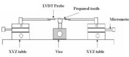
Figure 1 Configuration of the instrument for measuring cusp deflection.
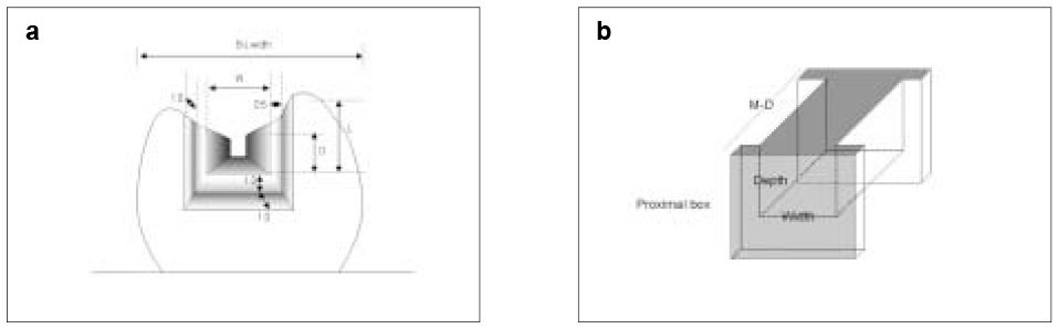
Figure 2 Diagram of prepared cavity. a. The dimension of occlusal cavity and proximal box (W: Width of cavity, D: Depth of cavity, L: Length of cavity wall) b. Simplified cavity shape for calculation of the C-factor.

Figure 3 Representative curves of cusp deflection as a function of time. a. Bulk cure (Group 1-4) b. Bulk cure vs. Incremental cure and Indirect Resin Inlay (Group 4, 5 and 6)
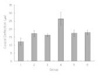
Figure 4 Mean cusp deflections for each tested group.

Figure 5 a. A positive correlationship was present between the L3/T3 and the cusp deflection. b. A positive correlationship was present between the C-factor and the %flexure.
Reference
-
1. de Gee AF, Feilzer AJ, Davidson CL. True linear polymerization shrinkage of unfilled resins and composites determined with a linometer. Dent Mater. 1993. 9:11–14.
Article2. Song YX, Inoue K. Linear shrinkage of photo-activated composite resins during setting. J Oral Rehabil. 2001. 28:335–341.
Article3. Lee IB. A new method-real time measurement of the initial dynamic volumetric shrinkage of composite resins during polymerization. J Korean Acad Conserv Dent. 2001. 26:134–140.4. Rosin M, Urban AD, Gartner C, Bernhardt O, Splieth C, Meyer G. Polymerization shrinkage-strain and microleakage in dentin-bordered cavities of chemically and light-cured restorative materials. Dent Mater. 2002. 18:521–528.
Article5. Goldman M. Polymerization shrinkage of resin-based restorative materials. Aust Dent J. 1983. 28:156–161.
Article6. Rees JS, Jacobsen PH. The polymerization shrinkage of composite resins. Dent Mater. 1989. 5:41–44.
Article7. Lai JH, Johnson AE. Measuring polymerization shrinkage of photo-activated restorative materials by a water-filled dilatometer. Dent Mater. 1993. 9:139–143.
Article8. Opdam NJ, Roeters FJ, Feilzer AJ, Verdonschot EH. Marginal integrity and postoperative sensitivity in Class 2 resin composite restorations in vivo. J Dent. 1998. 26:555–562.
Article9. Carvalho RM, Pereira JC, Yoshiyama M, Pashley DH. A review of polymerization contraction: the influence of stress development versus stress relief. Oper Dent. 1996. 21:17–24.10. Segura A, Donly KJ. In vitro posterior composite polymerization recovery following hygroscopic expansion. J Oral Rehabil. 1993. 20:495–499.
Article11. Alomari QD, Reinhardt JW, Boyer DB. Effect of liners on cusp deflection and gap formation in composite restorations. Oper Dent. 2001. 26:406–411.12. Suliman AH, Boyer DB, Lakes RS. Polymerization shrinkage of composite resins: comparison with tooth deformation. J Prosthet Dent. 1994. 71:7–12.
Article13. Meredith N, Setchell DJ. In vitro measurement of cuspal strain and displacement in composite restored teeth. J Dent. 1997. 25:331–337.
Article14. McCullock AJ, Smith BG. In vitro studies of cusp reinforcement with adhesive restorative material. Br Dent J. 1986. 161:450–452.
Article15. Suliman AA, Boyer DB, Lakes RS. Interferometric measurements of cusp deformation of teeth restored with composites. J Dent Res. 1993. 72:1532–1536.
Article16. Pearson GJ, Hegarty SM. Cusp movement of molar teeth with composite filling materials in conventional and modified MOD cavities. Br Dent J. 1989. 166:162–165.
Article17. Jantarat J, Panitvisai P, Palamara JE, Messer HH. Comparison of methods for measuring cuspal deformation in teeth. J Dent. 2001. 29:75–82.
Article18. Hood JA. Biomechanics of the intact, prepared and restored tooth: some clinical implications. Int Dent J. 1991. 41:25–32.19. Davidson CL, de Gee AJ. Relaxation of polymerization contraction stresses by flow in dental composites. J Dent Res. 1984. 63:146–148.
Article20. Unterbrink GL, Liebenberg WH. Flowable resin composites as "filled adhesives": literature review and clinical recommendations. Quintessence Int. 1999. 30:249–257.21. Versluis A, Douglas WH, Cross M, Sakaguchi RL. Does an incremental filling technique reduce polymerization shrinkage stresses? J Dent Res. 1996. 75:871–878.
Article22. Abbas G, Fleming GJ, Harrington E, Shortall AC, Burke FJ. Cuspal movement and microleakage in premolar teeth restored with a packable composite cured in bulk or in increments. J Dent. 2003. 31:437–444.
Article23. Rees JS, et al. A reappraisal of the incremental packing technique for light cured composite resins. J Oral Rehabil. 2004. 31:81–84.
Article24. Rees JS, Jacobsen PH. Stresses generated by luting resins during cementation of composite and ceramic inlays. J Oral Rehabil. 1992. 19:115–122.
Article25. Ericson D, Paulsson L, Sowiak H, Derand T. Reduction of cusp deflection resulting from composite polymerization shrinkage, using a light-transmitting cone. Scand J Dent Res. 1994. 102:244–248.
Article26. Davidson CL, de Gee AJ. Light-curing units, polymerization, and clinical implications. J Adhes Dent. 2000. 2:167–173.27. Feilzer AJ, De Gee AJ, Davidson CL. Increased wall-to-wall curing contraction in thin bonded resin layers. J Dent Res. 1989. 68:48–50.
Article28. Feilzer AJ, De Gee AJ, Davidson CL. Setting stress in composite resin in relation to configuration of the restoration. J Dent Res. 1987. 66:1636–1639.
Article29. Davidson CL, Feilzer AJ. Polymerization shrinkage and polymerization shrinkage stress in polymer-based restoratives. J Dent. 1997. 25:435–440.
Article30. Lee IB, Son HH, Kwon HC, Um CM, Cho BH. The effect of viscosity, specimen geometry and adhesion on the linear polymerization shrinkage measurement of light cured composites. J Korean Acad Conserv Dent. 2003. 28:457–466.
Article31. Davidson CL, Van Zeghbroeck L, Feilzer AJ. Destructive stresses in adhesive luting cements. J Dent Res. 1991. 70:880–882.
Article32. Alster D, Feilzer AJ, de Gee AJ, Davidson CL. Polymerization contraction stress in thin resin composite layers as a function of layer thickness. Dent Mater. 1997. 13:146–150.
Article33. Craig RG, Powers JM. Restorative dental materials. 2002. 11th ed. Mosby, Inc;87–88.34. Blank JT. Scientifically based rationale and protocol for use of modern indirect resin inlays and onlays. J Esthet Dent. 2000. 12:195–208.
Article35. Watts DC, Cash AJ. Determination of polymerization shrinkage kinetics in visible-light-cured materials: methods development. Dent Mater. 1991. 7:281–287.
Article
- Full Text Links
-
- Actions
-
Cited
- CITED
-
- Close
- Share
-
- Similar articles
-
- Cuspal deflection in class V cavities restored with composite resins
- Effect of the restorative technique on load-bearing capacity, cusp deflection, and stress distribution of endodontically-treated premolars with MOD restoration
- The influence of combining composite resins with different elastic modulus on the stress distribution of Class V restoration: a three-dimensional finite element study
- Stress distribution of Class V composite resin restorations: A three-dimensional finite element study
- The effect of restorative materials on the stress distribution of class V composite resin restorations: a 3D finite element investigation
