A Malignant Melanoma Associated with a Blue Nevus of the Lip
- Affiliations
-
- 1Department of Dermatology, Gachon University of Medicine and Science, Gil Hospital, Incheon, Korea. jyroh@gachon.ac.kr
- KMID: 2172056
- DOI: http://doi.org/10.5021/ad.2010.22.1.119
Abstract
- Blue nevi are characterized by a collection of pigment-producing melanocytes in the dermis. These lesions clinically present as well demarcated cerulean-blue or bluish black colored papules or plaques that usually measure less than 1 cm in diameter. They are typically found on the dorsal surface of the hands and feet or in the head and neck region; however, they are rarely found in the oral cavity. These lesions are usually benign and stable over time. However, malignant melanomas developing in or associated with a blue nevus (which is also called malignant blue nevus) have been only rarely reported. A malignant blue nevus might develop in a common blue or cellular blue nevus, a giant congenital nevus or in a nevus of Ota, or it may be malignant from the start. Malignant blue nevi most commonly are found on the scalp. A malignant blue nevus of the lip has not been previously reported in the medical literature. We report here on a patient with a malignant melanoma associated with a blue nevus of the lip. The malignant melanoma was presumed to have developed from a blue nevus that was present on the upper lip of a 50-year-old male.
MeSH Terms
Figure
-
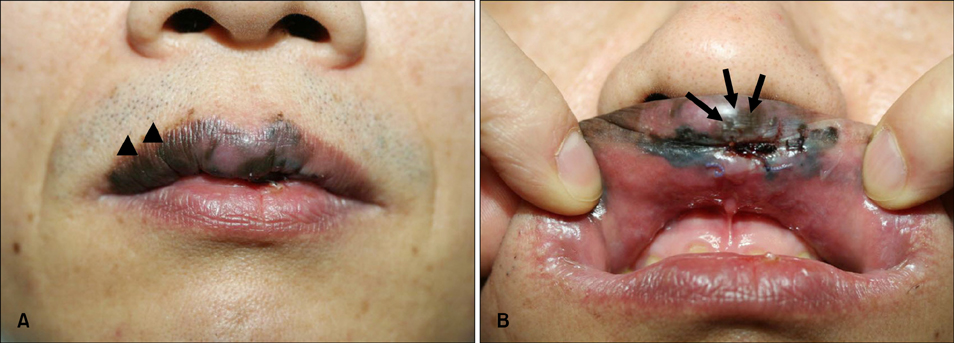
Fig. 1 (A) Bluish black macules (arrow head) with irregular borders and (B) ulcerative nodules (arrow) localized on the upper lip with the development of satellite lesions.
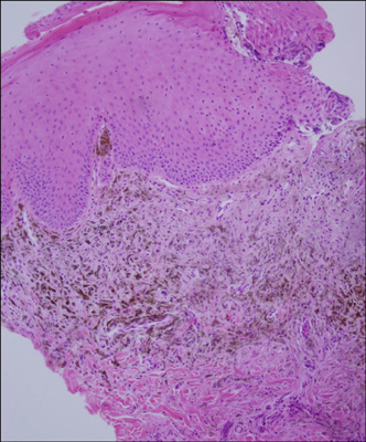
Fig. 2 The histopathological findings of the bluish black macule showed pigmented spindle-shaped and dendritic melanocytes among thickened collagen bundles from the reticular dermis which were close to the epidermis (H&E, ×100).
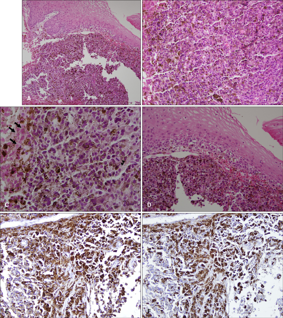
Fig. 3 (A) The histopathologic findings of the bluish black ulcerative nodule showed atypical epithelioid tumor cells and scattered dark pigmented spindle-shaped cells in the dermis. The findings shown include (B) pleomorphic epothelioid tumor cells intermingled with pigmented spindle shaped cells, and (C) necrotic tumor cells (arrow) and mitosis (arrow head). (D) Focal atypical large melanocytes were arranged as single cells on the basal layer of the epidermis without pagetoid spread. The immunohistochemical stains were positive for HMB45 (E) and S-100 (F) (A: H&E, ×100, B: H&E, ×200, C: H&E, ×400, D: H&E, ×200, E: HMB45, ABC method, ×200, F: S-100, ABC method, ×200).
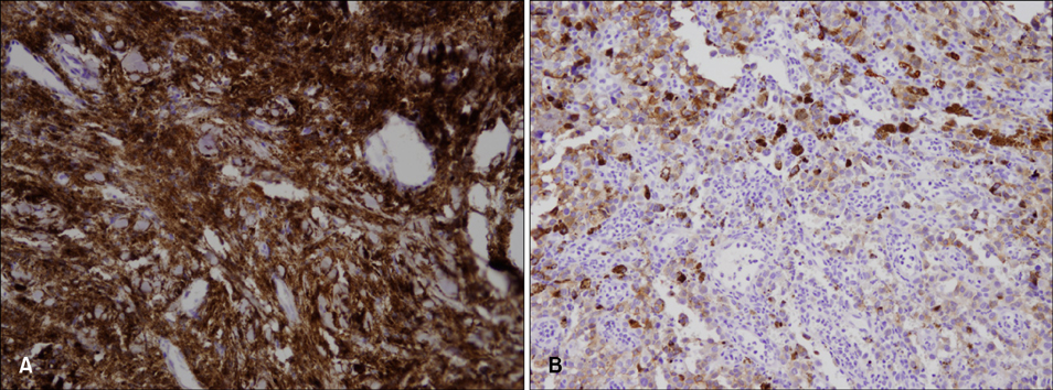
Fig. 4 CD117 (c-kit) staining of the blue nevus (from the bluish black macule) (A) showed strong positivity and (B) there was a decreased c-kit expression on the deep invasive part of the malignant blue nevus (from the ulcerative nodule) (A, B: CD 117 stain, ABC method, ×200).
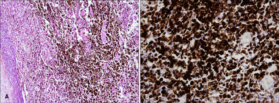
Fig. 5 (A) Atypical dark pigmented tumor cells have infiltrated the lymph node. (B) The infiltrated tumor cells in the lymph node showed a strong c-kit expression (A: H&E, ×100, B: CD 117 stain, ×200).
Reference
-
1. Fistarol SK, Itin PH. Plaque-type blue nevus of the oral cavity. Dermatology. 2005. 211:224–233.
Article2. Granter SR, McKee PH, Calonje E, Mihm MC Jr, Busam K. Melanoma associated with blue nevus and melanoma mimicking cellular blue nevus: a clinicopathologic study of 10 cases on the spectrum of so-called 'malignant blue nevus'. Am J Surg Pathol. 2001. 25:316–323.
Article3. Meleti M, Mooi WJ, Casparie MK, van der Waal I. Melanocytic nevi of the oral mucosa - no evidence of increased risk for oral malignant melanoma: an analysis of 119 cases. Oral Oncol. 2007. 43:976–981.
Article4. Buchner A, Leider AS, Merrell PW, Carpenter WM. Melanocytic nevi of the oral mucosa: a clinicopathologic study of 130 cases from northern California. J Oral Pathol Med. 1990. 19:197–201.
Article5. Scotto J, Fraumeni JF Jr, Lee JA. Melanomas of the eye and other noncutaneous sites: epidemiologic aspects. J Natl Cancer Inst. 1976. 56:489–491.
Article6. Saida T, Kawachi S, Takata M, Kurita H, Kurashina K, Kageshita T, et al. Histopathological characteristics of malignant melanoma affecting mucous membranes: a unifying concept of histogenesis. Pathology. 2004. 36:404–413.
Article7. Hu W, Nelson JE, Mohney CA, Willen MD. Malignant melanoma arising in a pregnant African American woman with a congenital blue nevus. Dermatol Surg. 2004. 30:1530–1532.
Article8. Pathy AL, Helm TN, Elston D, Bergfeld WF, Tuthill RJ. Malignant melanoma arising in a blue nevus with features of pilar neurocristic hamartoma. J Cutan Pathol. 1993. 20:459–464.
Article9. Hagiwara T, Kaku T, Kobayashi H, Hirakawa T, Nakano H. Coexisting vulvar malignant melanoma and blue nevus of the cervix. Gynecol Oncol. 2005. 99:519–520.
Article10. Barnhill RL, Argenyi Z, Berwick M, Duray PH, Erickson L, Guitart J, et al. Atypical cellular blue nevi (cellular blue nevi with atypical features): lack of consensus for diagnosis and distinction from cellular blue nevi and malignant melanoma ("malignant blue nevus"). Am J Surg Pathol. 2008. 32:36–44.
Article11. Mones JM, Ackerman AB. "Atypical" blue nevus, "malignant" blue nevus, and "metastasizing" blue nevus: a critique in historical perspective of three concepts flawed fatally. Am J Dermatopathol. 2004. 26:407–430.
Article12. Elder DE, Elenitsas R, Murphy GF, Xu X. Elder DE, Johnson BL, Elenitsas R, editors. Benign pigmented lesions and malignant melanoma. Lever's histopathology of the skin. 2009. 10th ed. Philadelphia: Lippincott Williams & Wilkins;701–704.13. Zyrek-Betts J, Micale M, Lineen A, Chaudhuri PK, Keil S, Xue J, et al. Malignant blue nevus with lymph node metastases. J Cutan Pathol. 2008. 35:651–657.
Article14. Gontier E, Cario-Andre M, Vergnes P, Bizik J, Surleve-Bazeille JE, Taieb A. The 'Abtropfung phenomenon' revisited: dermal nevus cells from congenital nevi cannot activate matrix metalloproteinase 2 (MMP-2). Pigment Cell Res. 2003. 16:366–373.
Article15. Isabel Zhu Y, Fitzpatrick JE. Expression of c-kit (CD117) in Spitz nevus and malignant melanoma. J Cutan Pathol. 2006. 33:33–37.
Article16. Natali PG, Nicotra MR, Winkler AB, Cavaliere R, Bigotti A, Ullrich A. Progression of human cutaneous melanoma is associated with loss of expression of c-kit proto-oncogene receptor. Int J Cancer. 1992. 52:197–201.
Article17. Montone KT, van Belle P, Elenitsas R, Elder DE. Protooncogene c-kit expression in malignant melanoma: protein loss with tumor progression. Mod Pathol. 1997. 10:939–944.18. Willmore-Payne C, Holden JA, Tripp S, Layfield LJ. Human malignant melanoma: detection of BRAF- and c-kit-activating mutations by high-resolution amplicon melting analysis. Hum Pathol. 2005. 36:486–493.
Article19. Connelly J, Smith JL Jr. Malignant blue nevus. Cancer. 1991. 67:2653–2657.
Article20. Aloi F, Pich A, Pippione M. Malignant cellular blue nevus: a clinicopathological study of 6 cases. Dermatology. 1996. 192:36–40.
- Full Text Links
-
- Actions
-
Cited
- CITED
-
- Close
- Share
-
- Similar articles
-
- Two Cases of Common Blue Nevus with Satellite Lesions
- A Case of Common Blue Nevus with Malignant Melanoma-Like Satellite Lesions
- Three Cases of Malignant Melanoma Possibly Arising in a Long Standing Melanocytic Nevus
- Comments to "A Case of Epithelioid Blue Nevus Developing on the Lower Lip"
- Deep Penetrating Nevus
