A repeatedly recurrent desmoplastic ameloblastoma after removal and allobone graft: Radiographic features compared with histological changes
- Affiliations
-
- 1Department of Oral and Maxillofacial Radiology, School of Dentistry, Chosun University, Gwangju, Korea. hidds@chosun.ac.kr
- 2Department of Oral Pathology, School of Dentistry, Chosun University, Gwangju, Korea.
- KMID: 2167459
- DOI: http://doi.org/10.5624/isd.2013.43.3.201
Abstract
- A 40-year-old man suffered from a repeatedly recurrent desmoplastic ameloblastoma in the right maxillary anterior and premolar regions. During the first visit, the patient was provisionally histopathologically diagnosed with a developmental cyst, and it was confirmed to be unicystic ameloblastoma and resected. Four years later, the lesion recurred, and was diagnosed as a desmoplastic type of ameloblastoma and removed again. Then, 5 years after the second surgery, the lesion recurred again, and was diagnosed as a type containing a follicular pattern, recurrent ameloblastoma. A panoramic radiograph showed a multilocular and mixed radiolucent/radiopaque expansile lesion at the first visit, a unilocular cystic lesion confined to the premolar area at the second visit, and a small soap bubble appearance in the molar area in the final visit. Cone-beam computed tomographic images of the final recurrence of the tumor revealed multiple small cyst-like structures in the right maxillary anterior and posterior regions.
MeSH Terms
Figure
-
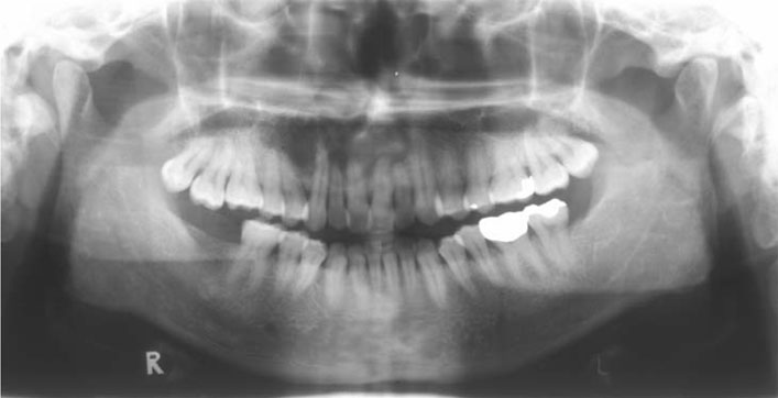
Fig. 1 Panoramic radiograph reveals a multilocular and mixed radiolucent/radiopaque expansile lesion extending from the right second premolar to the left canine of the maxilla, showing a soap bubble appearance in the middle third of the lesion, and an ill-defined radiopacity in the left third of the lesion.

Fig. 2 A. The incisional biopsy shows a cystic structure lined by epithelium (H&E stain, 200×). B. The histopathologic examination after the first surgery on the tumor shows peripheral palisading of hyperchromatic epithelium and loose fibrous stroma (H&E stain, 200×).
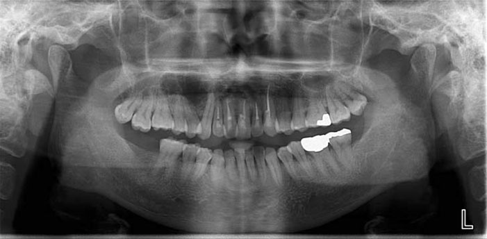
Fig. 3 A panoramic radiograph reveals a unilocular radiolucent cystic area confined to the premolar region of the right maxilla, 4 years after the first surgery.
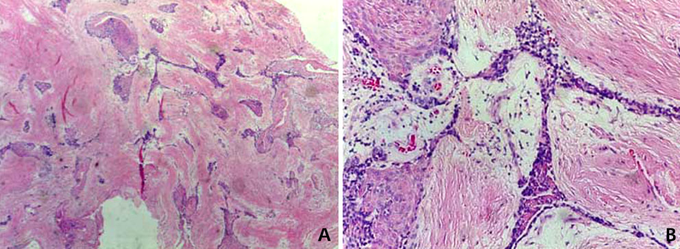
Fig. 4 A. Histopathologic examination 4 years after the first surgery shows the desmoplastic type of ameloblastoma (H&E stain, 40×). B. The histopathologic features contain small islands and thin cords of ameloblastic epithelium within a dense fibrous connective tissue stroma (H&E stain, 200×).
Fig. 5 Five years after the second surgery. The panoramic radiograph reveals a soap bubble appearance in the periapical area of the right first and second molars of the maxilla.
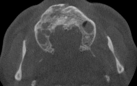
Fig. 6 An axial view of the CBCT image shows multiple small cyst-like structures in the right anterior and posterior regions. They are separated by normal bone. The lesion in the molar region is well demarcated, and separated from the maxillary antrum.
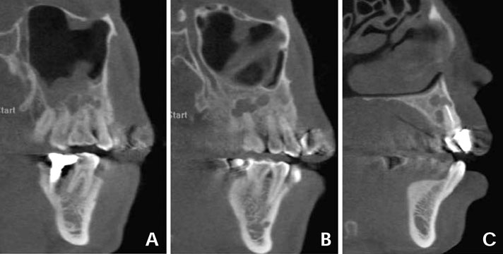
Fig. 7 Sagittal CT images (A; molar area B; premolar area C: anterior area) show small loculations in the molar and anterior region of the maxilla, respectively, separated by normal bone. The tumor shows a relatively defined border, and it is adjacent to the maxillary sinus.
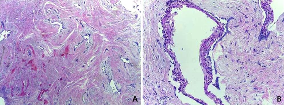
Fig. 8 A. The histopathologic examination 5 years after the second surgery reveals small cystic tumor islands and thin cords of ameloblastic epithelium within connective tissue stroma (H&E stain, 40×). B. A follicular pattern next to the desmoplastic type of ameloblastoma (H&E stain, 200×).
Reference
-
1. White SC, Pharoah MJ. Oral radiology: principles and interpretation. 6th ed. St. Louis: Mosby Elsevier;2009. p. 373–375.2. Becelli R, Carboni A, Cerulli G, Perugini M, Iannetti G. Mandibular ameloblastoma: analysis of surgical treatment carried out in 60 patients between 1977 and 1998. J Craniofac Surg. 2002; 13:395–400.
Article3. Hertog D, Schulten EA, Leemans CR, Winters HA, Van der Waal I. Management of recurrent ameloblastoma of the jaws: a 40-year single institution experience. Oral Oncol. 2011; 47:145–146.
Article4. Rapidis AD, Andressakis DD, Stavrianos SD, Faratzis G, Arnogiannaki-Liappi N, Lagogiannis GA, et al. Ameloblastomas of the jaws: clinico-pathological review of 11 patients. Eur J Surg Oncol. 2004; 30:998–1002.
Article5. Siar CH, Lau SH, Ng KH. Ameloblastoma of the jaws: a retrospective analysis of 340 cases in a Malaysian population. J Oral Maxillofac Surg. 2012; 70:608–615.
Article6. Dissanayake RK, Jayasooriya PR, Siriwardena DJ, Tilakaratne WM. Review of metastasizing (malignant) ameloblastoma (METAM): pattern of metastasis and treatment. Oral Surg Oral Med Oral Pathol Oral Radiol Endod. 2011; 111:734–741.
Article7. Mendenhall WM, Werning JW, Fernandes R, Malyapa RS, Mendenhall NP. Ameloblastoma. Am J Clin Oncol. 2007; 30:645–648.
Article8. Ladeinde AL, Ogunlewe MO, Bamgbose BO. Ameloblastoma: analysis of 207 cases in a Nigerian teaching hospital. Quintessence Int. 2006; 37:69–74.9. Zemann W, Feichtinger M, Kowatsch E, Kärcher H. Extensive ameloblastoma of the jaws: surgical management and immediate reconstruction using microvascular flaps. Oral Surg Oral Med Oral Pathol Oral Radiol Endod. 2007; 103:190–196.
Article10. Reichart PA, Philipsen HP, Sonner S. Ameloblastoma: biological profile of 3677 cases. Eur J Cancer B Oral Oncol. 1995; 31B:86–99.
Article11. Kim SG, Jang HS. Ameloblastoma: a clinical, radiographic, and histopathologic analysis of 71 cases. Oral Surg Oral Med Oral Pathol Oral Radiol Endod. 2001; 91:649–653.
Article12. Waldron CA, El-mofty SK. A histopathologic study of 116 ameloblastomas with special reference to the desmoplastic variant. Oral Surg Oral Med Oral Pathol. 1987; 63:441–451.
Article13. Kaffe I, Buchner A, Taicher S. Radiologic features of desmoplastic variant of ameloblastoma. Oral Surg Oral Med Oral Pathol. 1993; 76:525–529.
Article14. Ng KH, Siar CH. Desmoplastic variant of ameloblastomas in Malaysians. Br J Oral Maxillofac Surg. 1993; 31:299–303.15. Takata T, Miyauchi M, Ogawa I, Zhao M, Kudo Y, Sato S, et al. So-called 'hybrid' lesion of desmoplastic and conventional ameloblastoma: report of a case and review of the literature. Pathol Int. 1999; 49:1014–1018.
Article16. Philipsen HP, Reichart PA, Takata T. Desmoplastic ameloblastoma (including "hybrid" lesion of ameloblastoma). Biological profile based on 100 cases from the literature and own files. Oral Oncol. 2001; 37:455–460.
Article17. Eversole LR, Leider AS, Strub D. Radiographic characteristics of cystogenic ameloblastoma. Oral Surg Oral Med Oral Pathol. 1984; 57:572–577.
Article18. Sun ZJ, Wu YR, Cheng N, Zwahlen RA, Zhao YF. Desmoplastic ameloblastoma - A review. Oral Oncol. 2009; 45:752–759.
Article19. Huang CM, Chen JY, Chen CH, Huang CJ. Radiotherapy for a repeatedly recurrent ameloblastoma with malignant transformation. Head Neck. (in press).
Article20. Thompson IO, van Rensburg LJ, Phillips VM. Desmoplastic ameloblastoma: correlative histopathology, radiology and CT-MR imaging. J Oral Pathol Med. 1996; 25:405–410.
Article21. Bianchi S, Tarello F, Polastri F, Valente G. Ameloblastoma of the mandible involving an autogenous bone graft. J Oral Maxillofac Surg. 1998; 56:1187–1191.
Article22. Stea G. Recurrence of an ameloblastoma in an autogenous iliac bone graft. J Oral Maxillofac Surg. 1985; 43:374–377.
Article23. Wakoh M, Harada T, Inoue T. Follicular/desmoplastic hybrid ameloblastoma with radiographic features of concomitant fibro-osseous and solitary cystic lesions. Oral Surg Oral Med Oral Pathol Oral Radiol Endod. 2002; 94:774–780.
Article24. Philipsen HP, Ormiston IW, Reichart PA. The desmo- and osteoplastic ameloblastoma. Histologic variant or clinicopathologic entity? Case reports. Int J Oral Maxillofac Surg. 1992; 21:352–357.25. Effiom OA, Odukoya O. Desmoplastic ameloblastoma: analysis of 17 Nigerian cases. Oral Surg Oral Med Oral Pathol Oral Radiol Endod. 2011; 111:e27–e31.
Article26. Kawai T, Kishino M, Hiranuma H, Sasai T, Ishida T. A unique case of desmoplastic ameloblastoma of the mandible: report of a case and brief review of the English language literature. Oral Surg Oral Med Oral Pathol Oral Radiol Endod. 1999; 87:258–263.27. Gardner DG, Heikinheimo K, Shear M, Philipsen HP, Coleman H. Ameloblastomas. In : Barnes L, Eveson JW, Reichart P, Sidransky D, editors. World Health Organization classification of tumors: pathology and genetics of head and neck tumors. 3rd ed. Lyon: IARC Press;2005. p. 296–300.28. Keszler A, Paparella ML, Dominguez FV. Desmoplastic and non-desmoplastic ameloblastoma: a comparative clinicopathological analysis. Oral Dis. 1996; 2:228–231.
Article29. Ashman SG, Corio RL, Eisele DW, Murphy MT. Desmoplastic ameloblastoma. A case report and literature review. Oral Surg Oral Med Oral Pathol. 1993; 75:479–482.30. Zachariades N. Recurrences of ameloblastoma in bone grafts. Report of 4 cases. Int J Oral Maxillofac Surg. 1988; 17:316–318.31. Gold L, Upton GW, Marx RE. Standardized surgical terminology for the excision of lesions in bone: an argumentum for accuracy in reporting. J Oral Maxillofac Surg. 1991; 49:1214–1217.32. Philipsen HP, Reichart PA. Unicystic ameloblastoma. A review of 193 cases from the literature. Oral Oncol. 1998; 34:317–325.
Article
- Full Text Links
-
- Actions
-
Cited
- CITED
-
- Close
- Share
-
- Similar articles
-
- Desmoplastic variant of ameloblastoma of the maxilla: A case report
- Radiographic features of desmoplastic ameloblastoma: Report of 3 cases
- Comparison of the Result of Lumbar Posterolateral Bone Fusion using Autogenous Bone Graft with Allobone Graft
- A Case of Desmoplastic Trichoepithelioma, Showing Similar Histological Features to Microcystic Adnexal Carcinoma
- Desmoplastic Trichoepithelioma
