Desmoplastic variant of ameloblastoma of the maxilla: A case report
- Affiliations
-
- 1Department of Oral and Maxillofacial Radiology, School of Dentistry and Institute of Oral Bioscience, Chonbuk National University, Jeonju, Korea. kkj1512@jbnu.ac.kr
- KMID: 2132937
- DOI: http://doi.org/10.5624/isd.2015.45.4.241
Abstract
- The desmoplastic variant of ameloblastoma is a rare form of ameloblastoma characterized by unique radiographic and histologic features. A 46-year-old female was referred to our hospital, complaining of swelling in the left upper lip area. Radiographic findings revealed an ill-defined multilocular lesion with a large cystic lesion and thick sclerotic trabeculae on the left anterior maxilla. After the patient underwent partial osteotomy, histologic analysis revealed a desmoplastic ameloblastoma with no evidence of a hybrid lesion or cyst formation. The radiographic findings in the present case were different from those described in previous case reports. These findings are of special importance due to the unfamiliar radiographic and histologic features of this lesion.
Keyword
Figure
-
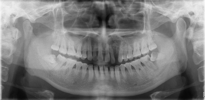
Fig. 1 A panoramic radiograph shows an ill-defined multilocular radiolucency with a large cystic lesion extending from the left upper central incisor to the premolar region and displacement of the left upper lateral incisor and canine.

Fig. 2 Periapical radiographs show a large cystic lesion on the apical region of the left upper central incisor area and an ill-defined multilocular radiolucency with thick sclerotic trabeculae on the left upper anterior maxilla with displacement of the adjacent lateral incisor and canine.

Fig. 3 The axial (A), coronal (B) computed tomographic (CT) images show an ill-defined multilocular lesion with a large cystic lesion, thick trabeculae, expansion, and perforation of the both buccal and palatal cortical plates. C. A sagittal CT image shows an ill-defined multilocular radiolucency with a large cystic lesion. D. A sagittal CT image shows bony expansion to the left maxillary sinus.
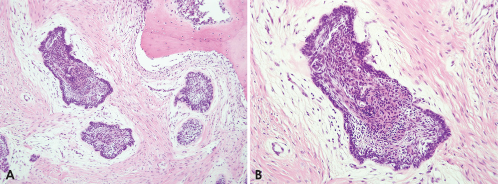
Fig. 4 Photomicrographs show small, scattered tumor nests and new bone formation, as well as desmoplastic changes in the stroma. A. H&E stain, 100×. B. H&E stain, 200×.
Reference
-
1. Lam KY, Chan AC, Wu PC, Chau KY, Tideman H, Wei W. Desmoplastic variant of ameloblastoma in Chinese patients. Br J Oral Maxillofac Surg. 1998; 36:129–134.
Article2. Kawai T, Kishino M, Hiranuma H, Sasai T, Ishida T. A unique case of desmoplastic ameloblastoma of the mandible: report of a case and brief review of the English language literature. Oral Surg Oral Med Oral Pathol Oral Radiol Endod. 1999; 87:258–263.3. Louis PJ, Fugler RC, August M. Mixed radiolucent/radiopaque lesion of the maxilla. J Oral Maxillofac Surg. 2000; 58:86–90.
Article4. Manuel S, Simon D, Rajendran R, Naik BR. Desmoplastic ameloblastoma: a case report. J Oral Maxillofac Surg. 2002; 60:1186–1188.
Article5. Pillai RS, Ongole R, Ahsan A, Radhakrishnan RA, Pai KM. Recurrent desmoplastic ameloblastoma of the maxilla: a case report. J Can Dent Assoc. 2004; 70:100–104.6. Waldron CA, El-Mofty SK. A histopathologic study of 116 ameloblastomas with special reference to the desmoplastic variant. Oral Surg Oral Med Oral Pathol. 1987; 63:441–451.
Article7. Kaffe I, Buchner A, Taicher S. Radiologic features of desmoplastic variant of ameloblastoma. Oral Surg Oral Med Oral Pathol. 1993; 76:525–529.
Article8. Ng KH, Siar CH. Desmoplastic variant of ameloblastoma in Malaysians. Br J Oral Maxillofac Surg. 1993; 31:299–303.
Article9. Wakoh M, Harada T, Inoue T. Follicular/desmoplastic hybrid ameloblastoma with radiographic features of concomitant fibro-osseous and solitary cystic lesions. Oral Surg Oral Med Oral Pathol Oral Radiol Endod. 2002; 94:774–780.
Article10. Black CC, Addante RR, Mohila CA. Intraosseous ameloblastoma. Oral Surg Oral Med Oral Pathol Oral Radiol Endod. 2010; 110:585–592.
Article11. Eversole LR, Leider AS, Hansen LS. Ameloblastomas with pronounced desmoplasia. J Oral Maxillofac Surg. 1984; 42:735–740.
Article12. Effiom OA, Odukoya O. Desmoplastic ameloblastoma: analysis of 17 Nigerian cases. Oral Surg Oral Med Oral Pathol Oral Radiol Endod. 2011; 111:e27–e31.
Article13. Reichart PA, Philipsen HP, Sonner S. Ameloblastoma: biological profile of 3677 cases. Eur J Cancer B Oral Oncol. 1995; 31B:86–99.
Article14. MacDonald-Jankowski DS, Yeung R, Lee KM, Li TK. Ameloblastoma in the Hong Kong Chinese. Part 2: systematic review and radiological presentation. Dentomaxillofac Radiol. 2004; 33:141–151.
Article15. Poon CS, Wu PC, So MK. Ameloblastoma in Hong Kong Chinese. Hong Kong Med J. 1996; 2:172–176.16. Li B, Long X, Wang S, Cheng Y, Chen X. Clinical and radiologic features of desmoplastic ameloblastoma. J Oral Maxillofac Surg. 2011; 69:2173–2185.
Article17. Lo Muzio L, Nocini P, Favia G, Procaccini M, Mignogna MD. Odontogenic myxoma of the jaws: a clinical, radiologic, immunohistochemical, and ultrastructural study. Oral Surg Oral Med Oral Pathol Oral Radiol Endod. 1996; 82:426–433.18. Takata T, Miyauchi M, Ito H, Ogawa I, Kudo Y, Zhao M, et al. Clinical and histopathological analyses of desmoplastic ameloblastoma. Pathol Res Pract. 1999; 195:669–675.
Article19. Thompson IO, van Rensburg LJ, Phillips VM. Desmoplastic ameloblastoma: correlative histopathology, radiology and CTMR imaging. J Oral Pathol Med. 1996; 25:405–410.
Article20. Iida S, Kogo M, Kishino M, Matsuya T. Desmoplastic ameloblastoma with large cystic change in the maxillary sinus: report of a case. J Oral Maxillofac Surg. 2002; 60:1195–1198.
Article21. Takata T, Miyauchi M, Ogawa I, Zhao M, Kudo Y, Sato S, et al. So-called 'hybrid' lesion of desmoplastic and conventional ameloblastoma: report of a case and review of the literature. Pathol Int. 1999; 49:1014–1018.
Article22. Philipsen HP, Reichart PA, Takata T. Desmoplastic ameloblastoma (including "hybrid" lesion of ameloblastoma). Biological profile based on 100 cases from the literature and own files. Oral Oncol. 2001; 37:455–460.
Article
