External apical root resorption in maxillary incisors in orthodontic patients: associated factors and radiographic evaluation
- Affiliations
-
- 1Department of Dentistry, Overbrook Hospital, Chiang Rai, Thailand. kamo_nan@hotmail.com
- 2Division of Orthodontics, Faculty of Dentistry, Chiang Mai University, Chiang Mai, Thailand.
- 3Division of Oral and Maxillofacial Radiology, Faculty of Dentistry, Chiang Mai University, Chiang Mai, Thailand.
- 4Division of Community Dentistry, Faculty of Dentistry, Chiang Mai University, Chiang Mai, Thailand.
- KMID: 2167432
- DOI: http://doi.org/10.5624/isd.2012.42.3.147
Abstract
- PURPOSE
This study was performed to evaluate the incidence and degree of external apical root resorption of maxillary incisors after orthodontic treatment and to evaluate particular associated factors related to external apical root resorption.
MATERIALS AND METHODS
The records and maxillary incisor periapical radiographs of 181 patients were investigated. Crown and root lengths were measured and compared on the pre- and post-treatment periapical radiographs. Crown length was measured from the center of the incisal edge to the midpoint of the cemento-enamel junction (CEJ). Root length was measured from the CEJ midpoint to the root apex. A correction factor for the enlargement difference was used to calculate root resorption.
RESULTS
The periapical radiographs of 564 teeth showed that the average root resorption was 1.39+/-1.27 (8.24+/-7.22%) and 1.69+/-1.14 mm (10.16+/-6.78%) for the maxillary central and lateral incisors, respectively. The results showed that the dilacerated or pointed roots, maxillary premolar extraction cases, and treatment duration were highly significant factors for root resorption (p<0.001). Allergic condition was a significant factor at p<0.01. Age at the start of treatment, large overjet, and history of facial trauma were also factors significantly associated with root resorption (p<0.05). There was no statistically significant difference in root resorption among the factors of gender, overbite, tongue-thrusting habit, types of malocclusion, and types of bracket.
CONCLUSION
These results suggested that orthodontic treatment should be carefully performed in pre-treatment extraction patients who have pointed or dilacerated roots and need long treatment duration.
Keyword
MeSH Terms
Figure
-
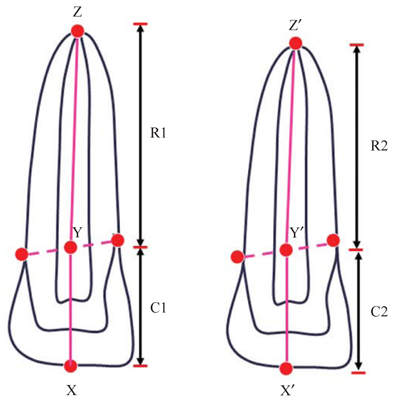
Fig. 1 The points and distance measured on the examination of periapical radiographs. (A) Pre-treatment: X=mid-incisal point of crown, Y=midpoint of CEJ, Z=most apical point of root, C1=crown length before treatment, R1=root length before treatment. (B) post-treatment: X'=mid-incisal point of crown, Y'=midpoint of CEJ, Z'=most apical point of root, C2=crown length after treatment, R2=root length after treatment.
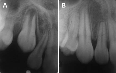
Fig. 2 A. Dilacerated maxillary lateral incisor before orthodontic treatment. B. Moderate external apical root resorption of dilacerated maxillary lateral incisor after orthodontic treatment.
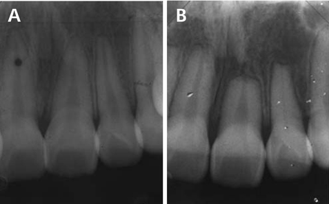
Fig. 3 A. Traumatized maxillary teeth before orthodontic treatment. B. Severe external apical root resorption of the traumatized maxillary incisors after orthodontic treatment.
Cited by 3 articles
-
Cone-beam computed tomography for the assessment of root–crown ratios of the maxillary and mandibular incisors in a Korean population
Sung-Hwan Choi, Jung-Suk Kim, Cheol-Soon Kim, Hyung-Seog Yu, Chung-Ju Hwang
Korean J Orthod. 2017;47(1):39-49. doi: 10.4041/kjod.2017.47.1.39.A posteriori registration and subtraction of periapical radiographs for the evaluation of external apical root resorption after orthodontic treatment
Eliane Maria Kreich, Ana Cláudia Chibinski, Ulisses Coelho, Letícia Stadler Wambier, Rosário de Arruda Moura Zedebski, Mari Eli Leonelli de Moraes, Luiz Cesar de Moraes
Imaging Sci Dent. 2016;46(1):17-24. doi: 10.5624/isd.2016.46.1.17.Effect of micro-osteoperforations on external apical root resorption: A randomized controlled trial
Azaitun Akma Shahrin, Sarah Haniza Abdul Ghani, Noraina Hafizan Norman
Korean J Orthod. 2021;51(2):86-94. doi: 10.4041/kjod.2021.51.2.86.
Reference
-
1. Brezniak N, Wasserstein A. Root resorption after orthodontic treatment: Part 1. Literature review. Am J Orthod Dentofacial Orthop. 1993. 103:62–66.
Article2. Brezniak N, Wasserstein A. Root resorption after orthodontic treatment: Part 2. Literature review. Am J Orthod Dentofacial Orthop. 1993. 103:138–146.
Article3. Brezniak N, Wasserstein A. Orthodontically induced inflammatory root resorption. Part I: The basic science aspects. Angle Orthod. 2002. 72:175–179.4. Brezniak N, Wasserstein A. Orthodontically Induced Inflammatory root resorption. Part II: The clinical aspects. Angle Orthod. 2002. 72:180–184.5. Leach HA, Ireland AJ, Whaites EJ. Radiographic diagnosis of root resorption in relation to orthodontics. Br Dent J. 2001. 190:16–22.
Article6. Sameshima GT, Asgarifar KO. Assessment of root resorption and root shape: periapical vs panoramic films. Angle Orthod. 2001. 71:185–189.7. Linge BO, Linge L. Apical root resorption in upper anterior teeth. Eur J Orthod. 1983. 5:173–183.
Article8. Blake M, Woodside DG, Pharoah MJ. A radiographic comparison of apical root resorption after orthodontic treatment with the edgewise and Speed appliances. Am J Orthod Dentofacial Orthop. 1995. 108:76–84.
Article9. Mavragani M, Vergari A, Selliseth NJ, Boe OE, Wisth PL. A radiographic comparison of apical root resorption after orthodontic treatment with a standard edgewise and a straight-wire edgewise technique. Eur J Orthod. 2000. 22:665–674.
Article10. Brezniak N, Goren S, Zoizner R, Shochat T, Dinbar A, Wasserstein A, et al. The accuracy of the cementoenamel junction identification on periapical films. Angle Orthod. 2004. 74:496–500.11. Brezniak N, Goren S, Zoizner R, Dinbar A, Arad A, Wasserstein A. A comparison of three methods to accurately measure root length. Angle Orthod. 2004. 74:786–791.12. Mirabella AD, Artun J. Prevalence and severity of apical root resorption of maxillary anterior teeth in adult orthodontic patients. Eur J Orthod. 1995. 17:93–99.
Article13. Sameshima GT, Sinclair PM. Predicting and preventing root resorption: Part I. Diagnostic factors. Am J Orthod Dentofacial Orthop. 2001. 119:505–510.
Article14. Mirabella AD, Artun J. Risk factors for apical root resorption of maxillary anterior teeth in adult orthodontic patients. Am J Orthod Dentofacial Orthop. 1995. 108:48–55.
Article15. Artun J, Smale I, Behbehani F, Doppel D, Van't Hof M, Kuijpers-Jagtman AM. Apical root resorption six and 12 months after initiation of fixed orthodontic appliance therapy. Angle Orthod. 2005. 75:919–926.16. Thilander B, Rygh P, Reitan K. Graber TM, Vanarsdall RL, editors. Tissue reactions in orthodontics. Orthodontics: current principles and techniques. 2000. 3rd ed. St. Louis: Mosby;117–125.17. Linge L, Linge BO. Patient characteristics and treatment variables associated with apical root resorption during orthodontic treatment. Am J Orthod Dentofacial Orthop. 1991. 99:35–43.
Article18. Baumrind S, Korn EL, Boyd RL. Apical root resorption in orthodontically treated adults. Am J Orthod Dentofacial Orthop. 1996. 110:311–320.
Article19. Levander E, Malmgren O. Evaluation of the risk of root resorption during orthodontic treatment: a study of upper incisors. Eur J Orthod. 1988. 10:30–38.
Article20. Brin I, Tulloch JF, Koroluk L, Philips C. External apical root resorption in Class II malocclusion: a retrospective review of 1- versus 2-phase treatment. Am J Orthod Dentofacial Orthop. 2003. 124:151–156.
Article21. Smale I, Artun J, Behbehani F, Doppel D, van't Hof M, Kuijpers-Jagtman AM. Apical root resorption 6 months after initiation of fixed orthodontic appliance therapy. Am J Orthod Dentofacial Orthop. 2005. 128:57–67.
Article22. Malmgren O, Goldson L, Hill C, Orwin A, Petrini L, Lundberg M. Root resorption after orthodontic treatment of traumatized teeth. Am J Orthod. 1982. 82:487–491.
Article23. Hamilton RS, Gutmann JL. Endodontic-orthodontic relationships: a review of integrated treatment planning challenges. Int Endod J. 1999. 32:343–360.
Article24. Andreasen JO, Andreasen FM. Root resorption following traumatic dental injuries. Proc Finn Dent Soc. 1992. 88:95–114.25. Nishioka M, Ioi H, Nakata S, Nakasima A, Counts A. Root resorption and immune system factors in the Japanese. Angle Orthod. 2006. 76:103–108.26. Owman-Moll P, Kurol J. Root resorption after orthodontic treatment in high- and low-risk patients: analysis of allergy as a possible predisposing factor. Eur J Orthod. 2000. 22:657–663.27. Krishnan V, Davidovitch Z. Cellular, molecular, and tissue-level reactions to orthodontic force. Am J Orthod Dentofacial Orthop. 2006. 129:469.e1. 469.e32.
Article28. Mohandesan H, Ravanmehr H, Valaei N. A radiographic analysis of external apical root resorption of maxillary incisors during active orthodontic treatment. Eur J Orthod. 2007. 29:134–139.
Article29. McFadden WM, Engstrom C, Engstrom H, Anholm JM. A study of the relationship between incisor intrusion and root shortening. Am J Orthod Dentofacial Orthop. 1989. 96:390–396.
Article
- Full Text Links
-
- Actions
-
Cited
- CITED
-
- Close
- Share
-
- Similar articles
-
- A radiographic study on root resorption in the malocclusion patients before orthodontic treatment
- Factors affecting external apical root resorption of maxillary incisors associated with microimplantassisted rapid palatal expansion
- Predisposing factors for external apical root resorption associated with orthodontic treatment
- A posteriori registration and subtraction of periapical radiographs for the evaluation of external apical root resorption after orthodontic treatment
- Cone-beam computed tomography-based diagnosis and treatment simulation for a patient with a protrusive profile and a gummy smile
