A posteriori registration and subtraction of periapical radiographs for the evaluation of external apical root resorption after orthodontic treatment
- Affiliations
-
- 1Department of Dental Radiology, School of Dentistry, Ponta Grossa State University, Ponta Grossa, Paraná, Brazil. elianekreich@gmail.com
- 2Department of Pediatric Dentistry, School of Dentistry, Ponta Grossa State University, Ponta Grossa, Paraná, Brazil.
- 3Department of Orthodontics, School of Dentistry, Ponta Grossa State University, Ponta Grossa, Paraná, Brazil.
- 4Department of Dental Radiology, School of Dentistry, State University of São Paulo, São José dos Campos, São Paulo, Brazil.
- KMID: 2160153
- DOI: http://doi.org/10.5624/isd.2016.46.1.17
Abstract
- PURPOSE
This study employed a posteriori registration and subtraction of radiographic images to quantify the apical root resorption in maxillary permanent central incisors after orthodontic treatment, and assessed whether the external apical root resorption (EARR) was related to a range of parameters involved in the treatment.
MATERIALS AND METHODS
A sample of 79 patients (mean age, 13.5±2.2 years) with no history of trauma or endodontic treatment of the maxillary permanent central incisors was selected. Periapical radiographs taken before and after orthodontic treatment were digitized and imported to the Regeemy software. Based on an analysis of the posttreatment radiographs, the length of the incisors was measured using Image J software. The mean EARR was described in pixels and relative root resorption (%). The patient's age and gender, tooth extraction, use of elastics, and treatment duration were evaluated to identify possible correlations with EARR.
RESULTS
The mean EARR observed was 15.44±12.1 pixels (5.1% resorption). No differences in the mean EARR were observed according to patient characteristics (gender, age) or treatment parameters (use of elastics, treatment duration). The only parameter that influenced the mean EARR of a patient was the need for tooth extraction.
CONCLUSION
A posteriori registration and subtraction of periapical radiographs was a suitable method to quantify EARR after orthodontic treatment, and the need for tooth extraction increased the extent of root resorption after orthodontic treatment.
Keyword
MeSH Terms
Figure
-
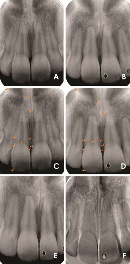
Fig. 1 Illustrative images of the post-processing procedures of periapical radiographs for the right maxillary central incisor. Periapical radiographs at baseline (A, Image 1) and after orthodontic treatment (B, Image 2) were digitalized. The same reference points are tagged in the pre-treatment (C) and post-treatment (D) radiographs to align the images and to generate the posteriori registration of Image 3(E). The quality of the registered image (E) is confirmed by the fact that the subtracted image (F) exhibited the least possible structural noise.

Fig. 2 External apical root resorption is evaluated by the measurement of the long axis of the maxillary central incisors at baseline (A, Image 1) and in the registered image (B, Image 3).
Reference
-
1. de Freitas JC, Lyra OC, de Alencar AH, Estrela C. Long-term evaluation of apical root resorption after orthodontic treatment using periapical radiography and cone beam computed tomography. Dental Press J Orthod. 2013; 18:104–112.2. Roscoe MG, Meira JB, Cattaneo PM. Association of orthodontic force system and root resorption: a systematic review. Am J Orthod Dentofacial Orthop. 2015; 147:610–626.
Article3. Tieu LD, Saltaji H, Normando D, Flores-Mir C. Radiologically determined orthodontically induced external apical root resorption in incisors after non-surgical orthodontic treatment of class II division 1 malocclusion: a systematic review. Prog Orthod. 2014; 15:48.
Article4. Weltman B, Vig KW, Fields HW, Shanker S, Kaizar EE. Root resorption associated with orthodontic tooth movement: a systematic review. Am J Orthod Dentofacial Orthop. 2010; 137:462–476.
Article5. Ono E, Medici Filho E, Faig Leite H, Tanaka JL, De Moraes ME, De Melo Castilho JC. Evaluation of simulated external root resorptions with digital radiography and digital subtraction radiography. Am J Orthod Dentofacial Orthop. 2011; 139:324–333.
Article6. Sunku R, Roopesh R, Kancherla P, Perumalla KK, Yudhistar PV, Reddy VS. Quantitative digital subtraction radiography in the assessment of external apical root resorption induced by orthodontic therapy: a retrospective study. J Contemp Dent Pract. 2011; 12:422–428.
Article7. Eraso FE, Parks ET, Roberts WE, Hohlt WF, Ofner S. Density value means in the evaluation of external apical root resorption: an in vitro study for early detection in orthodontic case simulations. Dentomaxillofac Radiol. 2007; 36:130–137.8. Gegler A, Fontanella V. In vitro evaluation of a method for obtaining periapical radiographs for diagnosis of external apical root resorption. Eur J Orthod. 2008; 30:315–319.
Article9. Maués CP, do Nascimento RR, Vilella Ode V. Severe root resorption resulting from orthodontic treatment: prevalence and risk factors. Dental Press J Orthod. 2015; 20:52–58.
Article10. Mahida K, Agrawal C, Baswaraj H, Tandur AP, Patel B, Chokshi H. Root resorption: an abnormal consequence of the orthodontic treatment. Int J Contemp Dent. 2015; 6:7–9.11. Sameshima GT, Asgarifar KO. Assessment of root resorption and root shape: periapical vs panoramic films. Angle Orthod. 2001; 71:185–189.12. Dudic A, Giannopoulou C, Leuzinger M, Kiliaridis S. Detection of apical root resorption after orthodontic treatment by using panoramic radiography and cone-beam computed tomography of super-high resolution. Am J Orthod Dentofacial Orthop. 2009; 135:434–437.
Article13. Chibinski AC, Reis A, Kreich EM, Tanaka JL, Wambier DS. Evaluation of primary carious dentin after cavity sealing in deep lesions: a 10- to 13-month follow-up. Pediatr Dent. 2013; 35:E107–E112.14. Patil SR, Prabhu A, Ranjan R. Quantitative digital subtraction radiography (DSR) as an approach for evaluating crestal alveolar bone density changes around teeth following orthodontic tooth movement. Int J Clin Dent Sci. 2011; 2:94–100.15. Goorabjavari NM, Talaeipour A, Ezoddini-Ardakani F, Safi Y, Shamloo N. Evaluation of diagnostic efficacy of digital subtraction radiography in the diagnosis of simulated external root resorption: an in vitro study. Health. 2015; 7:439–448.16. Eraso FE, Parks ET, Roberts WE, Hohlt WF, Van Dis ML. Digital subtraction evaluation for external apical root resorption in orthodontic case simulations. Oral Surg Oral Med Oral Pathol Oral Radiol Endod. 2006; 101:E6–E7.
Article17. Heo MS, Lee SS, Lee KH, Choi HM, Choi SC, Park TW. Quantitative analysis of apical root resorption by means of digital subtraction radiography. Oral Surg Oral Med Oral Pathol Oral Radiol Endod. 2001; 91:369–373.
Article18. Hintze H, Wenzel A, Andreasen FM, Swerin I. Digital subtraction radiography for assessment of simulated root resorption cavities. Performance of conventional and reverse contrast modes. Endod Dent Traumatol. 1992; 8:149–154.
Article19. Kravitz LH, Tyndall DA, Bagnell CP, Dove SB. Assessment of external root resorption using digital subtraction radiography. J Endod. 1992; 18:275–284.
Article20. Artun J, Van't Hullenaar R, Doppel D, Kuijpers-Jagtman AM. Identification of orthodontic patients at risk of severe apical root resorption. Am J Orthod Dentofacial Orthop. 2009; 135:448–455.21. Brezniak N, Wasserstein A. Orthodontically induced inflammatory root resorption. Part II: the clinical aspects. Angle Orthod. 2002; 72:180–184.22. Jacobs C, Gebhardt PF, Jacobs V, Hechtner M, Meila D, Wehrbein H. Root resorption, treatment time and extraction rate during orthodontic treatment with self-ligating and conventional brackets. Head Face Med. 2014; 10:2.
Article23. McNab S, Battistutta D, Taverne A, Symons AL. External apical root resorption following orthodontic treatment. Angle Orthod. 2000; 70:227–232.24. Walker SL, Tieu LD, Flores-Mir C. Radiographic comparison of the extent of orthodontically induced external apical root resorption in vital and root-filled teeth: a systematic review. Eur J Orthod. 2013; 35:796–802.
Article25. Jung YH, Cho BH. External root resorption after orthodontic treatment: a study of contributing factors. Imaging Sci Dent. 2011; 41:17–21.
Article26. Ioannidou-Marathiotou I, Papadopoulos MA, Kondylidou-Sidira A, Kokkas A, Karagiannis V. Digital subtraction radiography of panoramic radiographs to evaluate maxillary central incisor root resorption after orthodontic treatment. World J Orthod. 2010; 11:142–152.27. Ramanathan C, Hofman Z. Root resorption during orthodontic tooth movements. Eur J Orthod. 2009; 31:578–583.
Article28. de Freitas MR, Beltrão RT, Janson G, Henriques JF, Chiqueto K. Evaluation of root resorption after open bite treatment with and without extractions. Am J Orthod Dentofacial Orthop. 2007; 132:143.e15–143.e22.
Article29. Sharab LY, Morford LA, Dempsey J, Falcão-Alencar G, Mason A, Jacobson E, et al. Genetic and treatment-related risk factors associated with external apical root resorption (EARR) concurrent with orthodontia. Orthod Craniofac Res. 2015; 18:Suppl 1. 71–82.
Article30. Pereira SA, Lopez M, Lavado N, Abreu JM, Silva H. A clinical risk prediction model of orthodontic-induced external apical root resorption. Rev Port Estomatol Med Dent Cir Maxilofac. 2014; 55:66–72.
Article31. Mohandesan H, Ravanmehr H, Valaei N. A radiographic analysis of external apical root resorption of maxillary incisors during active orthodontic treatment. Eur J Orthod. 2007; 29:134–139.
Article32. Segal G, Schiffman P, Tuncay O. Meta analysis of the treatment-related factors of external apical root resorption. Orthod Craniofac Res. 2004; 7:71–78.
Article33. Mirabella AD, Årtun J. Risk factors for apical root resorption of maxillary anterior teeth in adult orthodontic patients. Am J Orthod Dentofacial Orthop. 1995; 108:48–55.
Article34. Linge BO, Linge L. Apical root resorption in upper anterior teeth. Eur J Orthod. 1983; 5:173–183.
Article35. Zahed Zahedani S, Oshagh M, Momeni Danaei Sh, Roeinpeikar S. A comparison of pical root resorption in incisors after fixed orthodontic treatment with standard edgewise and straight wire (MBT) method. J Dent (Shiraz). 2013; 14:103–110.36. Mavragani M, Vergari A, Selliseth NJ, Bøe OE, Wisth PL. A radiographic comparison of apical root resorption after orthodontic treatment with a standard edgewise and a straight-wire edgewise technique. Eur J Orthod. 2000; 22:665–674.
Article37. Motokawa M, Terao A, Kaku M, Kawata T, Gonzales C, Darendeliler MA, et al. Open bite as a risk factor for orthodontic root resorption. Eur J Orthod. 2013; 35:790–795.
Article38. Lempesi E, Pandis N, Fleming P, Mavragani M. A comparison of apical root resorption after orthodontic treatment with surgical exposure and traction of maxillary impacted canines versus that without impactions. Eur J Orthod. 2014; 36:690–697.
Article39. Sameshima GT, Sinclair PM. Predicting and preventing root resorption: Part I. Diagnostic factors. Am J Orthod Dentofacial Orthop. 2001; 119:505–510.
Article40. Nanekrungsan K, Patanaporn V, Janhom A, Korwanich N. External apical root resorption in maxillary incisors in orthodontic patients: associated factors and radiographic evaluation. Imaging Sci Dent. 2012; 42:147–154.
Article41. Mavragani M, Bøe OE, Wisth PJ, Selvig KA. Changes in root length during orthodontic treatment: advantages for immature teeth. Eur J Orthod. 2002; 24:91–97.
Article42. Jacobson A. A prospective study of apical root resorption during orthodontic treatment and into retention. Am J Orthod Dentofacial Orthop. 2001; 119:457.
Article
- Full Text Links
-
- Actions
-
Cited
- CITED
-
- Close
- Share
-
- Similar articles
-
- External apical root resorption in maxillary incisors in orthodontic patients: associated factors and radiographic evaluation
- A study on the affecting factors on root resorption
- Assessment of apical root resorption using digital subtraction radiography
- Effect of micro-osteoperforations on external apical root resorption: A randomized controlled trial
- Predisposing factors for external apical root resorption associated with orthodontic treatment
