Baseline Characteristics and Risk Factors of Retinal Vein Occlusion: A Study by the Korean RVO Study Group
- Affiliations
-
- 1Department of Ophthalmology, Asan Medical Center, University of Ulsan College of Medicine, Seoul, Korea. yhyoon@amc.seoul.kr
- 2Department of Ophthalmology, Kangnam Sacred Heart Hospital, Hallym University College of Medicine, Seoul, Korea.
- 3Sungmo Eye Hospital, Busan, Korea.
- 4Department of Ophthalmology, Samsung Medical Center, Sungkyunkwan University College of Medicine, Seoul, Korea.
- 5Department of Ophthalmology, Seoul National University College of Medicine and Bundang Hospital, Seongnam, Korea.
- 6Department of Ophthalmology, Chungnam National University Hospital, Daejeon, Korea.
- KMID: 2158013
- DOI: http://doi.org/10.3346/jkms.2013.28.1.136
Abstract
- We investigated the demographic characteristics and risk factors of Korean patients with naIve central or branch retinal vein occlusion (CRVO or BRVO). This study enrolled 41 clinical sites throughout Korea and included 557 consecutive patients with retinal vein occlusion (RVO) from May through November 2010. A total of 557 patients with new-onset RVO participated in this study. Two hundred and three (36.4%) patients were diagnosed with CRVO and 354 (63.6%) patients were diagnosed with BRVO. Comparisons between the two groups showed that the prevalence of diabetes mellitus was significantly higher in CRVO patients and hypertension was significantly higher in BRVO patients (P = 0.001 and 0.002, respectively). Poor baseline visual acuity was significantly associated with female and old age in BRVO patients (P = 0.002 and 0.013, respectively), whereas the wide intraretinal hemorrhage (CRVO, P = 0.029; BRVO, P < 0.001) and the macular ischemia (CRVO, P < 0.001; BRVO, P < 0.001) were associated with both groups. The study results show the clinical features of RVO in Korean patients. Hypertension is strongly associated with BRVO and diabetes mellitus is more strongly associated with CRVO in Korean patients with RVO. As the first nationwide study performed by the Korean Retinal Society, the results of this study can be applied to future studies on RVO.
MeSH Terms
-
Adolescent
Adult
Age Factors
Aged
Aged, 80 and over
Asian Continental Ancestry Group
Child
Child, Preschool
Demography
Diabetes Complications
Female
Humans
Hypertension/complications
Infant
Infant, Newborn
Male
Middle Aged
Regression Analysis
Republic of Korea
Retinal Hemorrhage/complications
Retinal Vein Occlusion/complications/*diagnosis
Risk Factors
Sex Factors
Young Adult
Figure
-
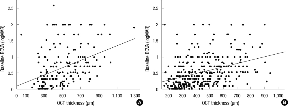
Fig. 1 Scatter plot of baseline OCT-measured central macular thickness and BCVA in (A) CRVO (correlation coefficient = 0.396, P < 0.001) and (B) BRVO patients (correlation coefficient = 0.390, P < 0.001). BCVA, best-corrected visual acuity; OCT, optical coherence tomography; CRVO, central retinal vein occlusion; BRVO, branch retinal vein occlusion.
Cited by 3 articles
-
Intraocular Pressure: Intravitreal Preservative-free Triamcinolone Injection in Diabetic Macular Edema and Branch Retinal Vein Occlusion
Chan Ho Lee, Young Seung Seo
J Korean Ophthalmol Soc. 2020;61(2):167-174. doi: 10.3341/jkos.2020.61.2.167.The Nationwide Incidence of Retinal Vein Occlusion Following Dialysis due to End-stage Renal Disease in Korea, 2004 through 2013
Tae Hwan Moon, Joung-Ho Han, Minseok Kang, Ji Soo Kim, Jin Young Kim, Ju Byung Chae, Soon Kil Kwon, Gilwon Kang, Dong Yoon Kim
J Korean Med Sci. 2021;36(30):e201. doi: 10.3346/jkms.2021.36.e201.Macular Ischemia Correlated with Final Visual Outcome in Retinal Vein Occlusion Patients
Gwang Myung Noh, Ji Eun Lee, Ki Yup Nam, Seung Uk Lee, Sang Joon Lee
J Korean Ophthalmol Soc. 2014;55(10):1493-1498. doi: 10.3341/jkos.2014.55.10.1493.
Reference
-
1. Hayreh SS. Prevalent misconceptions about acute retinal vascular occlusive disorders. Prog Retin Eye Res. 2005. 24:493–519.2. Klein R, Klein BE, Moss SE, Meuer SM. The epidemiology of retinal vein occlusion: the beaver dam eye study. Trans Am Ophthalmol Soc. 2000. 98:133–141.3. Mitchell P, Smith W, Chang A. Prevalence and associations of retinal vein occlusion in Australia: the blue mountains eye study. Arch Ophthalmol. 1996. 114:1243–1247.4. Wong TY, Larsen EK, Klein R, Mitchell P, Couper DJ, Klein BE, Hubbard LD, Siscovick DS, Sharrett AR. Cardiovascular risk factors for retinal vein occlusion and arteriolar emboli: the atherosclerosis risk in communities & cardiovascular health studies. Ophthalmology. 2005. 112:540–547.5. Varma R, Paz SH, Azen SP, Klein R, Globe D, Torres M, Shufelt C, Preston-Martin S. The Los Angeles Latino Eye Study: design, methods, and baseline data. Ophthalmology. 2004. 111:1121–1131.6. Cheung N, Klein R, Wang JJ, Cotch MF, Islam AF, Klein BE, Cushman M, Wong TY. Traditional and novel cardiovascular risk factors for retinal vein occlusion: the multiethnic study of atherosclerosis. Invest Ophthalmol Vis Sci. 2008. 49:4297–4302.7. Lim LL, Cheung N, Wang JJ, Islam FM, Mitchell P, Saw SM, Aung T, Wong TY. Prevalence and risk factors of retinal vein occlusion in an Asian population. Br J Ophthalmol. 2008. 92:1316–1319.8. Liu W, Xu L, Jonas JB. Vein occlusion in Chinese subjects. Ophthalmology. 2007. 114:1795–1796.9. Arakawa S, Yasuda M, Nagata M, Ninomiya T, Hirakawa Y, Doi Y, Kiyohara Y, Ishibashi T. Nine-year incidence and risk factors for retinal vein occlusion in a general Japanese population: the Hisayama Study. Invest Ophthalmol Vis Sci. 2011. 52:5905–5909.10. Retinal Physician. Normative databases in SD-OCT: a status report. accessed on 1 April 2012. Available at http://www.retinalphysician.com/articleviewer.aspx?articleID=104438.11. Statistics Korea. Korean statistical information service. accessed on 1 April 2012. Available at http://kosis.kr/abroad/abroad_01List.jsp.12. Ministry of Health & Welfare [Rep.of Korea]. Statistical information by Ministry of Health and Welfare. accessed on 1 April 2012. Available at http://www.mw.go.kr/front/jb/sjb0601vw.jsp?PAR_MENU_ID=03&MENU_ID=03160501&page=1&CONT_SEQ=238954&SEARCHKEY=TITLE&SEARCHVALUE=%C1%BE%C7%D5%BA%B4%BF%F8.13. Korea Centers for Disease Control & Prevention. Korea National Health & Nutrition Examination Survey. accessed on 1 April 2012. Available at http://knhanes.cdc.go.kr/knhanes/sub04/sub04_03.do?classType=7.14. Appiah AP, Greenidge KC. Factors associated with retinal-vein occlusion in Hispanics. Ann Ophthalmol. 1987. 19:307–309. 31215. Appiah AP, Trempe CL. Risk factors associated with branch vs. central retinal vein occlusion. Ann Ophthalmol. 1989. 21:153–155. 15716. Elman MJ, Bhatt AK, Quinlan PM, Enger C. The risk for systemic vascular diseases and mortality in patients with central retinal vein occlusion. Ophthalmology. 1990. 97:1543–1548.17. Johnston RL, Brucker AJ, Steinmann W, Hoffman ME, Holmes JH. Risk factors of branch retinal vein occlusion. Arch Ophthalmol. 1985. 103:1831–1832.18. McGrath MA, Wechsler F, Hunyor AB, Penny R. Systemic factors contributory to retinal vein occlusion. Arch Intern Med. 1978. 138:216–220.19. Rath EZ, Frank RN, Shin DH, Kim C. Risk factors for retinal vein occlusions: a case-control study. Ophthalmology. 1992. 99:509–514.20. Sperduto RD, Hiller R, Chew E, Seigel D, Blair N, Burton TC, Farber MD, Gragoudas ES, Haller J, Seddon JM, et al. Risk factors for hemiretinal vein occlusion: comparison with risk factors for central and branch retinal vein occlusion: the eye disease case-control study. Ophthalmology. 1998. 105:765–771.21. Seitz R. The retinal vessels: comparative ophthalmoscopic and histologic studies on healthy and diseased eyes. 1964. Saint Louis: CV Mosby;102–146.22. Frangieh GT, Green WR, Barraquer-Somers E, Finkelstein D. Histopathologic study of nine branch retinal vein occlusions. Arch Ophthalmol. 1982. 100:1132–1140.23. Suzuki Y, Nakazawa M, Suzuki K, Yamazaki H, Miyagawa Y. Expression profiles of cytokines and chemokines in vitreous fluid in diabetic retinopathy and central retinal vein occlusion. Jpn J Ophthalmol. 2011. 55:256–263.24. Klein R, Klein BE, Moss SE, Linton KL. The beaver dam eye study: Retinopathy in adults with newly discovered and previously diagnosed diabetes mellitus. Ophthalmology. 1992. 99:58–62.25. Jahn CE, Töpfner von Schutz K, Richter J, Boller J, Kron M. Improvement of visual acuity in eyes with diabetic macular edema after treatment with pars plana vitrectomy. Ophthalmologica. 2004. 218:378–384.26. Pendergast SD, Hassan TS, Williams GA, Cox MS, Margherio RR, Ferrone PJ, Garretson BR, Trese MT. Vitrectomy for diffuse diabetic macular edema associated with a taut premacular posterior hyaloid. Am J Ophthalmol. 2000. 130:178–186.27. Goebel W, Kretzchmar-Gross T. Retinal thickness in diabetic retinopathy: a study using optical coherence tomography (OCT). Retina. 2002. 22:759–767.28. Shah SP, Patel M, Thomas D, Aldington S, Laidlaw DA. Factors predicting outcome of vitrectomy for diabetic macular oedema: results of a prospective study. Br J Ophthalmol. 2006. 90:33–36.29. Otani T, Kishi S. A controlled study of vitrectomy for diabetic macular edema. Am J Ophthalmol. 2002. 134:214–219.
- Full Text Links
-
- Actions
-
Cited
- CITED
-
- Close
- Share
-
- Similar articles
-
- Protein C and Protein S as a Risk Factor for Retinal Vein Occlusion
- Lamina Cribrosa Thickness in the Fellow Eyes of Patients with Unilateral Retinal Vein Occlusion
- Activation of Protein Tyrosine Kinase Pathways After Rat Retinal Vein Occlusion
- A Case of Bilateral Central Retinal Vein Occlusion
- Comparison between Glaucomatous and Non-glaucomatous Eyes with Unilateral Retinal Vein Occlusion in the Fellow Eye
