Investig Magn Reson Imaging.
2015 Dec;19(4):248-251. 10.13104/imri.2015.19.4.248.
Solid Cerebellar Hemangioblastoma with Peritumoral Edema: 5-Years Follow up
- Affiliations
-
- 1Department of Neurology, School of Medicine, Kyung Hee University, Seoul, Korea. ricash@hanmail.net
- KMID: 2151773
- DOI: http://doi.org/10.13104/imri.2015.19.4.248
Abstract
- Hemangioblastomas are angioblastic tumors of the central nervous system. Cerebellar hemangioblastomas are traditionally classified into two morphologic types-cystic and solid. Cystic hemangioblastomas are associated with peritumoral edema, but solid hemangioblastomas are not. We report a case of solid cerebellar hemangioblastoma with massive peritumoral edema. An 83-year-old female visited our hospital due to a sudden headache. Five years ago, she had been admitted to our hospital with similar headache and diagnosed with cerebellar hemangioblastoma. Follow-up brain MRI 5 years later showed an increased size of a homogeneous enhancing mass with aggravated peritumoral edema in the left lower cerebellar hemisphere. Cerebral angiography showed a highly vascularized mass in the cerebellum, which was compatible with a solid-type hemangioblastoma.
Keyword
MeSH Terms
Figure
-
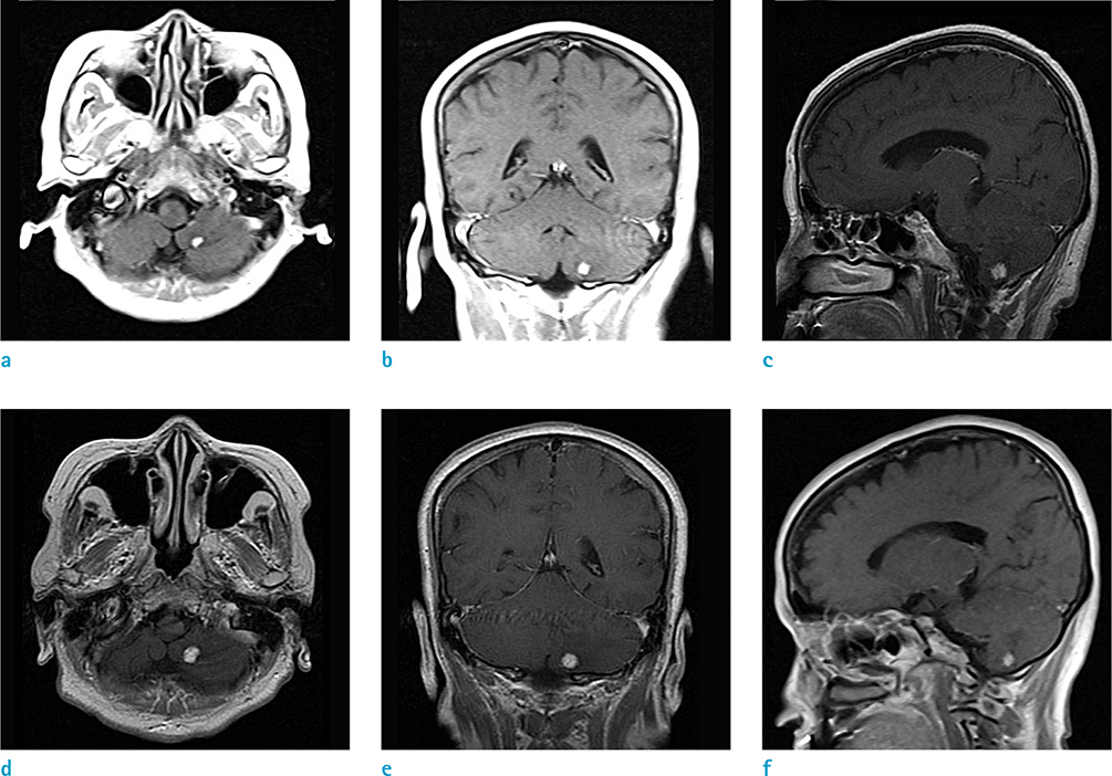
Fig. 1 T1 weighted postcontrast MR images. Brain MRI at the time of first admission (a-c) shows a homogeneous enhancing mass (~7.2 × 5.9 mm in size) in the left lower cerebellum. Follow-up brain MRI (d-f) 5 years later showed an increased size of the homogeneous enhancing mass (~9.0 × 7.4 mm) in the left lower cerebellar hemisphere.

Fig. 2 Fluid-attenuated inversion recovery MR images. Brain MRI at the time of the first admission (a-d) shows edematous changes in the left lower cerebellum. Follow-up brain MRI (e-h) 5 years later shows aggravated peritumoral edema in the left lower cerebellar hemisphere.
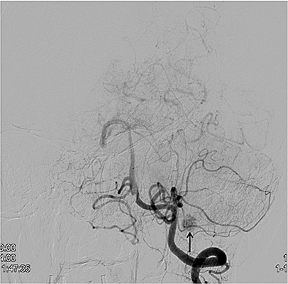
Fig. 3 Cerebral angiography shows a hypervascular mass (arrow) supplied by the left posterior inferior cerebellar artery.
Reference
-
1. Hussein MR. Central nervous system capillary haemangioblastoma: the pathologist's viewpoint. Int J Exp Pathol. 2007; 88:311–324.2. Lonser RR, Vortmeyer AO, Butman JA, et al. Edema is a precursor to central nervous system peritumoral cyst formation. Ann Neurol. 2005; 58:392–399.3. Richard S, Martin S, David P, Decq P. Von Hippel-Lindau disease and central nervous system hemangioblastoma. Progress in genetics and clinical management. Neurochirurgie. 1998; 44:258–266.4. Lee SR, Sanches J, Mark AS, Dillon WP, Norman D, Newton TH. Posterior fossa hemangioblastomas: MR imaging. Radiology. 1989; 171:463–468.5. Rachinger J, Buslei R, Prell J, Strauss C. Solid haemangioblastomas of the CNS: a review of 17 consecutive cases. Neurosurg Rev. 2009; 32:37–47. discussion 47-486. Slater A, Moore NR, Huson SM. The natural history of cerebellar hemangioblastomas in von Hippel-Lindau disease. AJNR Am J Neuroradiol. 2003; 24:1570–1574.7. Nie Q, Guo P, Shen L, Li X, Qiu Y. Early-stage hemangioblastoma presenting as a small lesion with significant edema in the cerebellum. J Craniofac Surg. 2015; 26:e119–e121.8. Girmens JF, Erginay A, Massin P, Scigalla P, Gaudric A, Richard S. Treatment of von Hippel-Lindau retinal hemangioblastoma by the vascular endothelial growth factor receptor inhibitor SU5416 is more effective for associated macular edema than for hemangioblastomas. Am J Ophthalmol. 2003; 136:194–196.
- Full Text Links
-
- Actions
-
Cited
- CITED
-
- Close
- Share
-
- Similar articles
-
- Cerebellar Hemangioblastoma:Hemorrhage as an Initial Presentation: Case Report
- MRImaging of Solid Cerebellar Tumors in Adult
- Computed tomographic features of cerebellar hemangioblastoma
- A Case Report of Recurrent and Disseminated Cerebellar Hemangioblastoma
- A Case of Posterior Medullary Hemangioblastoma
