Engagement of CD99 Reduces AP-1 Activity by Inducing BATF in the Human Multiple Myeloma Cell Line RPMI8226
- Affiliations
-
- 1Cell Dysfunction Research Center, University of Ulsan College of Medicine, Seoul 05505, Korea. csikpark@amc.seoul.kr
- 2Department of Pathology, Asan Medical Center, University of Ulsan College of Medicine, Seoul 05505, Korea.
- 3SIS Immunology Research Center, Sookmyung Women's University, Seoul 04310, Korea.
- 4Institute for Life Sciences, Asan Medical Center, University of Ulsan College of Medicine, Seoul 05505, Korea.
- 5Department of Oncology, Asan Medical Center, University of Ulsan College of Medicine, Seoul 05505, Korea.
- 6Department of Otolaryngology, Asan Medical Center, University of Ulsan College of Medicine, Seoul 05505, Korea.
- KMID: 2150855
- DOI: http://doi.org/10.4110/in.2015.15.5.260
Abstract
- CD99 signaling is crucial to a diverse range of biological functions including survival and proliferation. CD99 engagement is reported to augment activator protein-1 (AP-1) activity through mitogen-activated protein (MAP) kinase pathways in a T-lymphoblastic lymphoma cell line Jurkat and in breast cancer cell lines. In this study, we report that CD99 differentially regulated AP-1 activity in the human myeloma cell line RPMI8226. CD99 was highly expressed and the CD99 engagement led to activation of the MAP kinases, but suppressed AP-1 activity by inducing the expression of basic leucine zipper transcription factor, ATF-like (BATF), a negative regulator of AP-1 in RPMI8226 cells. By contrast, engagement of CD99 enhanced AP-1 activity and did not change the BATF expression in Jurkat cells. CD99 engagement reduced the proliferation of RPMI8226 cells and expression of cyclin 1 and 3. Overall, these results suggest novel CD99 functions in RPMI8226 cells.
Keyword
MeSH Terms
Figure
-
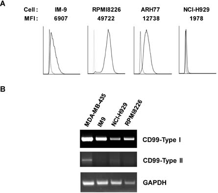
Figure 1 Expression of CD99 in B-cell lines. (A) Surface expression of CD99 in B-cell lines. Cells were stained using an anti-CD99 Ab (DN16-PE, solid line) or isotype control Ab (dotted line) and measured using flow cytometry. (B) PCR analysis with primers designed to specifically amplify CD99 type-I and -II. As described previously, the 583 bp fragment is characteristic of CD99 type-I and the 515 bp fragment is characteristic of CD99 type-II (513). Levels of GAPDH mRNA were used to normalize expression.
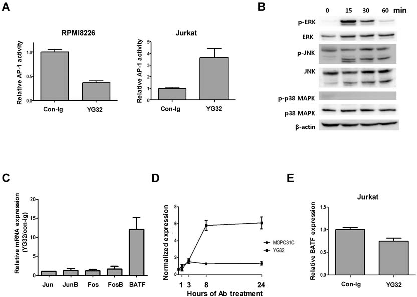
Figure 2 CD99 engagement reduces AP-1 activity and induces BATF expression in RPMI8226 cells. (A) AP-1 activity in RPMI8226 and Jurkat cells upon CD99 engagement. Both AP-1 luciferase constructs and a β-galactosidase expression plasmid were transfected into RPMI8226 and Jurkat cells. Luciferase activity was measured 24 hours after CD99 engagement and normalized against β-galactosidase activity. (B) Activation of ERK and JNK upon CD99 engagement in RPMI8226 cells. RPMI8226 cells were stimulated with 10 µg/ml of an anti-CD99 Ab (YG32) at 37℃ for 15, 30, and 60 min. The activities of ERK, JNK, and p38 kinase were measured by western blotting with antibodies specific for the phosphorylated form of each kinase, as well as suitable controls. Results are from a representative experiment performed in triplicate. (C) Expression of AP-1 transcription factors was measured using real-time PCR at 72 hours after CD99 engagement. Expression levels of the transcript in CD99-engaged cells are shown relative to those in control Ab (mouse IgG)-treated cells. (D) BATF expression was measured using real-time PCR at 1, 3, 8, and 24 hours after CD99 engagement in RPMI8226 cells. (E) BATF expression in CD99-engaged and control Jurkat cells. BATF expression was measured 24 hours after CD99 engagement by real-time PCR.

Figure 3 BATF-mediated regulation of AP-1 activity in RPMI8226 cells. (A) Overexpression of BATF reduces AP-1 activity in RPMI8226 and 293T cells. Cells were transiently transfected with reporter plasmids and expression plasmids, and treated with YG32. AP-1 activity was measured using a luciferase assay. Luciferase activity in cells co-transfected with pcDNA3.1-BATF is shown relative to that in cells co-transfected with pCDNA3.1 (set to 1). (B) Expression of BATF in RPMI8226 cells was reduced by siRNA treatment. Expression of BATF was measured using real-time PCR at 2 days after electroporation of siRNA oligonucleotides. (C) BATF knockdown reverses the reduction in AP-1 activity upon CD99 engagement. Cells were transfected with reporter plasmids 2 days after electroporation with siRNA oligonucleotides. Luciferase activities were normalized with respect to -galactosidase activities. The values show this normalized activity in Ab-treated cells relative to that in cells treated with control IgG. The error bars indicate the SD of three independent experiments.
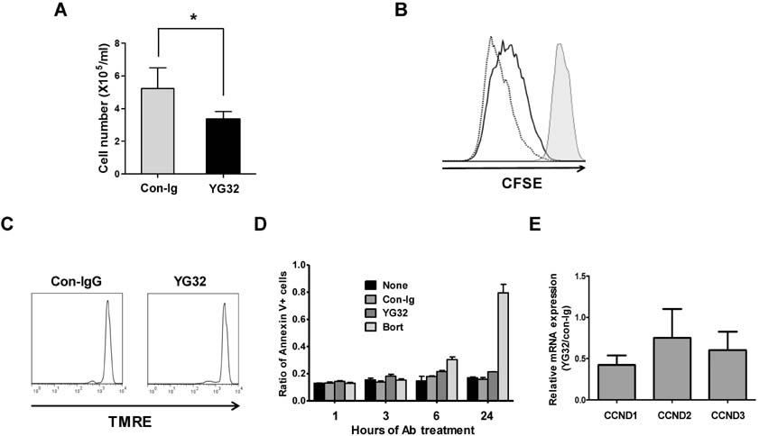
Figure 4 CD99 engagement reduces the proliferation of RPMI8226 cells. (A) The number of RPMI8226 cells was counted using a hemocytometer after 3 days of treatment with an anti-CD99 Ab (YG32) or control IgG. The asterisk denotes a significant difference (p<0.05) between the two groups. (B) CFSE-labeled RPMI8226 cells at day 0 (filled histogram) were incubated for 3 days with control IgG (dotted line) or YG32 (solid line), and analyzed by flow cytometry. (C) RPMI8226 cells were cultured with YG32 or control IgG for 24 hours, stained with TMRE, and quantified using flow cytometry. (D) Annexin V-positive cells were quantified using flow cytometry after culture for the indicated amount of time in the presence of control IgG, YG32, or bortezomib. The ratio of the number of annexin V-positive cells to the total number of cells is shown (E) Expression of cyclin D1, D2, and D3 was measured by performing real-time PCR using the cDNA of RPMI8226 cells harvested after 3 days of incubation with YG32.
Reference
-
1. Levy R, Dilley J, Fox RI, Warnke R. A human thymus-leukemia antigen defined by hybridoma monoclonal antibodies. Proc Natl Acad Sci U S A. 1979; 76:6552–6556.
Article2. Dworzak MN, Fritsch G, Buchinger P, Fleischer C, Printz D, Zellner A, Schollhammer A, Steiner G, Ambros PF, Gadner H. Flow cytometric assessment of human MIC2 expression in bone marrow, thymus, and peripheral blood. Blood. 1994; 83:415–425.
Article3. Park CK, Shin YK, Kim TJ, Park SH, Ahn GH. High CD99 expression in memory T and B cells in reactive lymph nodes. J Korean Med Sci. 1999; 14:600–606.
Article4. Bernard G, Zoccola D, Deckert M, Breittmayer JP, Aussel C, Bernard A. The E2 molecule (CD99) specifically triggers homotypic aggregation of CD4+ CD8+ thymocytes. J Immunol. 1995; 154:26–32.5. Hahn JH, Kim MK, Choi EY, Kim SH, Sohn HW, Ham DI, Chung DH, Kim TJ, Lee WJ, Park CK, Ree HJ, Park SH. CD99 (MIC2) regulates the LFA-1/ICAM-1-mediated adhesion of lymphocytes, and its gene encodes both positive and negative regulators of cellular adhesion. J Immunol. 1997; 159:2250–2258.6. Kasinrerk W, Tokrasinwit N, Moonsom S, Stockinger H. CD99 monoclonal antibody induce homotypic adhesion of Jurkat cells through protein tyrosine kinase and protein kinase C-dependent pathway. Immunol Lett. 2000; 71:33–41.
Article7. Jung KC, Kim NH, Park WS, Park SH, Bae Y. The CD99 signal enhances Fas-mediated apoptosis in the human leukemic cell line, Jurkat. FEBS Lett. 2003; 554:478–484.
Article8. Pettersen RD, Bernard G, Olafsen MK, Pourtein M, Lie SO. CD99 signals caspase-independent T cell death. J Immunol. 2001; 166:4931–4942.
Article9. Bernard G, Breittmayer JP, de MM, Trampont P, Hofman P, Senik A, Bernard A. Apoptosis of immature thymocytes mediated by E2/CD99. J Immunol. 1997; 158:2543–2550.10. Waclavicek M, Majdic O, Stulnig T, Berger M, Sunder-Plassmann R, Zlabinger GJ, Baumruker T, Stockl J, Ebner C, Knapp W, Pickl WF. CD99 engagement on human peripheral blood T cells results in TCR/CD3-dependent cellular activation and allows for Th1-restricted cytokine production. J Immunol. 1998; 161:4671–4678.11. Yoon SS, Kim HJ, Chung DH, Kim TJ. CD99 costimulation up-regulates T cell receptor-mediated activation of JNK and AP-1. Mol Cells. 2004; 18:186–191.12. Hahn MJ, Yoon SS, Sohn HW, Song HG, Park SH, Kim TJ. Differential activation of MAP kinase family members triggered by CD99 engagement. FEBS Lett. 2000; 470:350–354.
Article13. Byun HJ, Hong IK, Kim E, Jin YJ, Jeoung DI, Hahn JH, Kim YM, Park SH, Lee H. A splice variant of CD99 increases motility and MMP-9 expression of human breast cancer cells through the AKT-, ERK-, and JNK-dependent AP-1 activation signaling pathways. J Biol Chem. 2006; 281:34833–34847.
Article14. Choi EY, Park WS, Jung KC, Kim SH, Kim YY, Lee WJ, Park SH. Engagement of CD99 induces up-regulation of TCR and MHC class I and II molecules on the surface of human thymocytes. J Immunol. 1998; 161:749–754.15. Husak Z, Printz D, Schumich A, Potschger U, Dworzak MN. Death induction by CD99 ligation in TEL/AML1-positive acute lymphoblastic leukemia and normal B cell precursors. J Leukoc Biol. 2010; 88:405–412.
Article16. Yoon SS, Jung KI, Choi YL, Choi EY, Lee IS, Park SH, Kim TJ. Engagement of CD99 triggers the exocytic transport of ganglioside GM1 and the reorganization of actin cytoskeleton. FEBS Lett. 2003; 540:217–222.
Article17. Williams KL, Nanda I, Lyons GE, Kuo CT, Schmid M, Leiden JM, Kaplan MH, Taparowsky EJ. Characterization of murine BATF: a negative regulator of activator protein-1 activity in the thymus. Eur J Immunol. 2001; 31:1620–1627.
Article18. Iacobelli M, Wachsman W, McGuire KL. Repression of IL-2 promoter activity by the novel basic leucine zipper p21SNFT protein. J Immunol. 2000; 165:860–868.
Article19. Echlin DR, Tae HJ, Mitin N, Taparowsky EJ. B-ATF functions as a negative regulator of AP-1 mediated transcription and blocks cellular transformation by Ras and Fos. Oncogene. 2000; 19:1752–1763.
Article20. Dorsey MJ, Tae HJ, Sollenberger KG, Mascarenhas NT, Johansen LM, Taparowsky EJ. B-ATF: a novel human bZIP protein that associates with members of the AP-1 transcription factor family. Oncogene. 1995; 11:2255–2265.21. Thornton TM, Zullo AJ, Williams KL, Taparowsky EJ. Direct manipulation of activator protein-1 controls thymocyte proliferation in vitro. Eur J Immunol. 2006; 36:160–169.
Article22. Liao J, Humphrey SE, Poston S, Taparowsky EJ. Batf promotes growth arrest and terminal differentiation of mouse myeloid leukemia cells. Mol Cancer Res. 2011; 9:350–363.
Article23. Livak KJ, Schmittgen TD. Analysis of relative gene expression data using real-time quantitative PCR and the 2(-Delta Delta C(T)) Method. Methods. 2001; 25:402–408.
Article24. Bernard G, Raimondi V, Alberti I, Pourtein M, Widjenes J, Ticchioni M, Bernard A. CD99 (E2) up-regulates alpha4beta1-dependent T cell adhesion to inflamed vascular endothelium under flow conditions. Eur J Immunol. 2000; 30:3061–3065.
Article25. Gil MC, Lee MH, Seo JI, Choi YL, Kim MK, Jung KC, Park SH, Kim TJ. Characterization and epitope mapping of two monoclonal antibodies against human CD99. Exp Mol Med. 2002; 34:411–418.
Article26. Shaulian E, Karin M. AP-1 in cell proliferation and survival. Oncogene. 2001; 20:2390–2400.
Article27. Hess J, Angel P, Schorpp-Kistner M. AP-1 subunits: quarrel and harmony among siblings. J Cell Sci. 2004; 117:5965–5973.
Article28. Matthews CP, Colburn NH, Young MR. AP-1 a target for cancer prevention. Curr Cancer Drug Targets. 2007; 7:317–324.
Article29. Senga T, Iwamoto T, Humphrey SE, Yokota T, Taparowsky EJ, Hamaguchi M. Stat3-dependent induction of BATF in M1 mouse myeloid leukemia cells. Oncogene. 2002; 21:8186–8191.
Article30. Su ZZ, Lee SG, Emdad L, Lebdeva IV, Gupta P, Valerie K, Sarkar D, Fisher PB. Cloning and characterization of SARI (suppressor of AP-1, regulated by IFN). Proc Natl Acad Sci U S A. 2008; 105:20906–20911.
Article31. Quigley M, Pereyra F, Nilsson B, Porichis F, Fonseca C, Eichbaum Q, Julg B, Jesneck JL, Brosnahan K, Imam S, Russell K, Toth I, Piechocka-Trocha A, Dolfi D, Angelosanto J, Crawford A, Shin H, Kwon DS, Zupkosky J, Francisco L, Freeman GJ, Wherry EJ, Kaufmann DE, Walker BD, Ebert B, Haining WN. Transcriptional analysis of HIV-specific CD8+ T cells shows that PD-1 inhibits T cell function by upregulating BATF. Nat Med. 2010; 16:1147–1151.
Article32. Liu Y, Lu C, Shen Q, Munoz-Medellin D, Kim H, Brown PH. AP-1 blockade in breast cancer cells causes cell cycle arrest by suppressing G1 cyclin expression and reducing cyclin-dependent kinase activity. Oncogene. 2004; 23:8238–8246.
Article
- Full Text Links
-
- Actions
-
Cited
- CITED
-
- Close
- Share
-
- Similar articles
-
- The increased expression of CD99 in a differentiated neuroblastoma cell line
- The CXCR4 Antagonist AMD3100 Has Dual Effects on Survival and Proliferation of Myeloma Cells In Vitro
- Bortezomib inhibits the survival and proliferation of bone marrow stromal cells
- CD99 type II is a determining factor for the differentiation of primitive neuroectodermal cells
- A Case of Cutaneous Plasmacytoma Treated with Thalidomide
