Characterization of CTL Clones Specific for Single Antigen, H60 Minor Histocompatibility Antigen
- Affiliations
-
- 1Department of Biomedical Sciences, Seoul National University College of Medicine, Seoul 110-799, Korea. eycii@snu.ac.kr
- 2Graduate Program of Immunology, Seoul National University College of Medicine, Seoul 110-799, Korea.
- 3Division of Life and Pharmaceutical Sciences, Ewha Womans University, Seoul 120-750, Korea.
- KMID: 2150697
- DOI: http://doi.org/10.4110/in.2011.11.2.100
Abstract
- BACKGROUND
Disparities of Minor H antigens can induce graft rejection after MHC-matched transplantation. H60 has been characterized as a dominant antigen expressed on hematopoietic cells and considered to be an ideal model antigen for study on graft-versus-leukemia effect.
METHODS
Splenocytes from C57BL/6 mice immunized with H60 congenic splenocytes were used for establishment of H60-specific CTL clones. Then the clones were characterized for proliferation capacity and cytotoxicity after stimulation with H60. Clone #14, #15, and #23 were tested for the TCR binding avidity to H60-peptide/H-2Kb and analyzed for TCR sequences.
RESULTS
H60-specific CTL clones showed different levels of proliferation capacity and cytotoxic activity to H60-stimulation. Clones #14, #15, and #23 showed high proliferation activity, high cytotoxicity, and low activities on both aspects, respectively, and have TCRs with different binding avidities to H60-peptide/H-2Kb with t 1/2 values of 4.87, 6.92, and 13.03 minutes, respectively. The TCR usages were Valpha12D-3-01+Jalpha11-01 and Vbeta12-1-01+Dbeta1-01+J2-7-01 for clone #14, Valpha13D-1-02+Jalpha34-02 and Vbeta13-1-02+Dbeta2-01+Jbeta2-7-01 for clone #15, and Valpha16D+Jalpha45-01 and Vbeta12-1-01+Dbeta1-01+Jbeta2-5-01 for clone #23.
CONCLUSION
The results will be useful for modeling GVL and generation TCR transgenic mouse.
Keyword
MeSH Terms
Figure
-
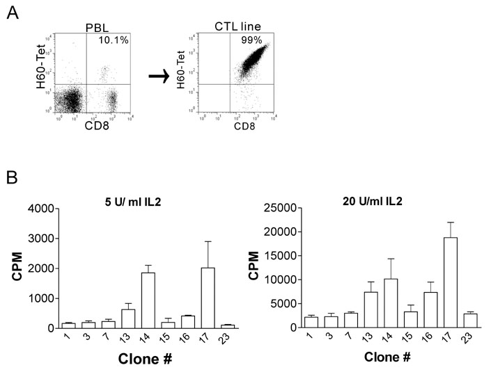
Figure 1 Establishment and characterization of H60-specific CTL clones. (A) Female C57Bl/6 (B6) mice were immunized with splenocytes from male H60 congenic mice, and, then, peripheral blood lymphocytes (PBLs) from the immunized mice were examined to see whether H60-specific response was induced by staining with H60 tetramer and subsequent flow cytometry on day 10 post-immunization (left). On day 14, splenocytes from the immunized mice were cultured with irradiated splenocytes from H60 congenic mouse and the mixed lymphocyte culture (MLC) was re-stimulated regularly on weekly basis to generate H60-specific CTL line (right). H60-specific CTL clones were derived from the CTL line on passage 5. (B) Proliferation assay of the H60-specific CTL clones. Established H60-specific CTL clones were examined for their proliferation capacity in reaction to the stimulation with H60 congenic cells in the presence of IL-2 at the concentrations of 5 U/ml (left) and 20 U/ml (right). 3H was added to the culture on 48 hr later and, after further induction for 18 hr, cells were harvested and 3H-incorporation was measured. The assay was performed in triplicates and the data represent three independent experiments.
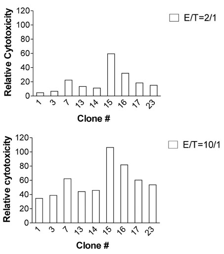
Figure 2 Cytotoxicity of H60-specific CTL clones. CTL clones were incubated with 51Cr-labeled T2-Kb cells after loading with H60 or VSV peptide at the E:T ratios of 2:1 and 10:1. 51Cr released into culture supernatant was measured and specific cytotoxicity was calculated in relation to the 51Cr released in the VSV-peptide-included wells. The assay was performed in triplicates and the mean values were plotted. The data represent three independent experiments.
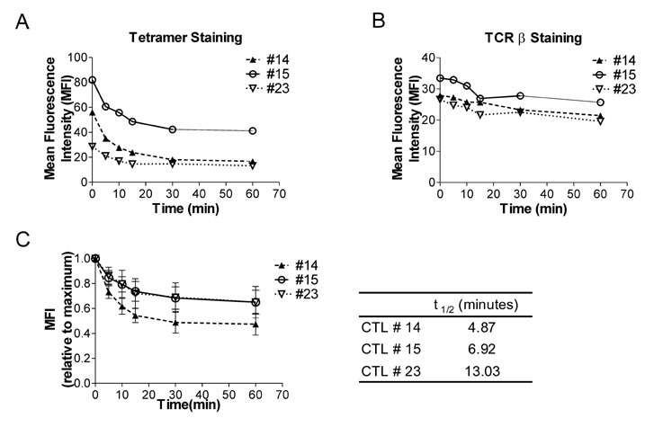
Figure 3 Biochemical features of the interaction of H60-peptide/H-2Kb-TCR of H60-specific CTL clones (A) CTL clones, #14, #15, and #23, were reacted with H60-tetramer-PE, washed extensively and incubated 37℃ for 1 hr. The dissociation of the tetramer from cells during the chase was monitored by flow cytometry. (B) Cells were also stained with anti-TCRβ antibody post-chase to detect TCR levels of the cells. The MFI values from the flow cytometry (A and B) were plotted. (C) Relative MFIs were MFI values at indicated time points fractionated by MFI value detected at the beginning of the chase. And the t1/2 was obtained based on this dissociation curve. The data represent two independent experiments.
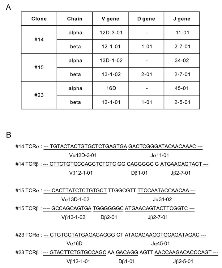
Figure 4 Sequence analysis of H60-specific CTL clones. (A) Annotation of TCR usages of H60-specific CTL clones, #14, #15, and #23, and (B) sequences of the CDR3 regions were shown.
Cited by 2 articles
-
Skewed Dendritic Cell Differentiation of MyD88-Deficient Donor Bone Marrow Cells, Instead of Massive Expansion as Myeloid-Derived Suppressor Cells, Aggravates GVHD
Young-Kwan Lee, Ji-Min Ju, Woo-Jeong Shon, Sehwa Oh, Chang-Ki Min, Myung-Soo Kang, Dong-Mi Shin, Eun Young Choi
Immune Netw. 2018;18(6):. doi: 10.4110/in.2018.18.e44.Selection of Thymocytes Expressing Transgenic TCR Specific for a Minor Histocompatibility Antigen, H60
Ji-Min Ju, Min Bum Kim, Su Jeong Ryu, Joo Young Kim, Jun Chang, Eun Young Choi
Immune Netw. 2015;15(5):222-231. doi: 10.4110/in.2015.15.5.222.
Reference
-
1. Simpson E. Minor histocompatibility antigens. Immunol Lett. 1991. 29:9–14.
Article2. Simpson E, Roopenian D. Minor histocompatibility antigens. Curr Opin Immunol. 1997. 9:655–661.
Article3. Wettstein PJ. Immunodominance in the T-cell response to multiple non-H-2 histocompatibility antigens. II. Observation of a hierarchy among dominant antigens. Immunogenetics. 1986. 24:24–31.
Article4. Choi EY, Yoshimura Y, Christianson GJ, Sproule TJ, Malarkannan S, Shastri N, Joyce S, Roopenian DC. Quantitative analysis of the immune response to mouse non-MHC transplantation antigens in vivo: the H60 histocompatibility antigen dominates over all others. J Immunol. 2001. 166:4370–4379.
Article5. Malarkannan S, Shih PP, Eden PA, Horng T, Zuberi AR, Christianson G, Roopenian D, Shastri N. The molecular and functional characterization of a dominant minor H antigen, H60. J Immunol. 1998. 161:3501–3509.6. Korngold R. Lethal graft-versus-host disease in mice directed to multiple minor histocompatibility antigens: features of CD8+ and CD4+ T cell responses. Bone Marrow Transplant. 1992. 9:355–364.7. van Lochem E, de Gast B, Goulmy E. In vitro separation of host specific graft-versus-host and graft-versus-leukemia cytotoxic T cell activities. Bone Marrow Transplant. 1992. 10:181–183.8. Goulmy E. Human minor histocompatibility antigens: new concepts for marrow transplantation and adoptive immunotherapy. Immunol Rev. 1997. 157:125–140.
Article9. Choi JH, Yoon H, Min CK, Choi EY. Effects of Pre-conditioning Dose on the Immune Kinetics and Cytokine Production in the Leukocytes Infiltrating GVHD Tissues after MHC-matched Transplantation. Immune Netw. 2011. 11:68–78.
Article10. Ryu SJ, Jung KM, Yoo HS, Kim TW, Kim S, Chang J, Choi EY. Cognate CD4 help is essential for the reactivation and expansion of CD8 memory T cells directed against the hematopoietic cell-specific dominant minor histocompatibility antigen, H60. Blood. 2009. 113:4273–4280.
Article11. Choi EY, Christianson GJ, Yoshimura Y, Sproule TJ, Jung N, Joyce S, Roopenian DC. Immunodominance of H60 is caused by an abnormally high precursor T cell pool directed against its unique minor histocompatibility antigen peptide. Immunity. 2002. 17:593–603.
Article12. Choi JH, Ryu SJ, Jung KM, Kim S, Chang J, Kim TW, Choi EY. TCR diversity of H60-specific CD8 T cells during the response evolution and influence of CD4 help. Transplantation. 2009. 87:1609–1616.
Article
- Full Text Links
-
- Actions
-
Cited
- CITED
-
- Close
- Share
-
- Similar articles
-
- Selection of Thymocytes Expressing Transgenic TCR Specific for a Minor Histocompatibility Antigen, H60
- Role for CD40 and CD40L Expression in Generating CD8 T Cell Response to Minor Histcompatibility Antigen, H60
- Subdominant H60 antigen-specific CD8 T-cell response precedes dominant H4 antigen-specific response during the initial phase of allogenic skin graft rejection
- Requirement of CD4 Help for Induction of CD8 T Cell Response Specific for Virally Derived H60
- Characterization of Dermatophagoides pteronyssinus-specific T cell clones from bronchial asthmatics
