Magnetic Resonance Imaging of Acute Cervical Cord Injuries (Clinical Correlation and Prognosis)
- Affiliations
-
- 1Department of Orthopaedic Surgery, Wonju College of Medicine, Yonsei University, Wonju, Korea. par73@wonju.yonsei.ac.kr
- KMID: 2097756
- DOI: http://doi.org/10.4184/jkss.2001.8.2.156
Abstract
-
STUDY DESIGN: Retrospective evaluation of MRI and clinical examinations in 60 acute cervical spine cord injury.
OBJECTIVES
To determine whether initial MRI appearances of the spinal cord in acute trauma correlate with clinical presentation and prognosis. SUMMARY OF LITERATURE REVIEW: Magnetic resonance imaging was known to be the best imaging modality to evaluate spinal cord injury. However, there was no sufficient report to correlate between clinical presentation, prognosis and findings of mag-netic resonance imaging.
METHODS
Sixty patients with cervical SCI were evaluated their clinical manifestations, prognosis and MRI findings. MRI was taken with 10 days after trauma in all patients. The patients initial and final neurologic status and functional outcome were evaluated and correlation with initial MRI findings.
RESULTS
Edema 37%, swelling 33%, contusion 20%, normal 10% was found at initial magnetic resonance imaging. The group of edema and swelling was more neurological deficit than other groups and low functional and neurological recovery was found at last follow up. The average length of the edema and swelling was each other 19.8, 20.4 mm. There was more neurological deficit, lower functional recovery in longer length of the edema and swelling.
CONCLUSION
There is a close correlation between initial magnetic resonance imaging and final neurological, functional recovery in acute spinal cord injury. Magnetic resonance imaging is useful in predicting the clinical outcome and prognosis.
Keyword
MeSH Terms
Figure
-
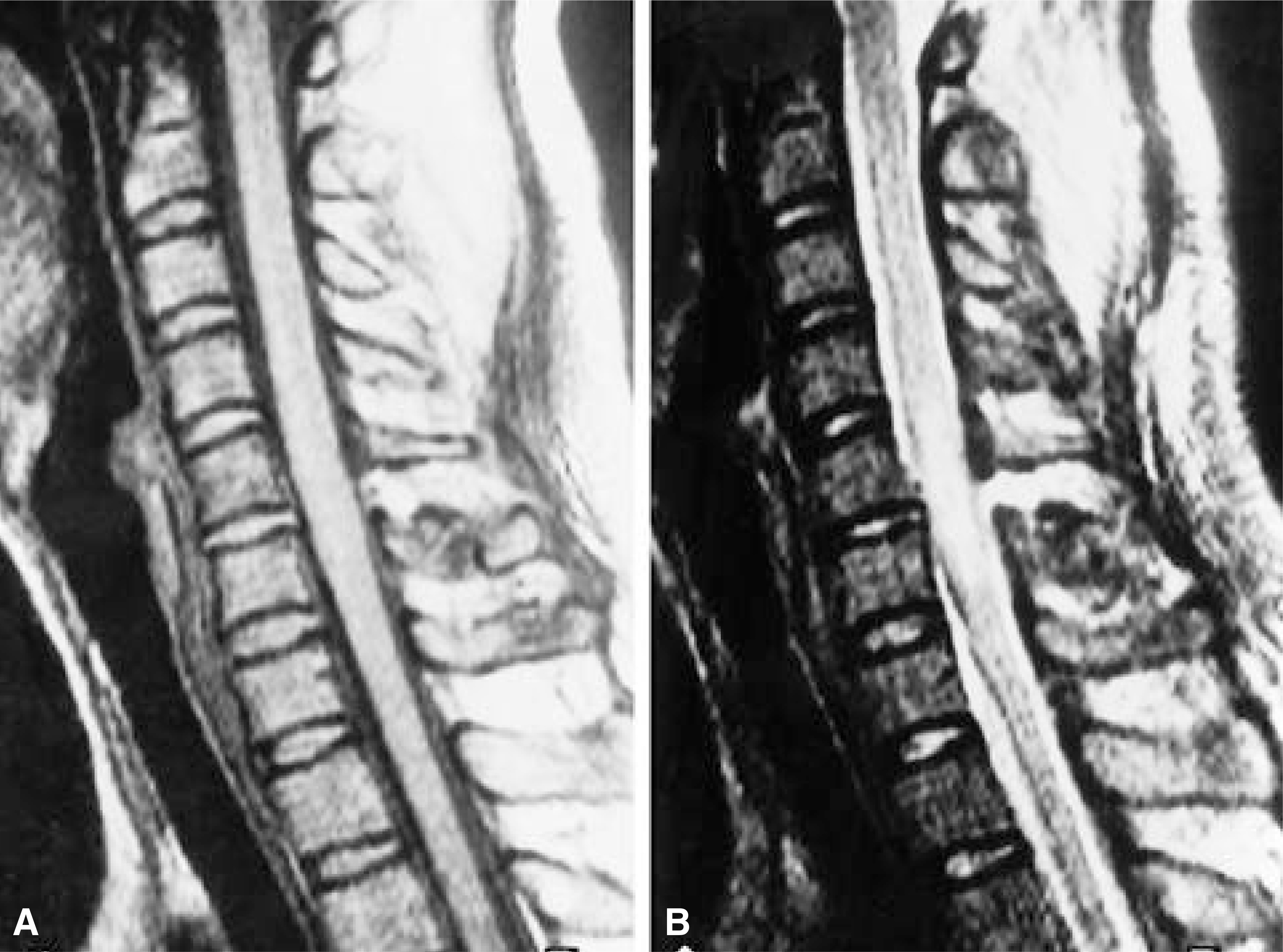
Fig. 1. Spinal cord edema; 35-year-old woman presenting Frankel grade D; MRI was performed 5 days from injury. Fig. A. Sagittal midline T1-weighted image demonstrated isointense signal in the cord opposite C5-6 associated with fracture C6 spinous process. Fig. B. T2-weighted image shows diffuse high signal in cord from C5 to C6

Fig. 2. Spinal cord swelling; 44-year-old man presenting Franke B; MRI was performed at 2 days from injury. Fig. A. Sagittal middle T1-weighted image demonstrates C4/5 subluxation and isointense signal with cord distension opposite C4-5. Fig. B. T2-weighted image shows diffuse hyperintense signal in cord from C2 to C6 with C4-5 interspinous and supraspinatus ligament rupture.

Fig. 3. Spinal cord contusion. 25-year-old man presenting Frankel B; MRI was performed 2 days from injury. Fig. A. Sagittal midline T1-weighted image demonstrates focal cord compression and isointense signal in the cord opposite C5 associated compression fracture of C5 vertebral body. Fig. B. T2-weighted image shows diffuse heterogenous hyperintense signal from C3 to C6.

Fig. 4. 41-year-old man presenting Frankel C; MRI was performed 2 days from injury. Fig. A. Preop T1-weighted image demonstrates isointense signal in cord opposite C4-5 associated with C4-5 disc rupture which migrate to the C4 vertebral body and C5-6 disc protrusion. Fig. B. Preop T2-weighted image shows diffuse hyperintense signal change in cord from C4 to C6. Fig. C. Postop T1-weighted image which performed 19 month from injury, was demonstrates focal hypointense signal change in the cord at C4-5 level with anterior cervical disectomy and fusion at C4-5-6. Fig. D. Postop T2-weighted image shows focal hyperintense signal in cord opposite C4-5.
Reference
-
1). Adam E. Flanders, Claire M. Spettell, David P. Fried-man, Ralph J. Marino and Gerald J. Herbison. The relationship between the functional abilities of patients with cervical spinal cord injury and the severity of dam -age revealed by MR Imaging. Am J Neuroradiol. 20:926–934. 1999.2). Bedbrook G. Recovery of spinal cord function. Paraplegia. 18:315–323. 1980.
Article3). Bondurant FJ, Cortler HB, Kulkarni MV. Acute spinal cord injury: a study using physical examination and magnetic resonance imaging. Spine. 15:161–168. 1990.4). Chakeres DW, Flickinger F, Bresnahan JC, Beattie MS, Weis KL, Miller C, Stokes BT. MR imiging of acute spinal cord trauma. AJNR. 8:5–10. 1987.5). Crozier KS, Graziani V, Ditunno JF, Herbison GJ. Spinal cord injury: Prognosis for ambulation based on sensory examination in patients who are initially motor complete. Arch Phys Med Rehabil. 72:119–121. 1991.6). Ducker TB, Russo GL, Bellegarrique R, Lucas JT. Complete sensorymotor paralysis after cord injury: Mortality, recovery, and therapeutic implication. Trauma. 19:837–840. 1979.7). Flanders AE, Schaefer DM, Doan HT, MIshkin MM, Gonzalez CF, Northrup BE. Acute cervical spine trauma: Correlation of MR imaging findings w ith degree of neurological deficit. Radiology. 177:25–33. 1990.8). Frankel HL, Hancock DO, Hyslop G, Melzak J, Michaelis LS, Ungar GH, Vernon JD, Walsh JJ. The value of postural reduction in the initial management of closed injuries of the spine with paraplegia and tetraplegia. Paralplegia. 7:179–192. 1969.
Article9). Gomori JM, Grossman RI, Goldberg HI, Zimmerman RA, Bilaniuk LT. Intracranial hematomas: imaging by high-field MR, Radiology. 157:87–93. 1985.10). Harris P, Karmi MZ, McClemont E, Matlhoko D, Paul KS. The prognosis of patients sustaining severe cervical spine injury. Paraplegia. 18:324–330. 1990.11). Hesselink JR. Spine imaging: History, achievements, remaining frontiers. Am J Roentgenol. 140:1223–1229. 1998.
Article12). Kalfas H, Wilverger J, Goldberg A, Prostoko KR. Magnetic resonance imaging in acute spinal cord trauma. 23:295–299. 1988.13). Kulkarni MV, Bondurant FJ, Rose SL, Narayana P A. 1.5 Tesla magnetic resonance imaging of acute spinal trauma, Radiographics. 8:1059–1082. 1988.14). Kulkarni MV, McArdle CB, Kopanicky D, Miner M, Cortler HB, Lee KF. A cute spinal cord injury: MR imaging at 1.5T. Radiology. 164:837–843. 1987.15). Lazar RB, Yakony GM, Ortolano D, Heinemann AW, Perlow E, Lovell L, Meyer PR. Prediction of functional outcome by motor capability after spinal cord injury, Arch Phys Med Rehabil. 70:819–822. 1989.16). Maynard FM, Reynolds GG, Fountain S, Wilmot C, Hamilton R. Neurological prognosis after traumatic quadriplegia: Three-year experience of California Regional Spinal Injury Care System, J Neurosurg. 50:611–616. 1979.17). Morry S, Brian M, and Oliver Hennessy: Prediction of neurologic outcome in acute spinal cord injury. The Role of CT and MR. AJNR. 13:1597–1608. 1982.18). Nathan RS, Douglas J, Quint , Nayna Ratel B.A., Hannah S, Stephen MP. Emergency magnetic resonance imaging of cervical spinal cord injuries: Clinical Correlation and prognosis. Neurosurgery. 44:785–793. 1999.19). Nordquist L. The sagittal diameter of the spinal cord and subarachnoid space in different age groups. Acta Radiol Diagn. 227:1–86. 1964.20). Schaefer DM, Flanders AE, Osterholm JL, Northrup BE. Prognositc significance of magnetic resonance imaging in the acute phase of cervical spine injury. J Neurosurg. 76:218–223. 1992.21). Sherman JL, Nassaux PY, Citrim CM. Measurements of the normal cervical spinal cord on MR imaging. AJNR. 11:369–372. 1990.22). Sato T, Kokubun S, Rijal K P, Ojima T, Moriari N, Hashimoto M, Hyodo H, Oonuma H. Prognosis of cervical spinal cord injury in correlation with magnetic resonance imaging. Paraplegia. 32:81–85. 1994.
Article23). Weirich SD, Cotler HB, Narayana DA, Hazle JD, Jackson EF, Coupe KJ, Mcdonald CL, Langford LA, Harris JH. Histopathologic correlation of magnetic resonance imaging signal pattern in spinal cord injury model. Spine. 15:630–638. 1990.
- Full Text Links
-
- Actions
-
Cited
- CITED
-
- Close
- Share
-
- Similar articles
-
- Magnetic Resonance Imaging of Traumatic Cervical Injury
- Solitary Osteochondroma of the Atlas with Cervical Cord Compresion: Case Report
- Neurologic Recovery According to Early Magnetic Resonance Imaging Findings in Traumatic Cervical Spinal Cord Injuries
- Vertebral Artery Dissection Presenting with Acute Infarction in Cervical Spinal Cord and Cerebellum
- Morphological Analysis of the Cervical Spinal Cord, Dural Tube, and Spinal Canal by Magnetic Resonance Imaging in Normal Korean Adults
