The Age-related Microstructural Changes of the Cortical Gray and White Matter Ratios on T2-, FLAIR and T1-weighted MR Images
- Affiliations
-
- 1Department of Radiology, College of Medicine, Dong-A University, Busan, Korea. sschoi317@yahoo.co.kr
- KMID: 2041225
- DOI: http://doi.org/10.13104/jksmrm.2011.15.1.32
Abstract
- PURPOSE
The purpose of this study was to investigate the microstructural changes according to aging on the thickness and signal intensity (SI) of the cortical gray matter (GM) and white matter (WM) on the T2-, fluid-attenuated inversion recovery (FLAIR) and T1-weighted MR images in normal subjects.
MATERIALS AND METHODS
The 10, 20, 30, 40, 50, 60, 70, 80 and 90 year age groups of men and women (each 10 individuals) who underwent routine brain MRI, including the T2-, FLAIR and T1-weighted images, were selected for this study. We measured the thickness and the SI of the cortical GM and WM at the postcentral gyrus, which has an even thickness at the level of centrum semiovale, on the axial scans and we calculated the mean values of the thickness ratio of the gray/white matter (TRGW) and the signal intensity ratio of the gray/white matter (SRGW), and we compared the ratios of each age group.
RESULTS
On the T2-weighted images, the TRGWs were 0.81 and 0.79 at the age of 10 and they were 0.73 and 0.71 at the age of 90 in the men and women, respectively. So, the GM thickness was decreased more than the WM thickness was with aging. On the FLAIR images, the TRGWs were 1.09 and 1.00 at the age of 10 and they were 1.11 and 0.95 at the age of 70 in the men and women, respectively. On the T1-weighted images, the TRGWs were 0.66 and 0.80 at the age of 10, and the ratio was changed to 0.90 and 0.78 at the age of 90 in the men and women, respectively. On the T2-weighted image, the SRGWs were 1.53 and 1.43 at the age of 10, and they were 1.23 and 1.27 at the age of 90 in the men and women, respectively. On the FLAIR images, the SRGWs were 1.23 and 1.25 at the age of 10 and they were 1.06 and 1.05 at the age of 90 in the men and women, respectively. On the T1-weighted images, the SRGWs were 0.86 and 0.85 at the age of 10, and they were 0.90 and 0.87 at the age of 90 in the men and women, respectively.
CONCLUSION
We suggest that the age-related microstructural changes of the thickness and the SI of the cortical GM and WM on the T2-, FLAIR and T1-weighted images are unique, and so this knowledge will be helpful to differentiate neurodegenerative disease from normal aging of the brain.
Figure
-
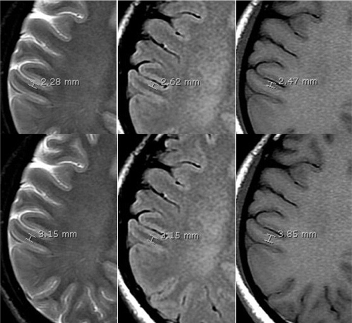
Fig. 1 Example of measuring the thicknesses of the cortical gray matter (upper row) and white matter (bottom row) on the T2-weighted (left), FLAIR (middle) and T1-weighted (right) images.
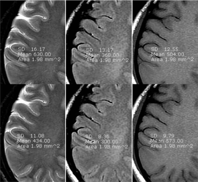
Fig. 2 Example of measuring the signal intensities of the cortical gray matter (upper row) and white matter (bottom row) on the T2-weighted (left), FLAIR (middle) and T1-weighted (right) images.
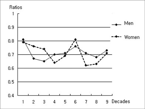
Fig. 3 The slopes of the thickness ratios on the T2-weighted images. The ratios decreased slightly with aging.
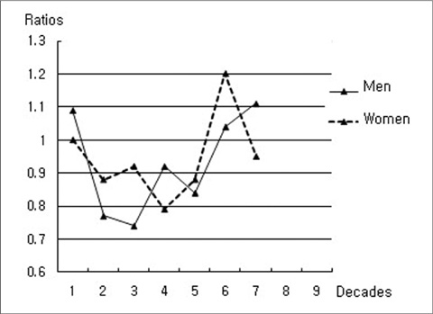
Fig. 4 The slopes of the thickness ratios on the FLAIR images. The ratios showed the lowest value at the age of 30 in men and at the age of 40 in women. The measurements of the GM and WM thickness separately were not possible in the aged brain, and so the 80 and 90 year old age groups were excluded.

Fig. 5 The slopes of the thickness ratios on the T1-weighted images. The ratios increased slightly with aging.
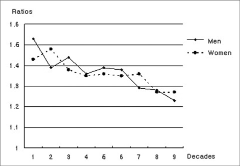
Fig. 6 The slopes of the signal intensity ratios on the T2-weighted images. The contrast between the GM and WM was decreased with aging.
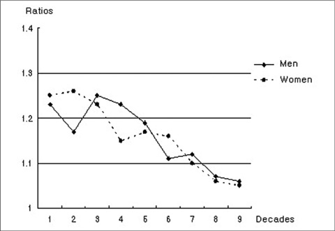
Fig. 7 The slopes of the signal intensity ratios on the FLAIR images. The ratios were decreased to 1 with aging, which means the contrast between the GM and WM had almost disappeared.
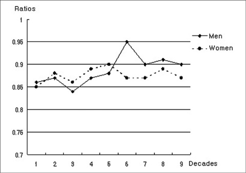
Fig. 8 The slopes of the signal intensity ratios on the T1-weighted images. The ratios were slightly increased with aging due to the decreased contrast between the GM and WM.
Reference
-
1. Fjell AM, Walhovd KB, Fennema-Notestine C, et al. One-year brain atrophy evident in healthy aging. J Neurosci. 2009. 29:15223–15231.2. Salat DH, Buckner RI, Snyder AZ, et al. Thinning of the cerebral cortex in aging. Cerebral Cortex. 2004. 14:721–730.3. Salat DH, Lee SY, Van der Kouwe AJ, Greve DN, Fischl B, Rosas HD. Age-associated alterations in cortical gray and white matter signal intensity and gray to white matter contrast. Neuro Image. 2009. 48:21–28.4. Coffey CE, Lucke JF, Saxton JA, et al. Sex differences in brain aging: a quantitative magnetic resonance imaging study. Arch Neurol. 1998. 55:169–179.5. Benedetti B, Charil A, Rovaris M, et al. Influence of aging on brain gray and white matter changes assessed by conventional. MT, and DT MRI. Neurology. 2006. 66:535–539.6. Rovaris M, Iannucci G, Cercignani M, et al. Age-related changes in conventional, magnetization transfer, and diffusion-tensor MR imaging findings: study with whole-brain tissue histogram analysis. Radiology. 2003. 227:731–738.7. Maezawa M, Seki T, Imura S, Akiyama K, Takikawa I, Yuasa Y. Magnetic resonance signal intensity ratio of gray/white matter in children. Quantitative assessment in developing brain. Brain Dev. 1993. 15:198–120.8. Kang EJ, Choi SS, Jo JH, Kang MJ, Ha DH, Nam KJ. Chronological changes of the signal intensities of white matter on the FLAIR Images of Infants. J Korean Soc Radiol. 2010. 62:411–419.9. Ashikaga R, Araki Y, Ono Y, Nishimura Y, Ishida O. Appearance of normal brain maturation on Fluid-attenuated inversion-recovery(FLAIR) MR images. AJNR Am J Neuroradiol. 1999. 20:427–431.10. Nagel BJ, Medina KL, Yoshii J, Schweinsburg AD, Moadab I, Tapert SF. Age-related changes in prefrontal white matter volume across adolescence. Neuroreport. 2006. 17:1427–1431.11. Fjell AM, Westlye LT, Amlien I, et al. Minute effects of sex on the aging brain: a multisample magnetic resonance imaging study of healthy aging and Alzheimer's disease. J Neurosci. 2009. 29:8774–8783.
- Full Text Links
-
- Actions
-
Cited
- CITED
-
- Close
- Share
-
- Similar articles
-
- Chronological Changes of the Signal Intensities of White Matter on the FLAIR Images of Infants
- Contrast-enhanced Fast Fluid-attenuated Inversion Recovery MR Imaging in Patients with Brain Tumors
- Relative Signal Intensity Changes of Frontal and Occipital White Matters on T2 Weighted Axial MR Image: Correlation with Age
- A Study on CT Attenuation and MR Signal Intensity of Protein Solution
- An experimental study on MRI signal intensity of albumin solution
