Association Factors for CT Angiography Spot Sign and Hematoma Growth in Korean Patients with Acute Spontaneous Intracerebral Hemorrhage : A Single-Center Cohort Study
- Affiliations
-
- 1Department of Neurosurgery, Incheon St. Mary's Hospital, College of Medicine, The Catholic University of Korea, Incheon, Korea. argus3620@gmail.com
- KMID: 2018079
- DOI: http://doi.org/10.3340/jkns.2014.56.4.295
Abstract
OBJECTIVE
This study was conducted to clarify the association factors and clinical significance of the CT angiography (CTA) spot sign and hematoma growth in Korean patients with acute intracerebral hemorrhage (ICH).
METHODS
We retrospectively collected the data of 287 consecutive patients presenting with acute ICH who arrived within 12 hours of ictus. Baseline clinical and radiological characteristics as well as the mortality rate within one month were assessed. A binary logistic regression was conducted to obtain association factors for the CTA spot sign and hematoma growth.
RESULTS
We identified a CTA spot sign in 40 patients (13.9%) and hematoma growth in 78 patients (27.2%). An elapsed time to CT scan of less than 3 hours (OR, 5.14; 95% CI, 1.76-15.02; p=0.003) was associated with the spot sign. A CTA spot sign (OR, 5.70; 95% CI, 2.70-12.01; p<0.001), elevated alanine transaminase (GPT) level >40 IU (OR, 2.01; 95% CI, 1.01-4.01; p=0.047), and an international normalized ratio > or =1.8 or warfarin medication (OR, 5.64; 95% CI, 1.29-24.57; p=0.021) were independent predictors for hematoma growth. Antiplatelet agent medication (OR, 4.92; 95% CI, 1.31-18.50; p=0.019) was significantly associated with hematoma growth within 6 hours of ictus.
CONCLUSION
As previous other populations, CTA spot sign was a strong predictor for hematoma growth especially in hyper-acute stage of ICH in Korea. Antithrombotics medication might also be associated with hyper-acute hematoma growth. In our population, elevated GPT was newly identified as a predictor for hematoma growth and its effect for hematoma growth is necessary to be confirmed through a further research.
Keyword
MeSH Terms
Figure
-
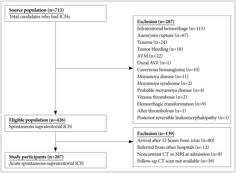
Fig. 1 Flowchart of patient selection. ICH : intracerebral hemorrhage, AVM : arteriovenous malformation, AVF : arteriovenous fistula.
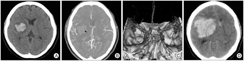
Fig. 2 Representative CT angiography (CTA) images obtained from a 50-year-old woman with a history of daily alcohol consumption who arrived at our hospital within 42 minutes of ictus. A : Non-contrast brain CT showing an acute hemorrhage in the right putamen. B : A small enhanced focus of contrast in the middle of the hematoma (black arrow). C : CTA image showing no intracranial vessel abnormality. D : Hematoma growth identified on a brain CT image acquired immediately after her neurologic deterioration 4 hours after arrival at our hospital.

Fig. 3 Among the 78 patients with hematoma growth, a comparison of the time from ictus to hematoma growth between the spot sign-positive group (n=25, 32.1%) and the spot sign-negative group (n=53, 67.9%). A Mann-Whitney U test was used to compare the two groups.
Cited by 1 articles
-
Management and Outcome of Spontaneous Cerebellar Hemorrhage
Jungin Han, Ho Kook Lee, Tack Geun Cho, Jae Gon Moon, Chang Hyun Kim
J Cerebrovasc Endovasc Neurosurg. 2015;17(3):185-193. doi: 10.7461/jcen.2015.17.3.185.
Reference
-
1. Anderson CS, Huang Y, Arima H, Heeley E, Skulina C, Parsons MW, et al. Effects of early intensive blood pressure-lowering treatment on the growth of hematoma and perihematomal edema in acute intracerebral hemorrhage : the Intensive Blood Pressure Reduction in Acute Cerebral Haemorrhage Trial (INTERACT). Stroke. 2010; 41:307–312. PMID: 20044534.
Article2. Becker KJ, Baxter AB, Bybee HM, Tirschwell DL, Abouelsaad T, Cohen WA. Extravasation of radiographic contrast is an independent predictor of death in primary intracerebral hemorrhage. Stroke. 1999; 30:2025–2032. PMID: 10512902.
Article3. Brouwers HB, Biffi A, McNamara KA, Ayres AM, Valant V, Schwab K, et al. Apolipoprotein E genotype is associated with CT angiography spot sign in lobar intracerebral hemorrhage. Stroke. 2012; 43:2120–2125. PMID: 22621984.
Article4. Brouwers HB, Goldstein JN, Romero JM, Rosand J. Clinical applications of the computed tomography angiography spot sign in acute intracerebral hemorrhage : a review. Stroke. 2012; 43:3427–3432. PMID: 23132779.
Article5. Cucchiara B, Messe S, Sansing L, Kasner S, Lyden P. CHANT Investigators. Hematoma growth in oral anticoagulant related intracerebral hemorrhage. Stroke. 2008; 39:2993–2996. PMID: 18703803.
Article6. Davis SM, Broderick J, Hennerici M, Brun NC, Diringer MN, Mayer SA, et al. Hematoma growth is a determinant of mortality and poor outcome after intracerebral hemorrhage. Neurology. 2006; 66:1175–1181. PMID: 16636233.
Article7. de Gans K, de Haan RJ, Majoie CB, Koopman MM, Brand A, Dijkgraaf MG, et al. PATCH : platelet transfusion in cerebral haemorrhage : study protocol for a multicentre, randomised, controlled trial. BMC Neurol. 2010; 10:19. PMID: 20298539.8. Delcourt C, Huang Y, Arima H, Chalmers J, Davis SM, Heeley EL, et al. Hematoma growth and outcomes in intracerebral hemorrhage : the INTERACT1 study. Neurology. 2012; 79:314–319. PMID: 22744655.
Article9. Delgado Almandoz JE, Yoo AJ, Stone MJ, Schaefer PW, Goldstein JN, Rosand J, et al. Systematic characterization of the computed tomography angiography spot sign in primary intracerebral hemorrhage identifies patients at highest risk for hematoma expansion : the spot sign score. Stroke. 2009; 40:2994–3000. PMID: 19574553.
Article10. Delgado Almandoz JE, Yoo AJ, Stone MJ, Schaefer PW, Oleinik A, Brouwers HB, et al. The spot sign score in primary intracerebral hemorrhage identifies patients at highest risk of in-hospital mortality and poor outcome among survivors. Stroke. 2010; 41:54–60. PMID: 19910545.
Article11. Demchuk AM, Dowlatshahi D, Rodriguez-Luna D, Molina CA, Blas YS, Dzialowski I, et al. Prediction of haematoma growth and outcome in patients with intracerebral haemorrhage using the CT-angiography spot sign (PREDICT) : a prospective observational study. Lancet Neurol. 2012; 11:307–314. PMID: 22405630.
Article12. Dowlatshahi D, Wasserman JK, Momoli F, Petrcich W, Stotts G, Hogan M, et al. Evolution of computed tomography angiography spot sign is consistent with a site of active hemorrhage in acute intracerebral hemorrhage. Stroke. 2014; 45:277–280. PMID: 24178918.
Article13. Ederies A, Demchuk A, Chia T, Gladstone DJ, Dowlatshahi D, Bendavit G, et al. Postcontrast CT extravasation is associated with hematoma expansion in CTA spot negative patients. Stroke. 2009; 40:1672–1676. PMID: 19286577.
Article14. Emlet LL, Crippen D. Early recombinant activated factor VII for intracerebral hemorrhage reduced hematoma growth and mortality, while improving functional outcomes. Crit Care. 2006; 10:304. PMID: 16420665.15. Feigin VL, Lawes CM, Bennett DA, Barker-Collo SL, Parag V. Worldwide stroke incidence and early case fatality reported in 56 population-based studies : a systematic review. Lancet Neurol. 2009; 8:355–369. PMID: 19233729.
Article16. Flaherty ML, Woo D, Haverbusch M, Sekar P, Khoury J, Sauerbeck L, et al. Racial variations in location and risk of intracerebral hemorrhage. Stroke. 2005; 36:934–937. PMID: 15790947.
Article17. Flibotte JJ, Hagan N, O'Donnell J, Greenberg SM, Rosand J. Warfarin, hematoma expansion, and outcome of intracerebral hemorrhage. Neurology. 2004; 63:1059–1064. PMID: 15452298.
Article18. Goldstein JN, Fazen LE, Snider R, Schwab K, Greenberg SM, Smith EE, et al. Contrast extravasation on CT angiography predicts hematoma expansion in intracerebral hemorrhage. Neurology. 2007; 68:889–894. PMID: 17372123.
Article19. Hallevi H, Abraham AT, Barreto AD, Grotta JC, Savitz SI. The spot sign in intracerebral hemorrhage : the importance of looking for contrast extravasation. Cerebrovasc Dis. 2010; 29:217–220. PMID: 20029193.
Article20. Huttner HB, Schellinger PD, Hartmann M, Köhrmann M, Juettler E, Wikner J, et al. Hematoma growth and outcome in treated neurocritical care patients with intracerebral hemorrhage related to oral anticoagulant therapy : comparison of acute treatment strategies using vitamin K, fresh frozen plasma, and prothrombin complex concentrates. Stroke. 2006; 37:1465–1470. PMID: 16675739.
Article21. Kim HC, Kang DR, Nam CM, Hur NW, Shim JS, Jee SH, et al. Elevated serum aminotransferase level as a predictor of intracerebral hemorrhage : Korea medical insurance corporation study. Stroke. 2005; 36:1642–1647. PMID: 16020763.
Article22. Kim HC, Oh SM, Pan WH, Ueshima H, Gu D, Chuang SY, et al. Association between alanine aminotransferase and intracerebral hemorrhage in East Asian populations. Neuroepidemiology. 2013; 41:131–138. PMID: 23880909.
Article23. Kim J, Smith A, Hemphill JC 3rd, Smith WS, Lu Y, Dillon WP, et al. Contrast extravasation on CT predicts mortality in primary intracerebral hemorrhage. AJNR Am J Neuroradiol. 2008; 29:520–525. PMID: 18065505.
Article24. Kim KH. Predictors of 30-day mortality and 90-day functional recovery after primary intracerebral hemorrhage : hospital based multivariate analysis in 585 patients. J Korean Neurosurg Soc. 2009; 45:341–349. PMID: 19609417.
Article25. Li N, Wang Y, Wang W, Ma L, Xue J, Weissenborn K, et al. Contrast extravasation on computed tomography angiography predicts clinical outcome in primary intracerebral hemorrhage : a prospective study of 139 cases. Stroke. 2011; 42:3441–3446. PMID: 21980207.
Article26. Martí-Fàbregas J, Borrell M, Silva Y, Delgado-Mederos R, Martínez-Ramírez S, de Juan-Delago M, et al. Hemostatic proteins and their association with hematoma growth in patients with acute intracerebral hemorrhage. Stroke. 2010; 41:2976–2978. PMID: 21115948.
Article27. Mayer SA, Brun NC, Begtrup K, Broderick J, Davis S, Diringer MN, et al. Efficacy and safety of recombinant activated factor VII for acute intracerebral hemorrhage. N Engl J Med. 2008; 358:2127–2137. PMID: 18480205.
Article28. Mendelow AD, Gregson BA, Rowan EN, Murray GD, Gholkar A, Mitchell PM, et al. Early surgery versus initial conservative treatment in patients with spontaneous supratentorial lobar intracerebral haematomas (STICH II) : a randomised trial. Lancet. 2013; 382:397–408. PMID: 23726393.
Article29. Moussouttas M, Malhotra R, Fernandez L, Maltenfort M, Holowecki M, Delgado J, et al. Role of antiplatelet agents in hematoma expansion during the acute period of intracerebral hemorrhage. Neurocrit Care. 2010; 12:24–29. PMID: 19844810.
Article30. Naidech AM, Jovanovic B, Liebling S, Garg RK, Bassin SL, Bendok BR, et al. Reduced platelet activity is associated with early clot growth and worse 3-month outcome after intracerebral hemorrhage. Stroke. 2009; 40:2398–2401. PMID: 19443791.
Article31. Park HS, Kang MJ, Huh JT. Recent epidemiological trends of stroke. J Korean Neurosurg Soc. 2008; 43:16–20. PMID: 19096539.
Article32. Park SY, Kong MH, Kim JH, Kang DS, Song KY, Huh SK. Role of 'spot sign' on CT angiography to predict hematoma expansion in spontaneous intracerebral hemorrhage. J Korean Neurosurg Soc. 2010; 48:399–405. PMID: 21286475.
Article33. Radmanesh F, Falcone GJ, Anderson CD, Battey TW, Ayres AM, Vashkevich A, et al. Risk factors for computed tomography angiography spot sign in deep and lobar intracerebral hemorrhage are shared. Stroke. 2014; 45:1833–1835. PMID: 24876264.
Article34. Rodriguez-Luna D, Piñeiro S, Rubiera M, Ribo M, Coscojuela P, Pagola J, et al. Impact of blood pressure changes and course on hematoma growth in acute intracerebral hemorrhage. Eur J Neurol. 2013; 20:1277–1283. PMID: 23647568.
Article35. Sansing LH, Messe SR, Cucchiara BL, Cohen SN, Lyden PD, Kasner SE, et al. Prior antiplatelet use does not affect hemorrhage growth or outcome after ICH. Neurology. 2009; 72:1397–1402. PMID: 19129506.
Article36. Takeda R, Ogura T, Ooigawa H, Fushihara G, Yoshikawa S, Okada D, et al. A practical prediction model for early hematoma expansion in spontaneous deep ganglionic intracerebral hemorrhage. Clin Neurol Neurosurg. 2013; 115:1028–1031. PMID: 23245855.
Article37. Toyoda K, Okada Y, Minematsu K, Kamouchi M, Fujimoto S, Ibayashi S, et al. Antiplatelet therapy contributes to acute deterioration of intracerebral hemorrhage. Neurology. 2005; 65:1000–1004. PMID: 16217049.
Article38. van Asch CJ, Luitse MJ, Rinkel GJ, van der Tweel I, Algra A, Klijn CJ. Incidence, case fatality, and functional outcome of intracerebral haemorrhage over time, according to age, sex, and ethnic origin : a systematic review and meta-analysis. Lancet Neurol. 2010; 9:167–176. PMID: 20056489.
Article
- Full Text Links
-
- Actions
-
Cited
- CITED
-
- Close
- Share
-
- Similar articles
-
- Role of 'Spot Sign' on CT Angiography to Predict Hematoma Expansion in Spontaneous Intracerebral Hemorrhage
- The Spot Sign Predicts Hematoma Expansion, Outcome, and Mortality in Patients with Primary Intracerebral Hemorrhage
- Which Emphasizing Factors Are Most Predictive of Hematoma Expansion in Spot Sign Positive Intracerebral Hemorrhage?
- Spot Sign on Initial Brain Computed Tomography Angiography Source Image to Predict Large Hemorrhagic Transformation after Middle Cerebral Artery Infarction
- Leak Sign on Dynamic-SusceptibilityContrast Magnetic Resonance Imaging in Acute Intracerebral Hemorrhage
