Which Emphasizing Factors Are Most Predictive of Hematoma Expansion in Spot Sign Positive Intracerebral Hemorrhage?
- Affiliations
-
- 1Department of Neurosurgery, Wonju Severance Christian Hospital, Yonsei University, Wonju, Korea. nsojw@yonsei.ac.kr
- 2Department of Neurosurgery, Severance Hospital, Yonsei University, Seoul, Korea.
- KMID: 1956508
- DOI: http://doi.org/10.3340/jkns.2014.56.2.86
Abstract
OBJECTIVE
The spot sign is related with the risk of hematoma expansion in spontaneous intracerebral hemorrhage (ICH). However, not all spot sign positive patients undergo hematoma expansion. Thus, the present study investigates the specific factors enhancing the spot sign positivity in predicting hematoma expansion.
METHODS
We retrospectively studied 316 consecutive patients who presented between March 2009 to March 2011 with primary ICH and whose initial computed tomography brain angiography (CTA) was performed at our Emergency Department. Of these patients, 47 primary ICH patients presented spot signs in their CTA. We classified these 47 patients into two groups based on the presence of hematoma expansion then analyzed them with the following factors : gender, age, initial systolic blood pressure, history of anti-platelet therapy, volume and location of hematoma, time interval from symptom onset to initial CTA, spot sign number, axial dimension, and Hounsfield Unit (HU) of spot signs.
RESULTS
Of the 47 spot sign positive patients, hematoma expansion occurred in 26 patients (55.3%) while the remaining 21 (44.7%) showed no expansion. The time intervals from symptom onset to initial CTA were 2.42+/-1.24 hours and 3.69+/-2.57 hours for expansion and no expansion, respectively (p=0.031). The HU of spot signs were 192.12+/-45.97 and 151.10+/-25.14 for expansion and no expansion, respectively (p=0.001).
CONCLUSIONS
The conditions of shorter time from symptom onset to initial CTA and higher HU of spot signs are the emphasizing factors for predicting hematoma expansion in spot sign positive patients.
MeSH Terms
Figure
-
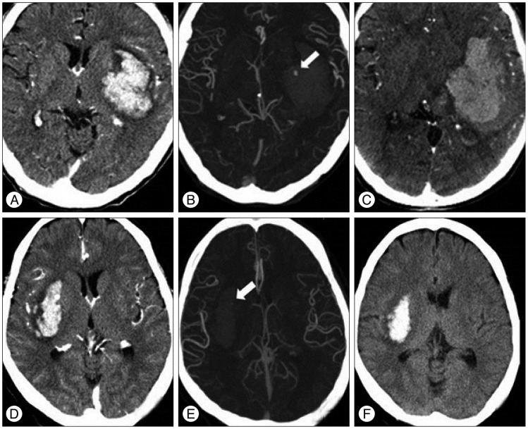
Fig. 1 CT angiograms of two spot sign positive patients with and without hematoma expansion. Brain CT scans demonstrate basal ganglia hemorrhage. The upper row CT images of a 47-year-old female display hematoma expansion 2 hours after presentation with a spot sign of 3.63 mm and a density of 194 Hounsfield units (HU) (A, B, and C). The lower row CT images of a 61-year-old male show no hematoma expansion 24 hours after presentation with a spot sign of 1.21 mm and a density of 117 HU (D, E, and F).
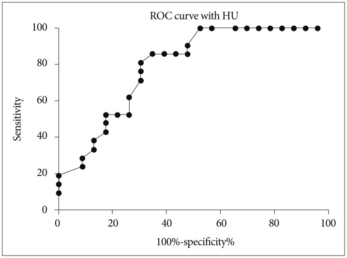
Fig. 2 Receiver operating characteristic (ROC) curve using the Hounsfield unit (HU). The cut-off HU value was 171 [sensitivity, 65.4%; specificity, 85.7%; area under the ROC curve (AUC)=0.7919]. If the cut-off HU value was set higher than 191.0, sensitivity was 100%.
Cited by 1 articles
-
Dual-Energy CT Angiography Improves Accuracy of Spot Sign for Predicting Hematoma Expansion in Intracerebral Hemorrhage
Michaël T.J. Peeters, Kim J.D. de Kort, Rik Houben, Wouter J.P. Henneman, Robert J. van Oostenbrugge, Julie Staals, Alida A. Postma
J Stroke. 2021;23(1):82-90. doi: 10.5853/jos.2020.03531.
Reference
-
1. Balami JS, Buchan AM. Complications of intracerebral haemorrhage. Lancet Neurol. 2012; 11:101–118. PMID: 22172625.
Article2. Broderick JP, Brott TG, Tomsick T, Barsan W, Spilker J. Ultra-early evaluation of intracerebral hemorrhage. J Neurosurg. 1990; 72:195–199. PMID: 2295917.
Article3. Broderick JP, Diringer MN, Hill MD, Brun NC, Mayer SA, Steiner T, et al. Determinants of intracerebral hemorrhage growth : an exploratory analysis. Stroke. 2007; 38:1072–1075. PMID: 17290026.4. Brott T, Broderick J, Kothari R, Barsan W, Tomsick T, Sauerbeck L, et al. Early hemorrhage growth in patients with intracerebral hemorrhage. Stroke. 1997; 28:1–5. PMID: 8996478.
Article5. Brouwers HB, Biffi A, Ayres AM, Schwab K, Cortellini L, Romero JM, et al. Apolipoprotein E genotype predicts hematoma expansion in lobar intracerebral hemorrhage. Stroke. 2012; 43:1490–1495. PMID: 22535266.
Article6. Brouwers HB, Chang Y, Falcone GJ, Cai X, Ayres AM, Battey TW, et al. Predicting hematoma expansion after primary intracerebral hemorrhage. JAMA Neurol. 2014; 71:158–164. PMID: 24366060.
Article7. Brouwers HB, Falcone GJ, McNamara KA, Ayres AM, Oleinik A, Schwab K, et al. CTA spot sign predicts hematoma expansion in patients with delayed presentation after intracerebral hemorrhage. Neurocrit Care. 2012; 17:421–428. PMID: 22878870.
Article8. Brouwers HB, Goldstein JN, Romero JM, Rosand J. Clinical applications of the computed tomography angiography spot sign in acute intracerebral hemorrhage : a review. Stroke. 2012; 43:3427–3432. PMID: 23132779.
Article9. Brouwers HB, Greenberg SM. Hematoma expansion following acute intracerebral hemorrhage. Cerebrovasc Dis. 2013; 35:195–201. PMID: 23466430.
Article10. Castellanos M, Leira R, Tejada J, Gil-Peralta A, Dávalos A, Castillo J, et al. Predictors of good outcome in medium to large spontaneous supratentorial intracerebral haemorrhages. J Neurol Neurosurg Psychiatry. 2005; 76:691–695. PMID: 15834028.
Article11. Castillo J, Dávalos A, Alvarez-Sabín J, Pumar JM, Leira R, Silva Y, et al. Molecular signatures of brain injury after intracerebral hemorrhage. Neurology. 2002; 58:624–629. PMID: 11865143.
Article12. Chakraborty S, Blacquiere D, Lum C, Stotts G. Dynamic nature of the CT angiographic "spot sign". Br J Radiol. 2010; 83:e216–e219. PMID: 20846980.
Article13. Davis SM, Broderick J, Hennerici M, Brun NC, Diringer MN, Mayer SA, et al. Hematoma growth is a determinant of mortality and poor outcome after intracerebral hemorrhage. Neurology. 2006; 66:1175–1181. PMID: 16636233.
Article14. Delgado Almandoz JE, Yoo AJ, Stone MJ, Schaefer PW, Goldstein JN, Rosand J, et al. Systematic characterization of the computed tomography angiography spot sign in primary intracerebral hemorrhage identifies patients at highest risk for hematoma expansion : the spot sign score. Stroke. 2009; 40:2994–3000. PMID: 19574553.
Article15. Dowlatshahi D, Demchuk AM, Flaherty ML, Ali M, Lyden PL, Smith Ee, et al. Defining hematoma expansion in intracerebral hemorrhage : relationship with patient outcomes. Neurology. 2011; 76:1238–1244. PMID: 21346218.
Article16. Dowlatshahi D, Wasserman JK, Momoli F, Petrcich W, Stotts G, Hogan M, et al. Evolution of computed tomography angiography spot sign is consistent with a site of active hemorrhage in acute intracerebral hemorrhage. Stroke. 2014; 45:277–280. PMID: 24178918.
Article17. Ederies A, Demchuk A, Chia T, Gladstone DJ, Dowlatshahi D, Bendavit G, et al. Postcontrast CT extravasation is associated with hematoma expansion in CTA spot negative patients. Stroke. 2009; 40:1672–1676. PMID: 19286577.
Article18. Gazzola S, Aviv RI, Gladstone DJ, Mallia G, Li V, Fox AJ, et al. Vascular and nonvascular mimics of the CT angiography "spot sign" in patients with secondary intracerebral hemorrhage. Stroke. 2008; 39:1177–1183. PMID: 18292380.
Article19. Greenberg CH, Frosch MP, Goldstein JN, Rosand J, Greenberg SM. Modeling intracerebral hemorrhage growth and response to anticoagulation. PLoS One. 2012; 7:e48458. PMID: 23119028.
Article20. Huttner HB, Schellinger PD, Hartmann M, Köhrmann M, Juettler E, Wikner J, et al. Hematoma growth and outcome in treated neurocritical care patients with intracerebral hemorrhage related to oral anticoagulant therapy : comparison of acute treatment strategies using vitamin K, fresh frozen plasma, and prothrombin complex concentrates. Stroke. 2006; 37:1465–1470. PMID: 16675739.
Article21. Huttner HB, Steiner T, Hartmann M, Köhrmann M, Juettler E, Mueller S, et al. Comparison of ABC/2 estimation technique to computer-assisted planimetric analysis in warfarin-related intracerebral parenchymal hemorrhage. Stroke. 2006; 37:404–408. PMID: 16373654.
Article22. Huynh TJ, Demchuk AM, Dowlatshahi D, Gladstone DJ, Krischek O, Kiss A, et al. Spot sign number is the most important spot sign characteristic for predicting hematoma expansion using first-pass computed tomography angiography : analysis from the PREDICT study. Stroke. 2013; 44:972–977. PMID: 23444309.
Article23. Huynh TJ, Flaherty ML, Gladstone DJ, Broderick JP, Demchuk AM, Dowlatshahi D, et al. Multicenter accuracy and interobserver agreement of spot sign identification in acute intracerebral hemorrhage. Stroke. 2014; 45:107–112. PMID: 24281226.
Article24. Kim J, Smith A, Hemphill JC 3rd, Smith WS, Lu Y, Dillon WP, et al. Contrast extravasation on CT predicts mortality in primary intracerebral hemorrhage. AJNR Am J Neuroradiol. 2008; 29:520–525. PMID: 18065505.
Article25. Mayer SA. Ultra-early hemostatic therapy for primary intracerebral hemorrhage : a review. Can J Neurol Sci. 2005; 32(Suppl 2):S31–S37. PMID: 16450806.26. Park SY, Kong MH, Kim JH, Kang DS, Song KY, Huh SK. Role of 'spot sign' on CT angiography to predict hematoma expansion in spontaneous intracerebral hemorrhage. J Korean Neurosurg Soc. 2010; 48:399–405. PMID: 21286475.
Article27. Qureshi AI, Mendelow AD, Hanley DF. Intracerebral haemorrhage. Lancet. 2009; 373:1632–1644. PMID: 19427958.
Article28. Rodríguez-Yáñez M, Castellanos M, Freijo MM, López-Fernández JC, Martí-Fàbregas J, Nombela F, et al. Clinical practice guidelines in intracerebral haemorrhage. Neurologia. 2013; 28:236–249. PMID: 21570742.
Article29. Skidmore CT, Andrefsky J. Spontaneous intracerebral hemorrhage : epidemiology, pathophysiology, and medical management. Neurosurg Clin N Am. 2002; 13:281–288. PMID: 12486918.
Article30. Sun SJ, Gao PY, Sui BB, Hou XY, Lin Y, Xue J, et al. "Dynamic spot sign" on CT perfusion source images predicts haematoma expansion in acute intracerebral haemorrhage. Eur Radiol. 2013; 23:1846–1854. PMID: 23508276.
Article31. van Asch CJ, Luitse MJ, Rinkel GJ, van der Tweel I, Algra A, Klijn CJ. Incidence, case fatality, and functional outcome of intracerebral haemorrhage over time, according to age, sex, and ethnic origin : a systematic review and meta-analysis. Lancet Neurol. 2010; 9:167–176. PMID: 20056489.
Article
- Full Text Links
-
- Actions
-
Cited
- CITED
-
- Close
- Share
-
- Similar articles
-
- Role of 'Spot Sign' on CT Angiography to Predict Hematoma Expansion in Spontaneous Intracerebral Hemorrhage
- The Spot Sign Predicts Hematoma Expansion, Outcome, and Mortality in Patients with Primary Intracerebral Hemorrhage
- Predictors of Hematoma Enlargement in Patients with Spontaneous Intracerebral Hemorrhage Treated with Rapid Administration of Antifibrinolytic Agents and Strict Conservative Management
- Spot Sign on Initial Brain Computed Tomography Angiography Source Image to Predict Large Hemorrhagic Transformation after Middle Cerebral Artery Infarction
- Leak Sign on Dynamic-SusceptibilityContrast Magnetic Resonance Imaging in Acute Intracerebral Hemorrhage
