The Spot Sign Predicts Hematoma Expansion, Outcome, and Mortality in Patients with Primary Intracerebral Hemorrhage
- Affiliations
-
- 1Department of Neurosurgery, Chonbuk National University Hospital and Medical School, Jeonju, Korea. nsjmlee@gmail.com
- KMID: 2018080
- DOI: http://doi.org/10.3340/jkns.2014.56.4.303
Abstract
OBJECTIVE
The purpose of this study was to retrospectively review cases of intracerebral hemorrhage (ICH) medically treated at our institution to determine if the CT angiography (CTA) 'spot sign' predicts in-hospital mortality and clinical outcome at 3 months in patients with spontaneous ICH.
METHODS
We conducted a retrospective review of all consecutive patients who were admitted to the department of neurosurgery. Clinical data of patients with ICH were collected by 2 neurosurgeons blinded to the radiological data and at the 90-day follow-up.
RESULTS
Multivariate logistic regression analysis identified predictors of poor outcome; we found that hematoma location, spot sign, and intraventricular hemorrhage were independent predictors of poor outcome. In-hospital mortality was 57.4% (35 of 61) in the CTA spot-sign positive group versus 7.9% (10 of 126) in the CTA spot-sign negative group. In multivariate logistic analysis, we found that presence of spot sign and presence of volume expansion were independent predictors for the in-hospital mortality of ICH.
CONCLUSION
The spot sign is a strong independent predictor of hematoma expansion, mortality, and poor clinical outcome in primary ICH. In this study, we emphasized the importance of hematoma expansion as a therapeutic target in both clinical practice and research.
Keyword
MeSH Terms
Figure
-
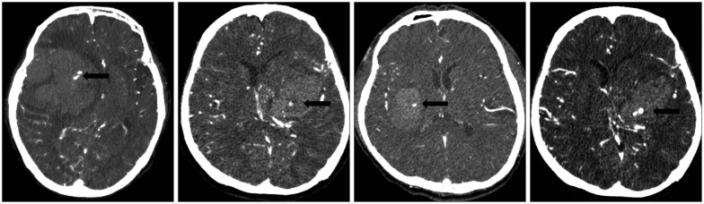
Fig. 1 The appearance of a spot sign on CT angiography in a patient with intracerebral hemorrhage. The spot sign (black arrow) assesses diameter and Hounsfield units. The spot sign is located within the hematoma, has no connection to any outside vessel, and is absent on baseline non-contrast CT.
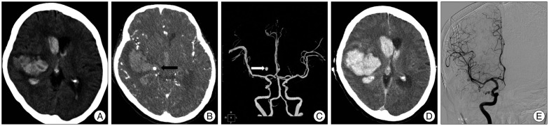
Fig. 2 A : A 61-year-old man underwent imaging 2 hours following onset of left-sided paralysis. NCCT demonstrates a right basal ganglia ICH (34 mL) with associated IVH (19 mL). B : Axial CTA source image in spot windows demonstrates 1 foci of contrast pooling within the ICH with an attenuation 176 HU (arrowheads), consistent with spot signs (a total of 4 spot signs were identified). The largest spot sign measured 5.7 mm in maximum axial dimension and had an attenuation of 245 HU (spot sign score, 4). C : Axial CTA image shows that the spot sign looks like aneurysmal sac. D : Non-contrast CT 4 hours after the baseline CTA demonstrates marked interval expansion of both the ICH (86 mL) and IVH (42 mL). E : Conventional angiographic image demonstrates absence of aneurysmal sac. NCCT : noncontrast CT, ICH : intracerebral hemorrhage, IVH : intraventricular hemorrhage, CTA : CT angiography, HU : Hounsfield units.
Cited by 2 articles
-
Management and Outcome of Spontaneous Cerebellar Hemorrhage
Jungin Han, Ho Kook Lee, Tack Geun Cho, Jae Gon Moon, Chang Hyun Kim
J Cerebrovasc Endovasc Neurosurg. 2015;17(3):185-193. doi: 10.7461/jcen.2015.17.3.185.Dual-Energy CT Angiography Improves Accuracy of Spot Sign for Predicting Hematoma Expansion in Intracerebral Hemorrhage
Michaël T.J. Peeters, Kim J.D. de Kort, Rik Houben, Wouter J.P. Henneman, Robert J. van Oostenbrugge, Julie Staals, Alida A. Postma
J Stroke. 2021;23(1):82-90. doi: 10.5853/jos.2020.03531.
Reference
-
1. Anderson CS, Huang Y, Arima H, Heeley E, Skulina C, Parsons MW, et al. Effects of early intensive blood pressure-lowering treatment on the growth of hematoma and perihematomal edema in acute intracerebral hemorrhage : the Intensive Blood Pressure Reduction in Acute Cerebral Haemorrhage Trial (INTERACT). Stroke. 2010; 41:307–312. PMID: 20044534.
Article2. Anderson CS, Huang Y, Wang JG, Arima H, Neal B, Peng B, et al. Intensive blood pressure reduction in acute cerebral haemorrhage trial (INTERACT) : a randomised pilot trial. Lancet Neurol. 2008; 7:391–399. PMID: 18396107.
Article3. Bhattathiri PS, Gregson B, Prasad KS, Mendelow AD. STICH Investigators. Intraventricular hemorrhage and hydrocephalus after spontaneous intracerebral hemorrhage : results from the STICH trial. Acta Neurochir Suppl. 2006; 65–68. PMID: 16671427.
Article4. Broderick JP, Brott TG, Duldner JE, Tomsick T, Huster G. Volume of intracerebral hemorrhage. A powerful and easy-to-use predictor of 30-day mortality. Stroke. 1993; 24:987–993. PMID: 8322400.
Article5. Brott T, Broderick J, Kothari R, Barsan W, Tomsick T, Sauerbeck L, et al. Early hemorrhage growth in patients with intracerebral hemorrhage. Stroke. 1997; 28:1–5. PMID: 8996478.
Article6. Davis SM, Broderick J, Hennerici M, Brun NC, Diringer MN, Mayer SA, et al. Hematoma growth is a determinant of mortality and poor outcome after intracerebral hemorrhage. Neurology. 2006; 66:1175–1181. PMID: 16636233.
Article7. Delgado Almandoz JE, Yoo AJ, Stone MJ, Schaefer PW, Goldstein JN, Rosand J, et al. Systematic characterization of the computed tomography angiography spot sign in primary intracerebral hemorrhage identifies patients at highest risk for hematoma expansion : the spot sign score. Stroke. 2009; 40:2994–3000. PMID: 19574553.
Article8. Delgado Almandoz JE, Yoo AJ, Stone MJ, Schaefer PW, Oleinik A, Brouwers HB, et al. The spot sign score in primary intracerebral hemorrhage identifies patients at highest risk of in-hospital mortality and poor outcome among survivors. Stroke. 2010; 41:54–60. PMID: 19910545.
Article9. Demchuk AM, Dowlatshahi D, Rodriguez-Luna D, Molina CA, Blas YS, Dzialowski I, et al. Prediction of haematoma growth and outcome in patients with intracerebral haemorrhage using the CT-angiography spot sign (PREDICT) : a prospective observational study. Lancet Neurol. 2012; 11:307–314. PMID: 22405630.
Article10. Flibotte JJ, Hagan N, O'Donnell J, Greenberg SM, Rosand J. Warfarin, hematoma expansion, and outcome of intracerebral hemorrhage. Neurology. 2004; 63:1059–1064. PMID: 15452298.
Article11. Godoy DA, Piñero G, Di Napoli M. Predicting mortality in spontaneous intracerebral hemorrhage : can modification to original score improve the prediction? Stroke. 2006; 37:1038–1044. PMID: 16514104.
Article12. Goldstein JN, Fazen LE, Snider R, Schwab K, Greenberg SM, Smith EE, et al. Contrast extravasation on CT angiography predicts hematoma expansion in intracerebral hemorrhage. Neurology. 2007; 68:889–894. PMID: 17372123.
Article13. Hauser CJ, Boffard K, Dutton R, Bernard GR, Croce MA, Holcomb JB, et al. Results of the CONTROL trial : efficacy and safety of recombinant activated Factor VII in the management of refractory traumatic hemorrhage. J Trauma. 2010; 69:489–500. PMID: 20838118.
Article14. Hemphill JC 3rd, Bonovich DC, Besmertis L, Manley GT, Johnston SC. The ICH score : a simple, reliable grading scale for intracerebral hemorrhage. Stroke. 2001; 32:891–897. PMID: 11283388.15. Kazui S, Naritomi H, Yamamoto H, Sawada T, Yamaguchi T. Enlargement of spontaneous intracerebral hemorrhage. Incidence and time course. Stroke. 1996; 27:1783–1787. PMID: 8841330.
Article16. Kim J, Smith A, Hemphill JC 3rd, Smith WS, Lu Y, Dillon WP, et al. Contrast extravasation on CT predicts mortality in primary intracerebral hemorrhage. AJNR Am J Neuroradiol. 2008; 29:520–525. PMID: 18065505.
Article17. Kothari RU, Brott T, Broderick JP, Barsan WG, Sauerbeck LR, Zuccarello M, et al. The ABCs of measuring intracerebral hemorrhage volumes. Stroke. 1996; 27:1304–1305. PMID: 8711791.
Article18. Leira R, Dávalos A, Silva Y, Gil-Peralta A, Tejada J, Garcia M, et al. Early neurologic deterioration in intracerebral hemorrhage : predictors and associated factors. Neurology. 2004; 63:461–467. PMID: 15304576.
Article19. Mayer SA, Brun NC, Begtrup K, Broderick J, Davis S, Diringer MN, et al. Efficacy and safety of recombinant activated factor VII for acute intracerebral hemorrhage. N Engl J Med. 2008; 358:2127–2137. PMID: 18480205.
Article20. Mayer SA, Brun NC, Begtrup K, Broderick J, Davis S, Diringer MN, et al. Recombinant activated factor VII for acute intracerebral hemorrhage. N Engl J Med. 2005; 352:777–785. PMID: 15728810.
Article21. Mendelow AD, Gregson BA, Fernandes HM, Murray GD, Teasdale GM, Hope DT, et al. Early surgery versus initial conservative treatment in patients with spontaneous supratentorial intracerebral haematomas in the International Surgical Trial in Intracerebral Haemorrhage (STICH) : a randomised trial. Lancet. 2005; 365:387–397. PMID: 15680453.
Article22. Morgenstern LB, Hemphill JC 3rd, Anderson C, Becker K, Broderick JP, Connolly ES Jr, et al. Guidelines for the management of spontaneous intracerebral hemorrhage : a guideline for healthcare professionals from the American Heart Association/American Stroke Association. Stroke. 2010; 41:2108–2129. PMID: 20651276.
Article23. Park SY, Kong MH, Kim JH, Kang DS, Song KY, Huh SK. Role of 'spot sign' on CT angiography to predict hematoma expansion in spontaneous intracerebral hemorrhage. J Korean Neurosurg Soc. 2010; 48:399–405. PMID: 21286475.
Article24. Qureshi AI, Mendelow AD, Hanley DF. Intracerebral haemorrhage. Lancet. 2009; 373:1632–1644. PMID: 19427958.
Article25. Qureshi AI, Palesch YY, Martin R, Novitzke J, Cruz-Flores S, Ehtisham A, et al. Effect of systolic blood pressure reduction on hematoma expansion, perihematomal edema, and 3-month outcome among patients with intracerebral hemorrhage : results from the antihypertensive treatment of acute cerebral hemorrhage study. Arch Neurol. 2010; 67:570–576. PMID: 20457956.
Article26. Rodriguez-Luna D, Rubiera M, Ribo M, Coscojuela P, Piñeiro S, Pagola J, et al. Ultraearly hematoma growth predicts poor outcome after acute intracerebral hemorrhage. Neurology. 2011; 77:1599–1604. PMID: 21998314.
Article27. Silva Y, Leira R, Tejada J, Lainez JM, Castillo J, Dávalos A, et al. Molecular signatures of vascular injury are associated with early growth of intracerebral hemorrhage. Stroke. 2005; 36:86–91. PMID: 15550687.
Article28. Steiner T, Diringer MN, Schneider D, Mayer SA, Begtrup K, Broderick J, et al. Dynamics of intraventricular hemorrhage in patients with spontaneous intracerebral hemorrhage : risk factors, clinical impact, and effect of hemostatic therapy with recombinant activated factor VII. Neurosurgery. 2006; 59:767–773. discussion 773-774. PMID: 17038942.29. Thompson AL, Kosior JC, Gladstone DJ, Hopyan JJ, Symons SP, Romero F, et al. Defining the CT angiography 'spot sign' in primary intracerebral hemorrhage. Can J Neurol Sci. 2009; 36:456–461. PMID: 19650356.
Article
- Full Text Links
-
- Actions
-
Cited
- CITED
-
- Close
- Share
-
- Similar articles
-
- Role of 'Spot Sign' on CT Angiography to Predict Hematoma Expansion in Spontaneous Intracerebral Hemorrhage
- Which Emphasizing Factors Are Most Predictive of Hematoma Expansion in Spot Sign Positive Intracerebral Hemorrhage?
- Leak Sign on Dynamic-SusceptibilityContrast Magnetic Resonance Imaging in Acute Intracerebral Hemorrhage
- Predictors of Hematoma Enlargement in Patients with Spontaneous Intracerebral Hemorrhage Treated with Rapid Administration of Antifibrinolytic Agents and Strict Conservative Management
- Association Factors for CT Angiography Spot Sign and Hematoma Growth in Korean Patients with Acute Spontaneous Intracerebral Hemorrhage : A Single-Center Cohort Study
