J Korean Acad Conserv Dent.
2006 Sep;31(5):359-370. 10.5395/JKACD.2006.31.5.359.
Effects of occlusal load on the stress distribution of four cavity configurations of noncarious cervical lesions: a three-dimensional finite element analysis study
- Affiliations
-
- 1Department of Conservative dentistry, College of Dentistry, Pusan National University, Busan, Korea. bhur@pusan.ac.kr
- 2Department of Mechanical design engineering, College of Engineering, Pusan National University, Busan, Korea.
- KMID: 1986879
- DOI: http://doi.org/10.5395/JKACD.2006.31.5.359
Abstract
- The objective of this study was to investigate the effect of excessive occlusal loading on stress distribution on four type of cervical lesion, using a three dimensional finite element analysis (3D FEA). The extracted maxillary second premolar was scanned serially with Micro-CT. The 3D images were processed by 3D-DOCTOR. ANSYS was used to mesh and analyze 3D FE model. Four different lesion configurations representative of the various types observed clinically for teeth were studied. A static point load of 500N was applied to the buccal and lingual cusp (Load A and B). The principal stresses in lesion apex, and vertical sectioned margin of cervical wall were analyzed. The results were as follows 1. The patterns of stress distribution were similar but the magnitude was different in four types of lesion. 2. The peak stress was observed at mesial corner and also stresses concentrated at lesion apex. 3. The compressive stress under load A and the tensile stress under load B were dominant stress. 4. Under the load, lesion can be increased and harmful to tooth structure unless restored.
Keyword
MeSH Terms
Figure
-
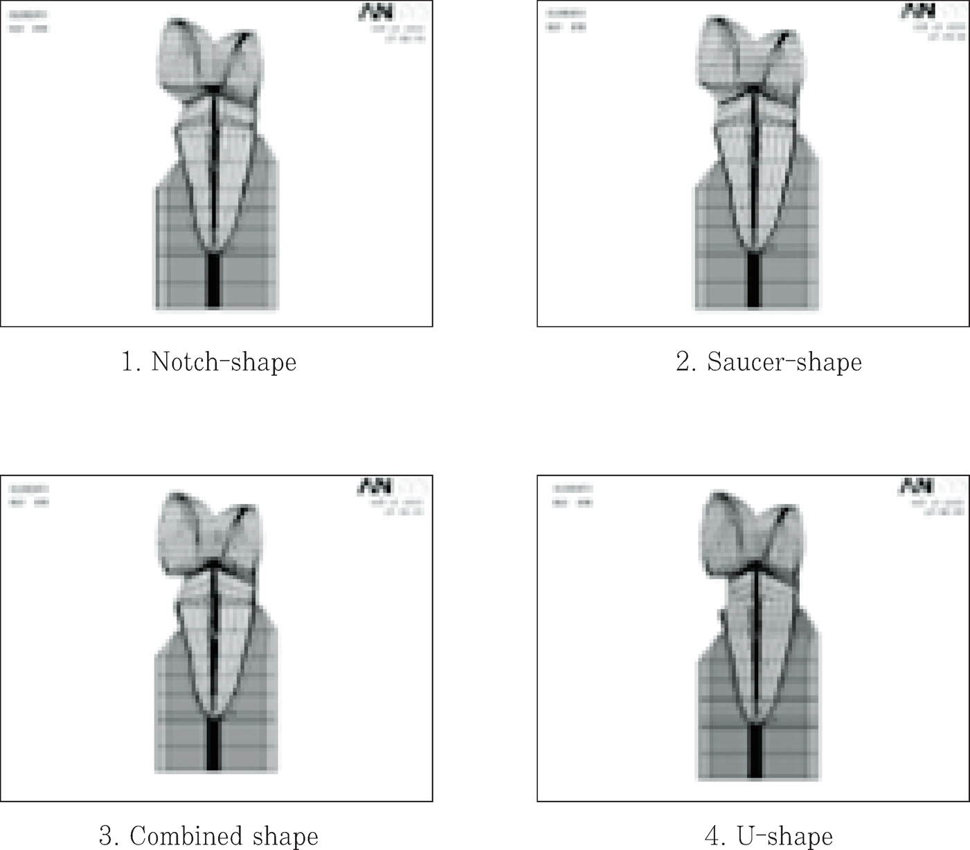
Figure 1. Four types of lesion configurations.
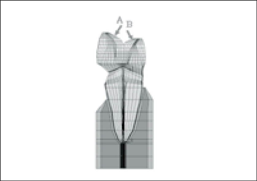
Figure 2. Design of loading conditions.

Figure 3. The Principal stress distribution of four cavities under Load A and B(Upper 2 rows: maximal and minimal principal stress under load A, Lower 2 rows; maximal and minimal principal stress under load B. From left line, notch-shape showed, followed by saucer, combined, U-shape lesion)

Figure 4. Principal stresses of lesion apex of 4 shape lesion.

Figure 5. Vertical distribution of principal stresses at cavity wall in 4 shape lesion. (If the value of one principal stress of an element was positive, the element was determined to be in the tensile condition, if it was negative, the element was determined to be in the compressive condition).
Reference
-
References
1. James D, Linda C, Levitch . How dentists classified and treated non-carious cervical lesions. J Am Dent Assoc. 124(5):46–54. 1993.2. Bradley T, William B Hancock. Examining the prevalence and characteristics of abfraction like cervical lesions in a population of U.S. veterans. J Am Dent Assoc. 132(12):1694–1701. 2001.3. Lee WC, Eakle WS. Stress-induced cervical lesions: Review of advances on the past 10 years. J Prosthet Dent. 75:487–494. 1996.4. Grippo JO. Abfractions: A new classification of hard tissue lesions of teeth. J Esthet Dent. 3(1):14–19. 1991.
Article5. Gallien GS, Kaplan I, Owen BM. A review of noncari-ous dental cervical lesions. Compend Contin Educ Dent. 15(11):1366–1372. 1994.6. Grippo JO, Schreiner S. Attrition, abrasion, corrosion and abfraction revisited-A new perspective on tooth surface lesion. J Am Dent Assoc. 135(8):1109–1117. 2004.7. Rees JS, Hammadeh M. Undermining of enamel as a mechanism of abfraction lesion formation: a finite element study. Eur J Oral Sci. 112:347–352. 2004.
Article8. Radentz WH, Barnes GP, Cutright DE. A survey of factors possibly associated with cervical abrasion of tooth surface. J Periodontol. 47:148–154. 1976.9. Kahn F, Young WG, Shahabi S, Daley TJ. Dental cervical lesions associated with occlusal erosion and attrition. Aust Dent J. 44:176–186. 1999.10. Levitch LC, Bader JD, Shugars DA, Heymann HO. Non-carious cervical lesions. J Dent. 22:195–207. 1994.
Article11. Leinfelder KF. Restoration of abfracted lesions. Compend Contin Educ Dent. 15(4):1396–1400. 1994.12. Park Jk, Hur B, Lee HJ. The effect of configuration on marginal leakage of class 5 restoration. J Kor Acad Cons Dent. 26(2):162–170. 2001.13. Kuroe T, Itoh H, Caputo AA, Konuma M. Biomechanics of cervical tooth structure lesions and their restoration. Quint Int. 31(4):267–274. 2000.14. Grippo JO. Noncarious cervical lesions: The decision to ignore or restore. J Esthet Dent. 4:118–132. 1992.
Article15. Litonjua LA, Andreana S, Patra AK, Cohen RE. An assessment of stress analyses in the theory of abfraction. Biomed Mater Eng. 14:311–321. 2004.16. Rees JS, Jacobsen PH. The effect of cuspal flexure on a buccal Class V restoration: a finite element study. J Dent. 26(4):361–367. 1998.
Article17. Lindehe J, Karring T. Textbook of Clinical Periodontology. 2nd edition. Munksgaard;Copenhagen: p. p19–69. 1989.18. Schroeder HE, Page RC. Periodontal Diseases. 2nd edition. Lea & Fabiger;Philadelphia: p. p 3–52. 1990.19. Rubin C, Krishnamurthy N, Capilouto E, Yi H. Stress analysis of the human tooth using a three-dimensional finite element model. J Dent Res. 62:82–86. 1983.20. Katona TR, Winkler MM. Stress analysis of a bulk-filled class V light-cured composite restoration. J Dent Res. 73(8):1470–1477. 1994.
Article21. Geramy A, Sharafoddin F. Abfraction: 3D analysis by means of the finite method. Quint Int. 34(7):526–533. 2003.22. AW TC, Lepe X, Johnson GH, Mancl L. Characteristics of noncarious cervical lesions: A clinical investigation. J Am Dent Assoc. 133(6):725–733. 2002.23. Widmalm SE, Ericsson SG. Maximal bite force with centric and eccentric load. J Oral Rehabil. 9:445–450. 1982.
Article24. Gibbs CH, Mahan PE, Lundeen HC, Brehnan K, Walsh EK, Holbrook WB. Occlusal forces during chewing and swallowing as measured by sound transmission. J Prosthet Dent. 46:443–449. 1981.
Article25. Caputo AA, Standlee JP. Biomechanics in Clinical Dentistry. Chicago Quintessence Int. 21–27. 1987.26. Ziemiecki TL, Dennison JB, Charbeneau GT. Clinical evaluation of cervical composite resin restorations placed without retention. Oper Dent. 12:27–33. 1987.27. Whitehead SA, Wilson NHF, Watts DC. Demonstration of “Vertical Barreling“using profilometry. Eur J Prosthodont Restor Dent. 7(4):131–134. 1999.28. Browning WD, Brackett WW, Gilpatrick RO. Two-year clinical comparison of a microfilled and hybrid resin based composite in noncarious class V lesions. Oper Dent. 25:46–50. 2000.29. Kubo S, Yokota H, Sata Y, Hayashi Y. The effect of flexural load cycling on the microleakage of cervical resin composites. Oper Dent. 26:451–459. 2001.
- Full Text Links
-
- Actions
-
Cited
- CITED
-
- Close
- Share
-
- Similar articles
-
- The effect of restorative materials on the stress distribution of class V composite resin restorations: a 3D finite element investigation
- Effects of occlusal load on the cervical stress distribution: A three-dimensional finite element study
- Stress distribution of Class V composite resin restorations: A three-dimensional finite element study
- The influence of composite resin restoration on the stress distribution of notch shaped noncarious cervical lesion; A three dimensional finite element analysis study
- The three dimensional finite element analysis of the stress distribution in the three treatment options of implants restorations for the posterior partial edentulism
