Elemental analysis of the fluoride varnish effects on root caries initiation
- Affiliations
-
- 1Department of Conservative Dentistry, Seoul National University School of Dentistry and Dental Research Institute, Seoul, Korea.
- 2Geochronology Team, Korea Basic Science Institute, Daejeon, Korea.
- 3Department of Dental Laboratory Science and Engineering, Korea University College of Health Science, Seoul, Korea.
- 4Clinic for Persons with Disabilities, Seoul National University Dental Hospital, Dental Research Institute, Seoul National University School of Dentistry, Seoul, Korea. juhchang@snu.ac.kr
- KMID: 1986691
- DOI: http://doi.org/10.5395/JKACD.2011.36.4.290
Abstract
OBJECTIVES
The usage of fluoride varnish for a moderate to low caries-risk group has not been well validated. This study aimed to evaluate the preventive and therapeutic efficacies of fluoride varnish on the initiated root caries.
MATERIALS AND METHODS
Ten premolars were sectioned into quarters, further divided into two windows, one of which was painted with Fluor Protector (1,000 ppm fluoride, Ivoclar Vivadent). An initial lesion with a well-preserved surface layer was produced by pH cycling. Scanned line analysis using energy dispersive spectrometry determined the weight percentages of Ca and P in the demineralized layer. Scanning Electron microscopy and confocal laser scanning microscopy (CLSM) evaluated the varnish-applied root surfaces.
RESULTS
The mean lesion depth (SD) was 12.3 (2.6) microm (single cycling) and 19.6 (3.8) microm (double cycling). Double cycling extended the lesion depth, but induced no more mineral loss than single cycling (p < 0.05). The mean weight percentages of Ca and P between groups with and without varnish were not significantly different (p < 0.05). A CLSM showed varnish remained within 15 microm of the surface layer.
CONCLUSIONS
When a mild acid challenge initiated root tissue demineralization, the application of low-concentration fluoride varnish did not influence the lesion depth or the mineral composition of the subsurface lesion.
Keyword
MeSH Terms
Figure
-

Figure 1 Specimen preparation. (a) The tooth was embedded in acrylic resin followed by crown removal. The coronal root was sectioned into quarters. (b) Two 4 mm × 1 mm windows were created by nail varnish coating. Fluoride varnish was applied to one window (v), while the other window (o) had no varnish application. (c) Specimens were embedded in epoxy resin. (d) The epoxy resin block was horizontally cross-sectioned in the midline of specimens and polished to expose the observation surface. The dark arrows were drawn on the surface of the specimen to distinguish the varnish applied windows (v) from the no-varnish applied windows (o).
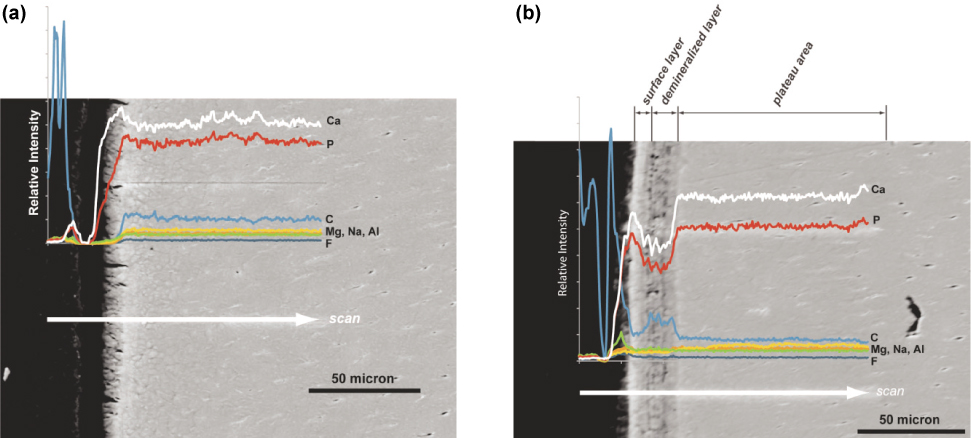
Figure 2 SEM images of 2 specimens produced from a single tooth (×600). Elemental analysis along the scan line illustrates the differentiated elemental composition. (a) Group Ao. No demineralization band appeared. (b) Group Do. Repeated demineralization bands were produced from a double pH cycling. Note that Ca and P levels dropped in the demineralized layer. C (carbon) level was elevated at the same area. It was assumed that diamond particles of polishing paste were impregnated in a porous structure. Other elements were detected by a small amount, which was at an insignificant level.

Figure 3 Two specimens from a single tooth in the thick cementum group (×600). Dark arrows indicate a junction between cementum (Cm) and dentin. While the sound specimen (group Ao) (a) shows no sign of demineralization, the varnish-applied and pH-cycled specimen (group Cv) (b) has the repeating bands of demineralization and remineralization in the outermost cementum layer (white arrow).
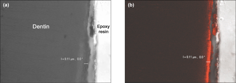
Figure 4 CLSM presents the cross-sectional image of the varnish impregnated root surface (×800). (a) Differential interference contrast image shows the varnish applied root surface without a fluorescent profile. (b) Laser fluorescence image shows the varnish layer as a band of red fluorescence. In the upper middle, free remnant of varnish was detached from the dentin and the superficially infiltrated varnish remained in the surface.
Cited by 2 articles
-
How to design in situ studies: an evaluation of experimental protocols
Young-Hye Sung, Hae-Young Kim, Ho-Hyun Son, Juhea Chang
Restor Dent Endod. 2014;39(3):164-171. doi: 10.5395/rde.2014.39.3.164.Evaluation of release of fluoride from dental varnishes marketed in Korea
Han-Na Kim, Myung-Su Jeong, Se-Yeon Kim, Jin-Bom Kim, Seung-Hwa Jeong
J Korean Acad Oral Health. 2014;38(3):131-137. doi: 10.11149/jkaoh.2014.38.3.131.
Reference
-
1. Heijnsbroek M, Paraskevas S, Van der Weijden GA. Fluoride interventions for root caries: a review. Oral Health Prev Dent. 2007. 5:145–152.2. Hoppenbrouwers PM, Driessens FC, Borggreven JM. The mineral solubility of human tooth roots. Arch Oral Biol. 1987. 32:319–322.
Article3. Beltran-Aguilar ED, Goldstein JW, Lockwood SA. Fluoride varnishes. A review of their clinical use, cariostatic mechanism, efficacy and safety. J Am Dent Assoc. 2000. 131:589–596.4. Strohmenger L, Brambilla E. The use of fluoride varnishes in the prevention of dental caries: a short review. Oral Dis. 2001. 7:71–80.
Article5. Petersson LG, Twetman S, Dahlgren H, Norlund A, Holm AK, Nordenram G, Lagerlöf F, Söder B, Källestål C, Mejàre I, Axelsson S, Lingström P. Professional fluoride varnish treatment for caries control: a systematic review of clinical trials. Acta Odontol Scand. 2004. 62:170–176.
Article6. Weintraub JA. Fluoride varnish for caries prevention: comparisons with other preventive agents and recommendations for a community-based protocol. Spec Care Dentist. 2003. 23:180–186.
Article7. Damen JJ, Buijs MJ, ten Cate JM. Fluoride-dependent formation of mineralized layers in bovine dentin during demineralization in vitro. Caries Res. 1998. 32:435–440.
Article8. Herkstroter FM, Witjes M, Arends J. Demineralization of human dentine compared with enamel in a pH-cycling apparatus with a constant composition during de- and remineralization periods. Caries Res. 1991. 25:317–322.
Article9. Ettinger RL, Olson RJ, Wefel JS, Asmussen C. In vitro evaluation of topical fluorides for overdenture abutments. J Prosthet Dent. 1997. 78:309–314.10. Hong L, Watkins CA, Ettinger RL, Wefel JS. Effect of topical fluoride and fluoride varnish on in vitro root surface lesions. Am J Dent. 2005. 18:182–187.11. Ole Fejerskov EK, Edwina Kidd, Benten Yvad, Vibeke Baelum. Dental caries: the disease and its clinical management. 2008. 2nd ed. Oxford, UK: Wiley-Blackwell.12. Arnold WH, Cerman M, Neuhaus K, Gaengler P. Volumetric assessment and quantitative element analysis of the effect of fluoridated milk on enamel demineralization. Arch Oral Biol. 2003. 48:467–473.
Article13. Rehder Neto FC, Maeda FA, Turssi CP, Serra MC. Potential agents to control enamel caries-like lesions. J Dent. 2009. 37:786–790.
Article14. Hara AT, Queiroz CS, Freitas PM, Giannini M, Serra MC, Cury JA. Fluoride release and secondary caries inhibition by adhesive systems on root dentine. Eur J Oral Sci. 2005. 113:245–250.
Article15. Kawasaki K, Ruben J, Tsuda H, Huysmans MC, Takagi O. Relationship between mineral distributions in dentine lesions and subsequent remineralization in vitro. Caries Res. 2000. 34:395–403.
Article16. Rex T, Kharbanda OP, Petocz P, Darendeliler MA. Physical properties of root cementum: Part 4. Quantitative analysis of the mineral composition of human premolar cementum. Am J Orthod Dentofacial Orthop. 2005. 127:177–185.
Article17. Lee C, Darling CL, Fried D. Polarization-sensitive optical coherence tomographic imaging of artificial demineralization on exposed surfaces of tooth roots. Dent Mater. 2009. 25:721–728.
Article18. Arends J, Christoffersen J. Nature and role of loosely bound fluoride in dental caries. J Dent Res. 1990. 69(Spec No):601–605. discussion 634-636.
Article19. Wefel JS. Root caries histopathology and chemistry. Am J Dent. 1994. 7:261–265.20. Smith PW, Preston KP, Higham SM. Development of an in situ root caries model. A. In vitro investigations. J Dent. 2005. 33:253–267.21. Arends J, Duschner H, Ruben JL. Penetration of varnishes into demineralized root dentine in vitro. Caries Res. 1997. 31:201–205.
Article22. Monticelli F, Osorio R, Tay FR, Sadek FT, Ferrari M, Toledano M. Resistance to thermo-mechanical stress of different coupling agents used as intermediate layer in resin-fiber post bonds. Am J Dent. 2007. 20:416–420.23. Scholtanus JD, Schuthof J, Arends J. Influence of fluoridating varnishes on dentine in vitro. Caries Res. 1986. 20:65–70.
Article
- Full Text Links
-
- Actions
-
Cited
- CITED
-
- Close
- Share
-
- Similar articles
-
- Effect of Silver Diamine Fluoride and Sodium Fluoride Varnish on Remineralization in Artificially Induced Enamel Caries: An in vitro Study
- Remineralization ability of fluoride varnish containing tricalcium phosphate by time
- In vitro fluoride release from five different fluoride varnishes
- Comparison of the remineralization effect of newly-developed fluoride agents according to the depth of early carious lesions
- Application of Fluoride for Dental Caries Prevention in Older Adults with Dry Mouth: a Clinical Review
