Micro-computed tomographic evaluation of single-cone obturation with three sealers
- Affiliations
-
- 1University of New England College of Dental Medicine, Portland, ME, USA
- 2Division of Endodontology, University of Connecticut School of Dental Medicine, Farmington, CT, USA
- 3Department of Clinical Sciences, University of Texas Southwestern Medical Center, Dallas, TX, USA
- 4Augusta University Dental College of Georgia, Augusta, GA, USA
- KMID: 2548069
- DOI: http://doi.org/10.5395/rde.2021.46.e25
Abstract
Objectives
This study used micro-computed tomography (µCT) to compare voids and interfaces in single-cone obturation among AH Plus, EndoSequence BC, and prototype surface pre-reacted glass ionomer (S-PRG) sealers and to determine the percentage of sealer contact at the dentin and gutta-percha (GP) interfaces.
Materials and Methods
Fifteen single-rooted human teeth were shaped using ProTaper NEXT size X5 rotary files using 2.5% NaOCl irrigation. Roots were obturated with a singlecone ProTaper NEXT GP point X5 with AH Plus, EndoSequence BC, or prototype S-PRG sealer (n = 5/group).
Results
The volumes of GP, sealer, and voids were measured in the region of 0–2, 2–4, 4–6, and 6–8 mm from the apex, using image analysis of sagittal µCT scans. GP volume percentages were: AH Plus (75.5%), EndoSequence BC (87.3%), and prototype S-PRG (94.4%). Sealer volume percentages were less: AH Plus (14.3%), EndoSequence BC (6.8%), and prototype S-PRG (4.6%). Void percentages were AH Plus (10.1%), EndoSequence BC (5.9%), and prototype S-PRG (1.0%). Dentin-sealer contact ratios of AH Plus, EndoSequence BC, and prototype S-PRG groups were 82.4% ± 6.8%, 71.6% ± 25.3%, and 70.2% ± 9.4%, respectively. GP-sealer contact ratios of AH Plus, EndoSequence BC, and prototype S-PRG groups were 65.6% ± 29.1%, 80.7% ± 25.8%, and 87.0% ± 8.6%, respectively.
Conclusions
Prototype S-PRG sealer created a low-void obturation, similar to EndoSequence BC sealer with similar dentin-sealer contact (> 70%) and GP-sealer contact (> 80%). Prototype S-PRG sealer presented comparable filling quality to EndoSequence BC sealer.
Figure
-
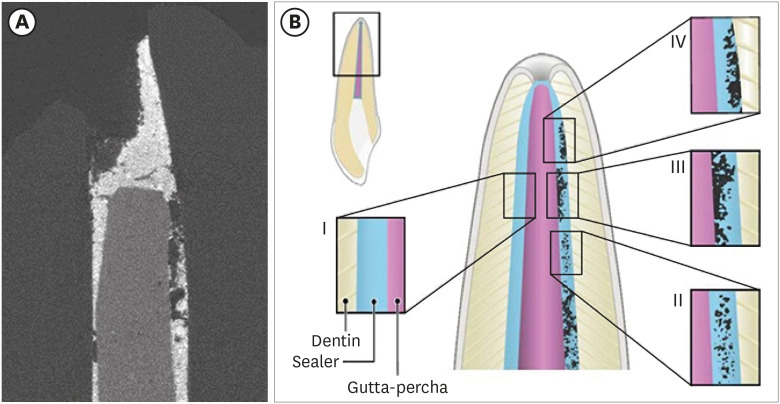
Figure 1 A sample micro-computed tomography image and schematic of 4 contact modes in endodontic obturation.(A) High resolution computed tomography sagittal image of tooth obturated by a gutta-percha (GP) point (center), AH Plus sealer (light grey), areas unfilled by AH Plus sealer (darkest grey), and dentin (dark grey). (B) Schematic of 4 contact modes in endodontic obturation.Type I: perfect contact mode (both the dentin-sealer and GP-sealer interface), type II: sealer porosity mode, type III: dentin-sealer interface contact mode (a perfect contact is present at the dentin-sealer interface, but pores exist at the GP-sealer interface), type IV: GP-sealer interface contact mode (pores exist at the dentin-sealer interface, but there is a perfect contact at the GP-sealer interface).
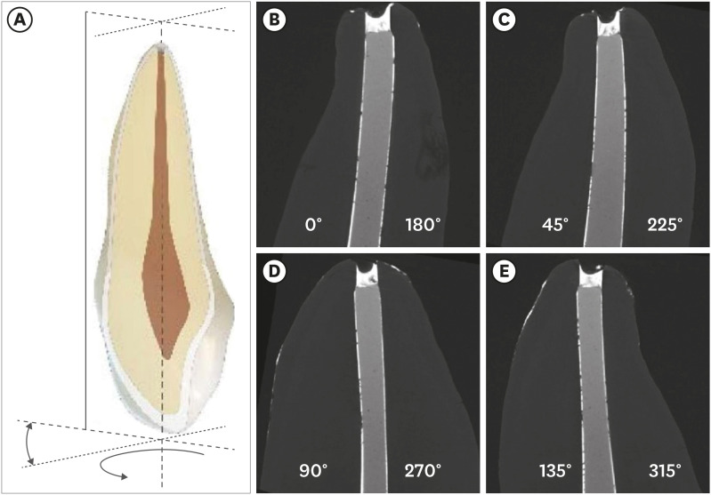
Figure 2 An innovative approach to rotate an image around the central axis of the tooth for interface observation. (A) Sample 2-D sagittal view from 3-D rotation of tooth around central axis. (B) Digital rotation of tooth around central axis every 45°. (B) 0°, (C) rotation 45°, (D) rotation 90°, (E) rotation 135°. Each image provides 2 views of sealer contact with dentin and 2 with gutta-percha (GP). For example, 1 image provides 2 slides, such as 0° (left) and 180° (right) in (B). The dentin-sealer and the GP-sealer interface contacts were evaluated every 100 μm (30 layers) from the apical tip of the GP, to evaluate voids (0: open) or contact (1: contact) on both sides.
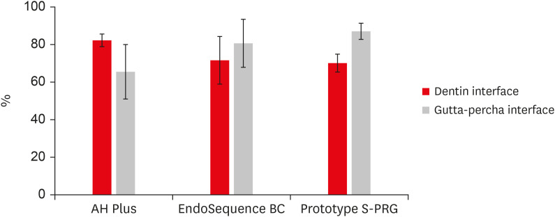
Figure 3 Average contact percentage at sealer interfaces for apical 3 mm (from gutta-percha [GP] tip) among 3 sealers.
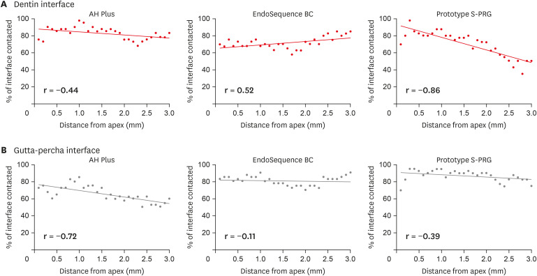
Figure 4 Contact percentage at sealer interface vs. distance from apex.
Cited by 1 articles
-
Calcium-doped zinc oxide nanocrystals as an innovative intracanal medicament: a pilot study
Gabriela Leite de Souza, Thamara Eduarda Alves Magalhães, Gabrielle Alves Nunes Freitas, Nelly Xiomara Alvarado Lemus, Gabriella Lopes de Rezende Barbosa, Anielle Christine Almeida Silva, Camilla Christian Gomes Moura
Restor Dent Endod. 2022;47(4):e38. doi: 10.5395/rde.2022.47.e38.
Reference
-
1. Sjogren U, Hagglund B, Sundqvist G, Wing K. Factors affecting the long-term results of endodontic treatment. J Endod. 1990; 16:498–504. PMID: 2084204.
Article2. Ng YL, Mann V, Rahbaran S, Lewsey J, Gulabivala K. Outcome of primary root canal treatment: systematic review of the literature -- Part 2. Influence of clinical factors. Int Endod J. 2008; 41:6–31. PMID: 17931388.
Article3. Komabayashi T, Colmenar D, Cvach N, Bhat A, Primus C, Imai Y. Comprehensive review of current endodontic sealers. Dent Mater J. 2020; 39:703–720. PMID: 32213767.
Article4. Lacey S, Pitt Ford TR, Watson TF, Sherriff M. A study of the rheological properties of endodontic sealers. Int Endod J. 2005; 38:499–504. PMID: 16011766.
Article5. Schäfer E, Schrenker C, Zupanc J, Bürklein S. Percentage of gutta-percha filled areas in canals obturated with cross-linked gutta-percha core-carrier systems, single-cone and lateral compaction technique. J Endod. 2016; 42:294–298. PMID: 26679718.
Article6. Ray HA, Trope M. Periapical status of endodontically treated teeth in relation to the technical quality of the root filling and the coronal restoration. Int Endod J. 1995; 28:12–18. PMID: 7642323.
Article7. Kataoka H, Yoshioka T, Suda H, Imai Y. Dentin bonding and sealing ability of a new root canal resin sealer. J Endod. 2000; 26:230–235. PMID: 11199725.
Article8. Zhou HM, Shen Y, Zheng W, Li L, Zheng YF, Haapasalo M. Physical properties of 5 root canal sealers. J Endod. 2013; 39:1281–1286. PMID: 24041392.
Article9. Chybowski EA, Glickman GN, Patel Y, Fleury A, Solomon E, He J. Clinical outcome of non-surgical root canal treatment using a single-cone technique with endosequence bioceramic sealer: a retrospective analysis. J Endod. 2018; 44:941–945. PMID: 29606401.
Article10. Miki S, Kitagawa H, Kitagawa R, Kiba W, Hayashi M, Imazato S. Antibacterial activity of resin composites containing surface pre-reacted glass-ionomer (S-PRG) filler. Dent Mater. 2016; 32:1095–1102. PMID: 27417376.
Article11. Yassen GH, Huang R, Al-Zain A, Yoshida T, Gregory RL, Platt JA. Evaluation of selected properties of a new root repair cement containing surface pre-reacted glass ionomer fillers. Clin Oral Investig. 2016; 20:2139–2148.
Article12. Miyaji H, Mayumi K, Miyata S, Nishida E, Shitomi K, Hamamoto A, Tanaka S, Akasaka T. Comparative biological assessments of endodontic root canal sealer containing surface pre-reacted glass-ionomer (S-PRG) filler or silica filler. Dent Mater J. 2020; 39:287–294. PMID: 31776316.
Article13. Kusaka Y. Influence of root canal sealer containing S-PRG filler on osteogenesis in tibia of rats. J Meikai Dent Med. 2018; 47:139–147.14. Keleş A, Alcin H, Kamalak A, Versiani MA. Micro-CT evaluation of root filling quality in oval-shaped canals. Int Endod J. 2014; 47:1177–1184. PMID: 24527697.15. Celikten B, Uzuntas CF, Orhan AI, Orhan K, Tufenkci P, Kursun S, Demiralp KÖ. Evaluation of root canal sealer filling quality using a single-cone technique in oval shaped canals: an in vitro micro-CT study. Scanning. 2016; 38:133–140. PMID: 26228657.
Article16. Jain S, Adhikari HD. Scanning electron microscopic evaluation of marginal adaptation of AH-plus, GuttaFlow, and RealSeal at apical one-third of root canals - part I: dentin-sealer interface. J Conserv Dent. 2018; 21:85–89. PMID: 29628654.17. Adhikari HD, Jain S. Scanning electron microscopic evaluation of marginal adaptation of AH-Plus, GuttaFlow, and RealSeal at apical one-third of root canals - part II: core-sealer interface. J Conserv Dent. 2018; 21:90–94. PMID: 29628655.18. Mohammadian F, Farahanimastary F, Dibaji F, Kharazifard MJ. Scanning electron microscopic evaluation of the sealer-dentine interface of three sealers. Iran Endod J. 2017; 12:38–42. PMID: 28179922.19. Eltair M, Pitchika V, Hickel R, Kühnisch J, Diegritz C. Evaluation of the interface between gutta-percha and two types of sealers using scanning electron microscopy (SEM). Clin Oral Investig. 2018; 22:1631–1639.
Article20. Polineni S, Bolla N, Mandava P, Vemuri S, Mallela M, Gandham VM. Marginal adaptation of newer root canal sealers to dentin: a SEM study. J Conserv Dent. 2016; 19:360–363. PMID: 27563187.
Article21. Ray H, Seltzer S. A new glass ionomer root canal sealer. J Endod. 1991; 17:598–603. PMID: 1820422.
Article22. McMichen FR, Pearson G, Rahbaran S, Gulabivala K. A comparative study of selected physical properties of five root-canal sealers. Int Endod J. 2003; 36:629–635. PMID: 12950578.
Article23. Lee JK, Kwak SW, Ha JH, Lee W, Kim HC. Physicochemical properties of epoxy resin-based and bioceramic-based root canal sealers. Bioinorg Chem Appl. 2017; 2017:2582849. PMID: 28210204.
Article24. Carrotte P. Endodontics: Part 8. Filling the root canal system. Br Dent J. 2004; 197:667–672. PMID: 15592542.
Article25. Naseri M, Kangarlou A, Khavid A, Goodini M. Evaluation of the quality of four root canal obturation techniques using micro-computed tomography. Iran Endod J. 2013; 8:89–93. PMID: 23922567.26. Wolf M, Küpper K, Reimann S, Bourauel C, Frentzen M. 3D analyses of interface voids in root canals filled with different sealer materials in combination with warm gutta-percha technique. Clin Oral Investig. 2014; 18:155–161.
Article27. Schäfer E, Köster M, Bürklein S. Percentage of gutta-percha-filled areas in canals instrumented with nickel-titanium systems and obturated with matching single cones. J Endod. 2013; 39:924–928. PMID: 23791265.
Article28. Moeller L, Wenzel A, Wegge-Larsen AM, Ding M, Kirkevang LL. Quality of root fillings performed with two root filling techniques. An in vitro study using micro-CT. Acta Odontol Scand. 2013; 71:689–696. PMID: 23145468.
Article
- Full Text Links
-
- Actions
-
Cited
- CITED
-
- Close
- Share
-
- Similar articles
-
- A micro-computed tomographic study of remaining filling materials of two bioceramic sealers and epoxy resin sealer after retreatment
- Bacterial leakage and micro-computed tomography evaluation in round-shaped canals obturated with bioceramic cone and sealer using matched single cone technique
- Micro-computed tomographic evaluation of a new system for root canal filling using calcium silicatebased root canal sealers
- Retreatability of two endodontic sealers, EndoSequence BC Sealer and AH Plus: a micro-computed tomographic comparison
- Perspective of endodontic sealers based on calcium silicate
