J Korean Acad Conserv Dent.
2005 Jan;30(1):7-15. 10.5395/JKACD.2005.30.1.007.
Comparison of apical seal with or without the use of dentin adhesive system
- Affiliations
-
- 1Department of Conservative Dentistry, Division of Dentistry, Graduate school of KyungHee University, Korea. gwchoi@khu.ac.kr
- KMID: 1986991
- DOI: http://doi.org/10.5395/JKACD.2005.30.1.007
Abstract
- The purpose of this study was to compare the sealing ability of root canal obturation with or without the use of dentin adhesive system. Forty extracted human teeth with one canal were selected and decoronated. The teeth were divided into two Groups. The obturation procedure of Group 1 was the same as that of Group 2 with the exception of dentin adhesive system. Group 2 were obturated with dentin adhesive system, AH-26, and gutta-percha. After obturation, the teeth were immersed in methylene blue solution for 84 hours. The teeth were sectioned horizontally at 1.5 mm (Level 1), 2.0 mm (Level 2), 2.5 mm (Level 3) from the root apex using a low speed microtome. Distance of dye-penetrated surface and total dentinal surface were measured using SigmaScan Pro 5.0, and the ratio of dye-penetrated distance to the total dentinal distance was analyzed statistically by Mann-Whitney U-test. 1. In both groups, the mean leakage ratio was decreased cervically. 2. At level 1, there was no significant difference between group 1 and grpup 2 (p > 0.05). 3. At level 2 and 3, group 1 showed significantly higher mean leakage ratio than group 2 (p < 0.05). The results suggest that using dentin adhesive system in root canal obturation procedure reduces the apical microleakage.
MeSH Terms
Figure
-
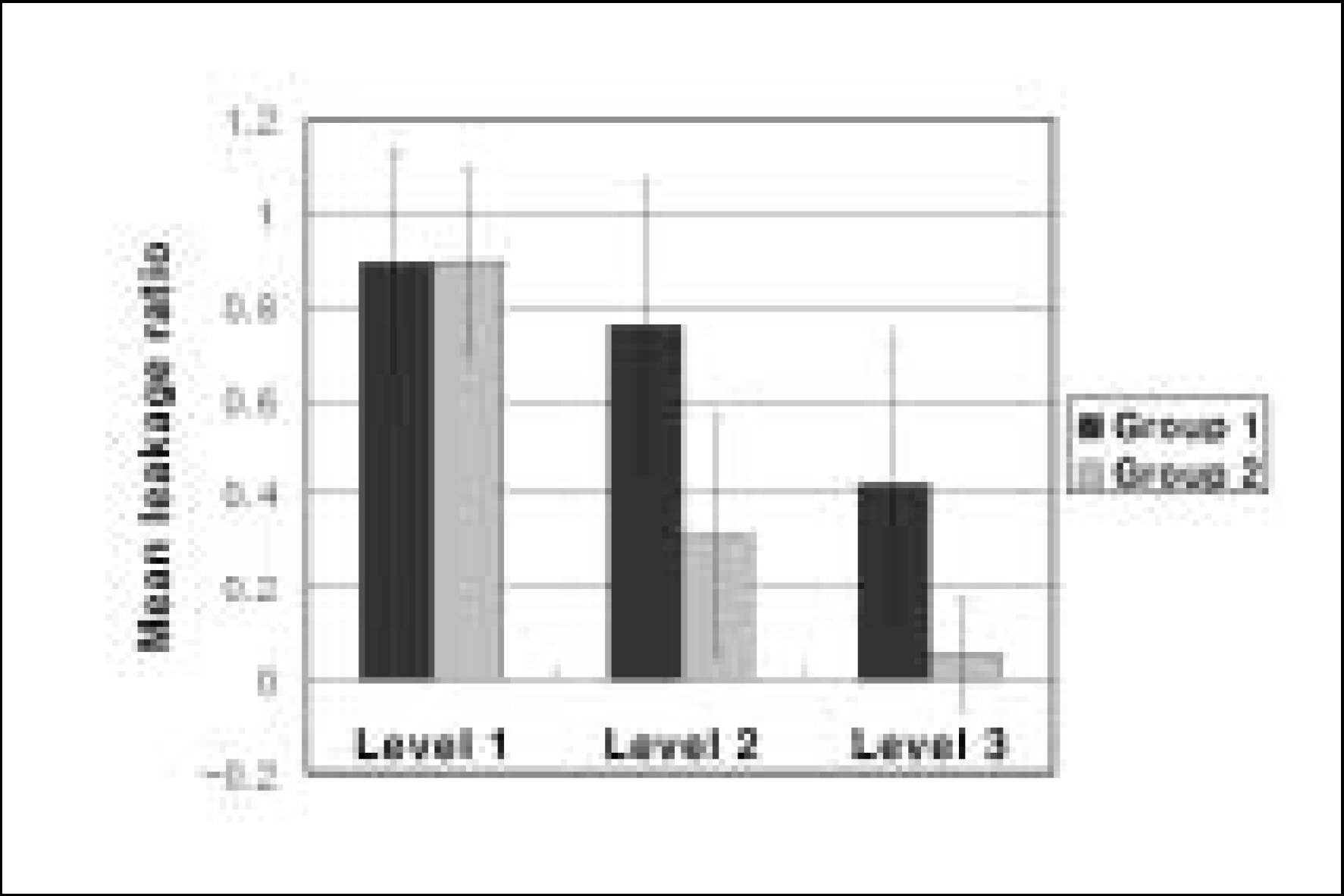
Figure 1. Mean leakage ratio in each group
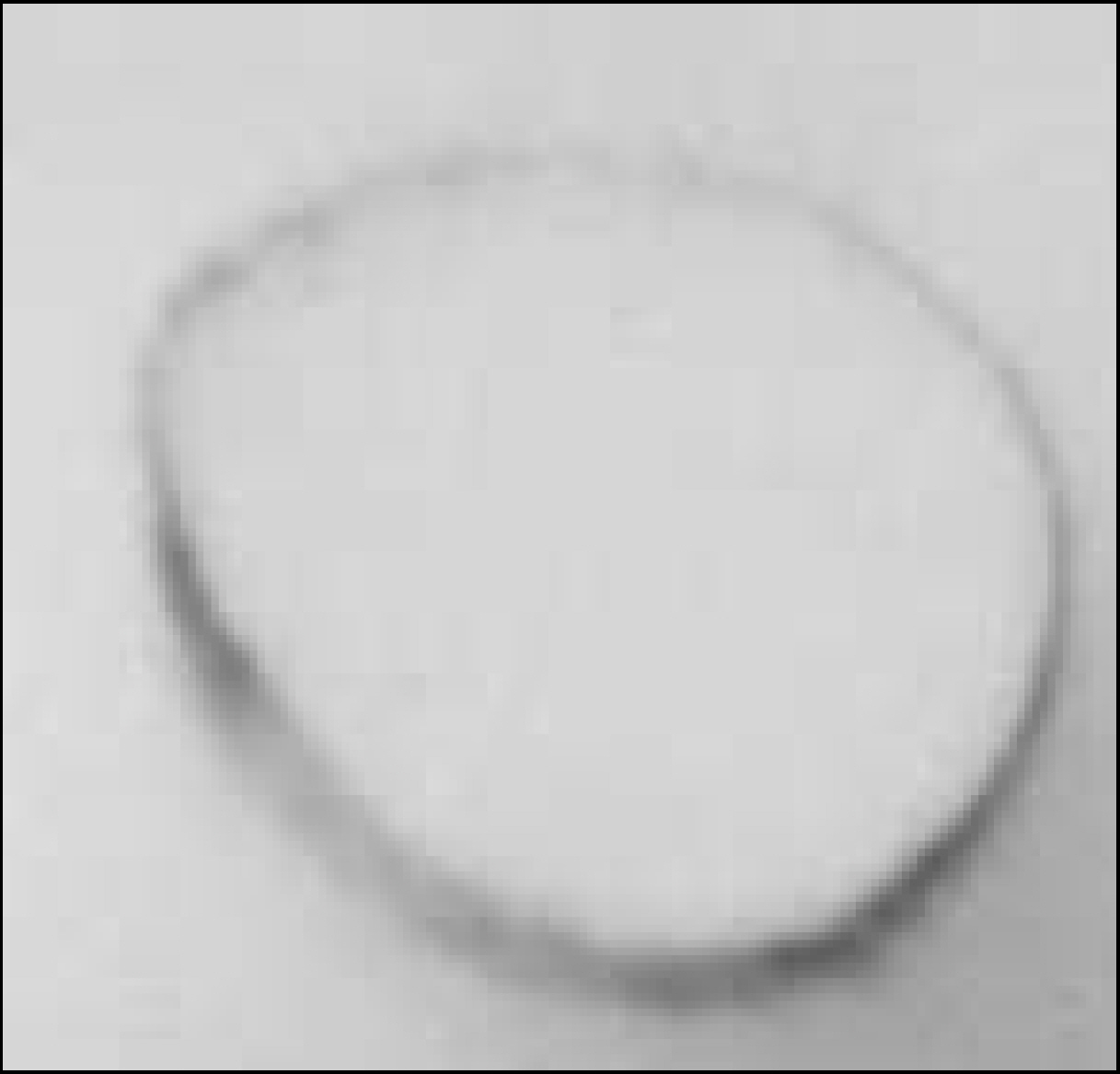
Figure 2. Representative photograph of the group without All-Bond 2. Level 1 (× 40)

Figure 3. Representative photograph of the group without All-Bond 2. Level 2 (× 40)
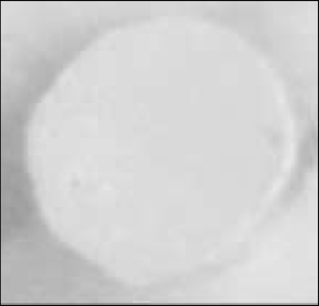
Figure 4. Representative photograph of the group without All-Bond 2. Level 3 (× 40)
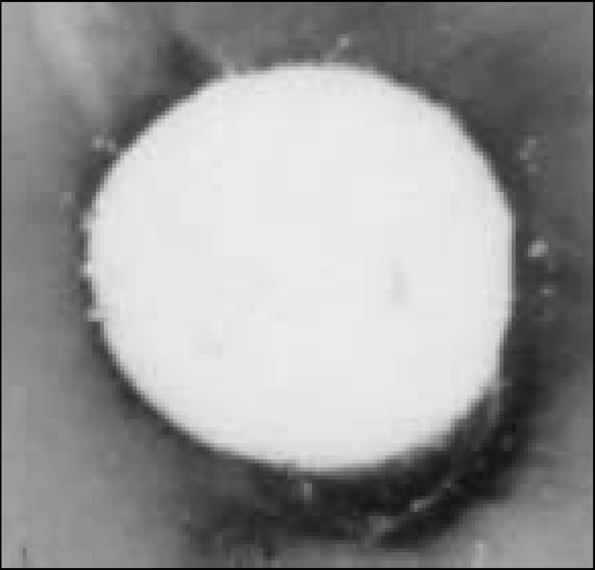
Figure 5. Representative photograph of the group with All-Bond 2. Level 1 (× 40)
Figure 6. Representative photograph of the group with All-Bond 2. Level 2 (× 40)

Figure 7. Representative photograph of the group with All-Bond 2. Level 3 (× 40)
Cited by 1 articles
-
Microleakage of resilon: Effects of several self-etching primer
Jong-Hyeon O, Se-Hee Park, Hye-Jin Shin, Kyung-Mo Cho, Jin-Woo Kim
J Korean Acad Conserv Dent. 2008;33(2):133-140. doi: 10.5395/JKACD.2008.33.2.133.
Reference
-
References
1. Stephen C, Richard CB. Obturation of the cleaned and shaped root canal system. Pathways of the pulp. 8th Edi. St Louis: Mosby Co.;p. p 293–295.2. Hovland EJ, Dumsha TC. Leakage evaluation in vitro of the root canal sealer cement Sealapex. J Endodon. 18:179–182. 1978.
Article3. Chong BS, Pitt Ford TR, Watson TF. The adaptation and sealing ability of light cured glass ionomer retrograde root fillings. Int Endodon J. 24:223–32. 1991.4. Dow PR, Ingle JI. Isotope determination of root canal failure. Oral Surg. 8:1100–1104. 1955.
Article5. Ingle JI, Lief KB. Obturation of the radicular space. Endodontics. 5th Edi. Hamilton: BC Decker;p. p571–6563.6. Mo¨ller AJ, Fabricius L, Dahlen G, O¨hman AE, Heyden G. Influence on periapical tissues of indigenous oral bacteria and necrotic pulp tissue in monkeys. Scand J Dent Res. 89:475–484. 1981.7. Oliver CM, Abbott PV. Correlation between clinical success and apical dye penetration. Int Endodon J. 34:637–644. 2001.
Article8. Kakehashi S, Stanley HR, Fitzgerald RJ. The effects of surgical exposures of dental pulps in germ-free and conventional laboratory rats. Oral Surg Oral Med Oral Pathol. 20:340–349. 1965.
Article9. Sjo¨gren U, Haggluand B, Sundqvist B, Wing K. Factors affecting the long term results of endodontic treatment. J Endodon. 16:498–504. 1990.10. Wu MK, Wesselink PR. Endodontic leakage studies reconsidered, Part Ⅰ. Methodology, application and relevance. Int Endodon J. 26:37–43. 1993.11. Ahlberg KMF, Assavanop P, Tay WM. A comparison of the apical dye penetration patterns shown by methylene blue and India ink in root-filled teeth. Int Endodon J. 28:30–34. 1995.
Article12. Oliver CM, Abbott PV. Entrapped air and its effect on dye penetration of voids. Endond Dent Traumatol. 135–138. 1991.13. Goldman M, Simmonds S, Rush R. The usefulness of dye penetration studies re-examined. Oral Surg Oral Medi Oral Pathol. 67:327–332. 1989.14. Spa¨ngberg LSW, Acierno TG, Yongbum CB. Influence of entrapped air on the accuracy of leakage studies using dye penetration methods. J of Endodon. 15:548–551. 1989.15. Wu MK, De Gee AJ, Wesselink PR. Fluid transport and dye penetration along root canal fillings. Int Endodon J. 27:233–238. 1994.
Article16. Peters LB, Harrison JW. A comparison of leakage of filling materials in demineralized and non-demineralized resected root ends under vacuum and non-vacuum conditions. Int Endodon J. 25:273–278. 1992.
Article17. Antonopoulos KG, Attin T, Hellwig E. Evaluation of the apical seal of root canal fillings with different methods. J Endodon. 24:655–658. 1998.
Article18. Dickson SS, Peters DD. Leakage evaluation with and without vacuum of two gutta-percha fill technique. J Endodon. 19:398–403. 1993.19. Masters J, Higa R, Torabinejad M. Effects of vacuuming on dye penetration patterns in root canals and glass tubes. J Endodon. 21:332–334. 1995.
Article20. Tamse A, Katz A, Kablan F. Comparison of apical leakage shown by four different dyes with two evaluating methods. Int Endodon J. 31:333–337. 1998.
Article21. Anic I, Shirasuka T, Matsumoto K. Scanning electron microscopic evaluation of two compaction techniques using a composite resin as a root canal filling material. J Endodon. 19:594–598. 1995.
Article22. Leonard JE, Gutmann JL, Guo IY. Apical and coronal seal of roots obturated with a dentin bonding agent and resin. Int Endodon J. 29:76–83. 1996.23. Mannocci F, Ferrari M. Apical seal of roots obturated with laterally condensed gutta-percha, epoxy resin cement, and dentin bonding agent. J Endodon. 24:41–44. 1998.
Article24. Leandro RB, Robert EB, Frank JV, James EH, Valeria VG. Comparison of the apical seal obtained by a dual-cure resin based cement of an epoxy resin sealer with or without the use of an acidic primer. J Endodon. 28:721–723. 2002.25. Kanca J Ⅲ. Improving bond strength through acid etching of dentin and bonding to wet dentin surfaces. J Am Dent Assoc. 123:35–43. 1992.
Article26. Gettleman BH, Messer HH, Eldeeb ME. Adhesion of sealer cements to dentin with and without the smear layer. J Endodon. 17:15–20. 1991.
Article27. Nakabayashi N. The hybrid layer: a resin-dentin composite. Proc Finn Dent Soc. 88(Suppl 1):321–329. 1992.28. Valdrighi L. The dimineralizing efficiency of EDTA solutions on dentin. Oral Surg. 16:446. 1981.29. Semra C, Ahmet S. Time-Dependent Effects of EDTA on Dentin Structures. J Endodon. 28:17–19. 2002.30. Rawlinson A. Sealing root canals with low-viscosity resins in vitro: a scanning electron microscopy study of canal cleansing and resin adaptation. Oral Surg Oral Med Oral Pathol. 68:330–338. 1989.
- Full Text Links
-
- Actions
-
Cited
- CITED
-
- Close
- Share
-
- Similar articles
-
- Adhesive systems applied to dentin substrate under electric current: systematic review
- Assessment of Fluoride Release through Dentin Adhesive in the Alkasite Restorative Material and Giomer
- The effect of multiple application on microtensile bond strength of all-in-one dentin adhesive systems
- In vitro micro-shear bond strength of five composite resins to dentin with five different dentin adhesives
- Effect of an aluminum chloride hemostatic agent on the dentin shear bond strength of a universal adhesive
