Single System Langerhans' Cell Histiocytosis with Multifocal Bone Lesions and Pathologic Fracture: A Case Report
- Affiliations
-
- 1Department of Orthopaedic Surgery, Guri Hospital, Hanyang University College of Medicine, Guri, Korea. hyparkys@hanyang.ac.kr
- 2Department of Pathology, Guri Hospital, Hanyang University College of Medicine, Guri, Korea.
- 3Department of Pediatrics, Hanyang University College of Medicine, Seoul, Korea.
- KMID: 1961762
- DOI: http://doi.org/10.5292/jkbjts.2013.19.2.78
Abstract
- Langerhans cell histiocytosis is known as one of the diseases related to excessive proliferation of normal monocytes and has the variety of clinical courses and treatment. Especially, in cases with the spine, it shows a feature of single or multiple osteolysis. According to the location, disease progression and concomitant symptom, variety of treatments (observation, radiotherapy, chemotherapy, surgery, etc.) have been attempted, however, appropriate treatment has not been established yet. The authors introduce the case of single system Langerhans cell histiocytosis which involves cervical and lumbar vertebrae simultaneously with bone marrow destruction and pathologic fracture.
MeSH Terms
Figure
-
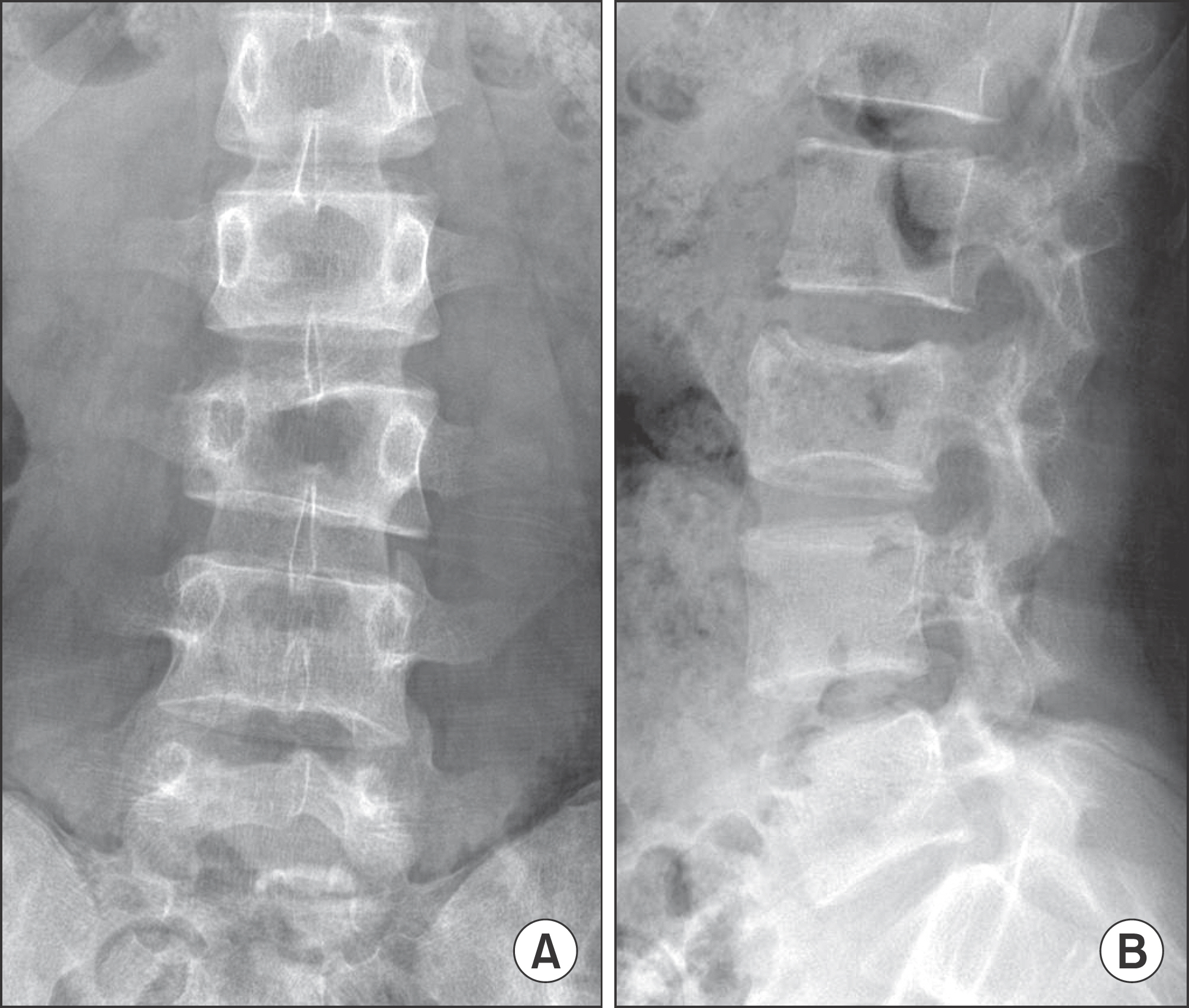
Figure 1. Initial plain radiographs show asymmetric collapse in anteroposterior view (A) and about 30% of collapse on L3 vertebral body in lateral view (B) of lumbar spine.
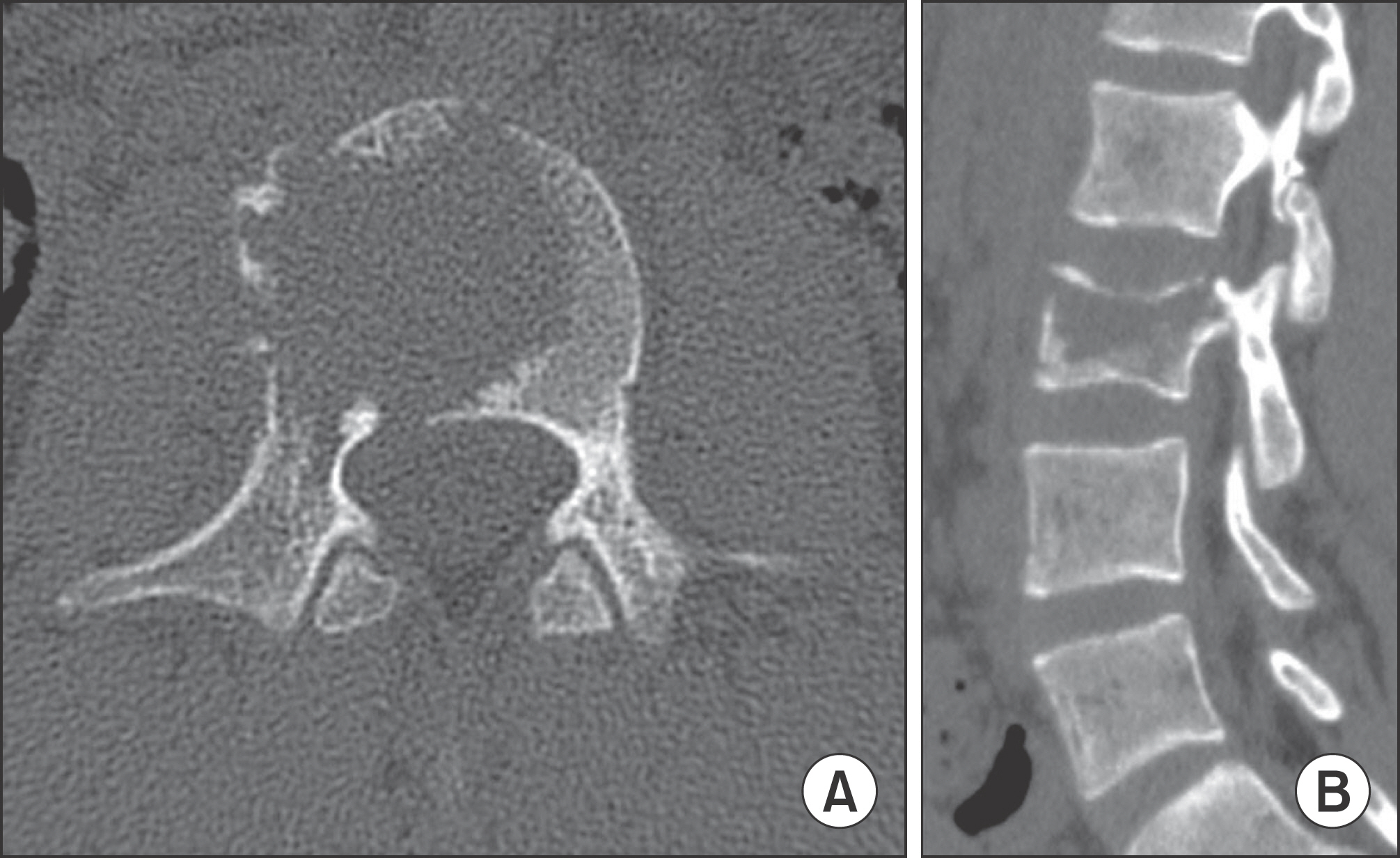
Figure 2. In preoperative computerized tomography scan, axial view shows osteolytic lesion in vertebral body and right pedicle (A). Sagittal view shows pathologic fracture with osteolytic lesion in L3 vertebral body (B).

Figure 3. (A) Contrast-enhanced T1WI MRI shows enhancing lesion in L3 vertebral body and paravertebral soft-tissue. (B) Fat suppression T1WI MRI shows high signal intensity in right superior articular process of C6 vertebra.

Figure 4. Staging study confirms that there was no evidence of other suspicious lesion except spinal lesion. PET-CT shows high uptake of 18fluorodeoxyglucose (FdG) in right superior articular process of C6 vertebra (A) and in right pedicle and body of L3 vertebra (B).

Figure 5. H&E stain (×100) shows nodular aggregation of histiocytes and variable inflammatory cells (A). Histiocytes (Langerhans cell) with oval nuclei as diagnostic hall marker are found in H & E stain (×500) (B). Immunohistochemically, the Langerhans’ cells are positive for S-100 protein (C) and CD1a (D).
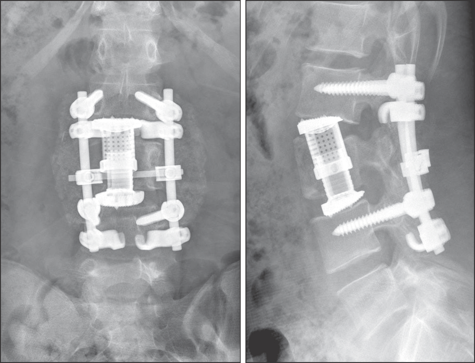
Figure 6. 12 months after treatment, plain radiographs show L3 corpectomy, and placement of an expandable anterior cage and L2-L4 posterior fusion using pedicle screws.
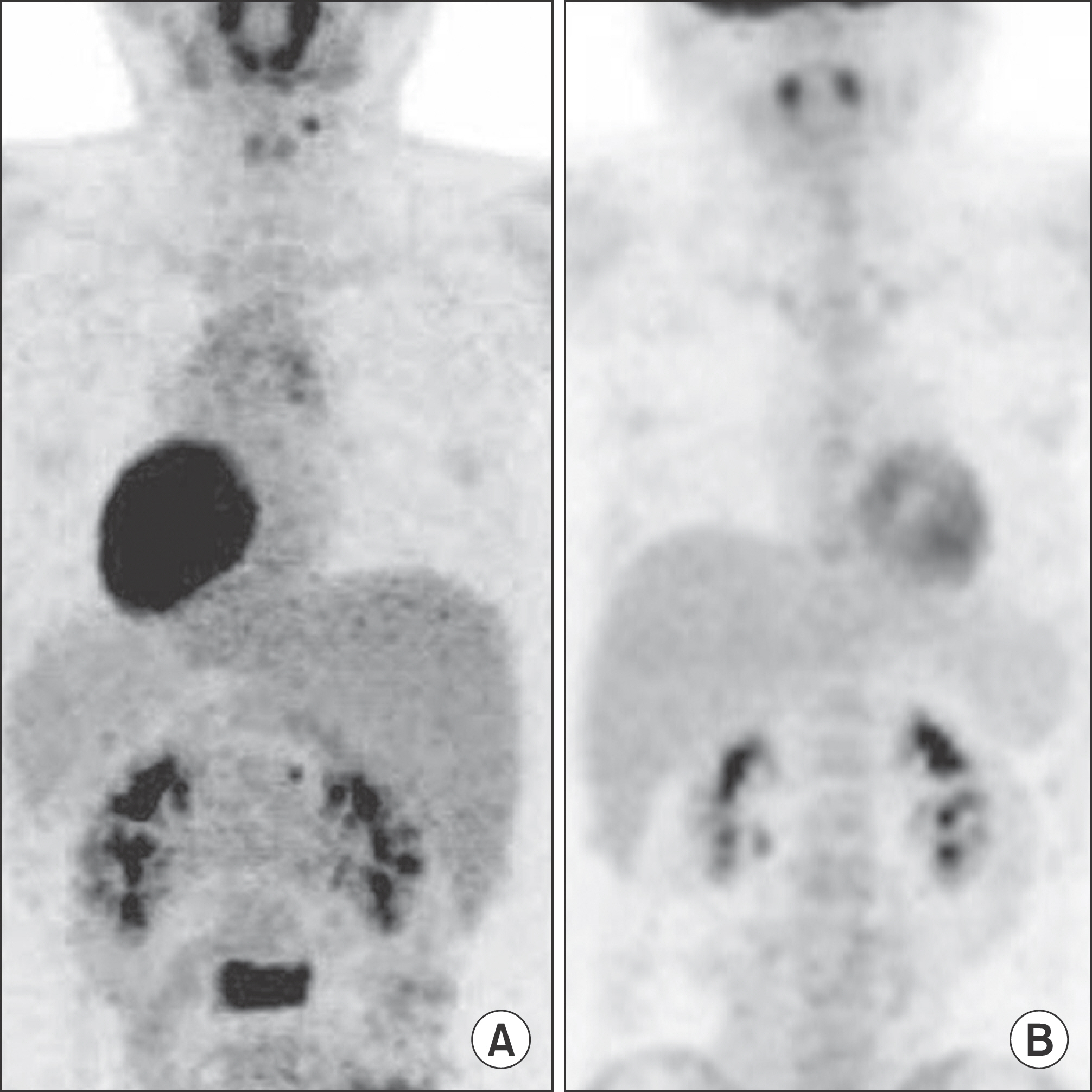
Figure 7. The lesions that were observed previously (A) disappeared in follow up PET-CT (B).
Reference
-
References
1. Lichtenstein L. Histiocytosis X; integration of eosinophilic granuloma of bone, Letterer-Siwe disease, and Schüller-Christian disease as related manifestations of a single nosologic entity. AMA Arch Pathol. 1953; 56:84–102.2. Cheyne C. Histiocytosis X. J Bone Joint Surg Br. 1971; 53:3663. Bertram C, Madert J, Eggers C. Eosinophilic granuloma of the cervical spine. Spine (Phila Pa 1976). 2002;27: 1408–13.
Article4. Yeom JS, Lee CK, Shin HY, Lee CS, Han CS, Chang H. Langerhans' cell histiocytosis of the spine. Analysis of twenty-three cases. Spine (Phila Pa 1976). 1999; 24:1740–9.5. Mammano S, Candiotto S, Balsano M. Cast and brace treatment of eosinophilic granuloma of the spine: longterm follow-up. J Pediatr Orthop. 1997; 17:821–7.
Article6. Garg S, Mehta S, Dormans JP. Langerhans cell histiocytosis of the spine in children. Long-term follow-up. J Bone Joint Surg Am. 2004; 86-A:1740–50.7. Haupt R, Minkov M, Astigarraga I, et al. Euro Histio Network. Langerhans cell histiocytosis (LCH): guidelines for diagnosis, clinical workup, and treatment for patients till the age of 18 years. Pediatr Blood Cancer. 2013; 60:175–84.
Article8. Floman Y, Bar-On E, Mosheiff R, Mirovsky Y, Robin GC, Ramu N. Eosinophilic granuloma of the spine. J Pediatr Orthop B. 1997; 6:260–5.
Article
- Full Text Links
-
- Actions
-
Cited
- CITED
-
- Close
- Share
-
- Similar articles
-
- Adult Scapular Langerhans Cell Histiocytosis Mistaken for Acute Osteomyelitis
- A Case of External Auditory Canal Stenosis in Langerhans Cell Histiocytosis
- Langerhans Cell Histiocytosis Misdiagnosed as Multifocall Osteomyelitis in an Old Patients: A Case Report
- Spontaneous Pneumothorax due to Pulmonary Invasion in Multisystemic Langerhans Cell Histiocytosis: A case report
- A Case of Eosinophilic Granuloma of the Temporal Bone
