p-Coumaric Acid Attenuates UVB-Induced Release of Stratifin from Keratinocytes and Indirectly Regulates Matrix Metalloproteinase 1 Release from Fibroblasts
- Affiliations
-
- 1Department of Molecular Medicine, Cell and Matrix Research Institute, BK21 Plus KNU Biomedical Convergence Program, Department of Biomedical Science, Kyungpook National University School of Medicine, Daegu 700-842, Korea. ycboo@knu.ac.kr
- KMID: 1791425
- DOI: http://doi.org/10.4196/kjpp.2015.19.3.241
Abstract
- Ultraviolet (UV) radiation-induced loss of dermal extracellular matrix is associated with skin photoaging. Recent studies demonstrated that keratinocyte-releasable stratifin (SFN) plays a critical role in skin collagen metabolism by inducing matrix metalloproteinase 1 (MMP1) expression in target fibroblasts. In the present study, we examined whether SFN released from UVB-irradiated epidermal keratinocytes increases MMP1 release from dermal fibroblasts, and whether these events are affected by p-coumaric acid (p-CA), a natural phenolic compound with UVB-shielding and antioxidant properties. HaCaT cells were exposed to UVB in the absence and presence of p-CA, and the conditioned medium was used to stimulate fibroblasts in medium transfer experiments. The cells and media were analyzed to determine the expressions/releases of SFN and MMP1. UVB exposure increased SFN release from keratinocytes into the medium. The conditioned medium of UVB-irradiated keratinocytes increased MMP1 release from fibroblasts. The depletion of SFN using a siRNA rendered the conditioned medium of UVB-irradiated keratinocytes ineffective at stimulating fibroblasts to release MMP1. p-CA mitigated UVB-induced SFN expression in keratinocytes, and attenuated the MMP1 release by fibroblasts in medium transfer experiments. In conclusion, the present study demonstrated that the use of UV absorbers such as p-CA would reduce UV-induced SFN-centered signaling events involved in skin photoaging.
Keyword
MeSH Terms
Figure
-
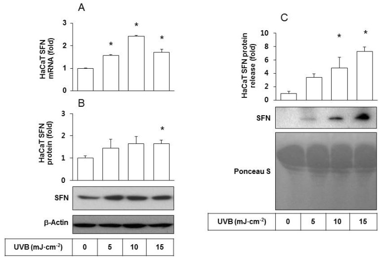
Fig. 1 Effects of UVB irradiation SFN expression/release of HaCaT cells. HaCaT cells were irradiated with UVB at 5~15 mJ cm-2 and incubated for 24 h. (A) SFN mRNA levels were determined by qRT-PCR analysis using GAPDH as a control. (B) SFN protein levels were determined by Western blot analysis of whole cell lysates using β-actin as a control. (C) The released SFN protein levels were determined by Western blot analysis of the conditioned media derived from HaCaT cells exposed to different doses of UVB. For detection of total protein, the transferred membrane was stained with Ponceau S. Data are presented as fold changes versus the non-irradiated control (Mean±SE, n=3). *p<0.05 versus non-irradiated controls.
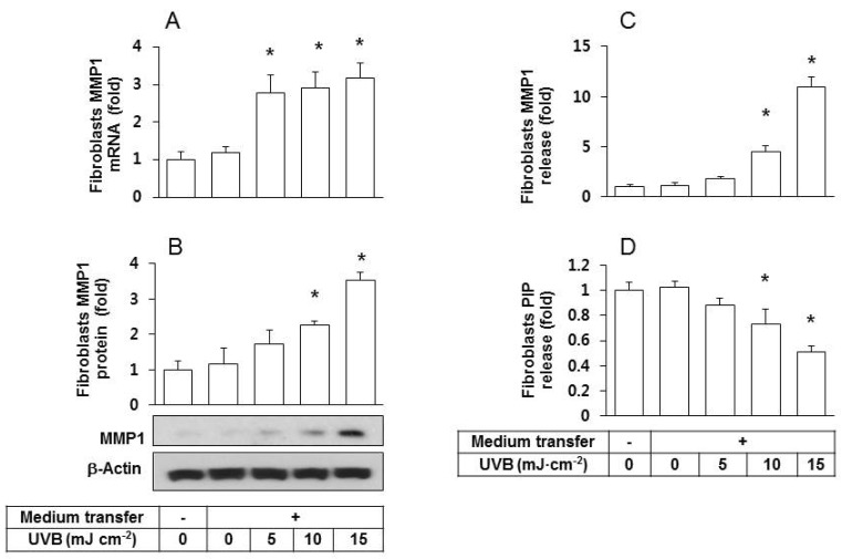
Fig. 2 Effects of the conditioned medium derived from UVB-irradiated HaCaT cells on fibroblast expression/release of MMP1 and PIP. Human epidermal fibroblasts were seeded and cultured in growth medium for 24 h then the medium was replaced with the conditioned medium derived from HaCaT cells exposed to UVB at 5~15 mJ cm-2. After a 24 h incubation period, MMP1 expression was determined at the mRNA by qRT-PCR (A), and at the protein levels by western blots (B). The levels of MMP1 (C) and PIP (D) in the conditioned medium of fibroblasts were determined using sandwich immunoassay kits. Data are presented as fold changes versus the control cells without medium transfer (mean±SE, n=3). *p<0.05 versus controls.
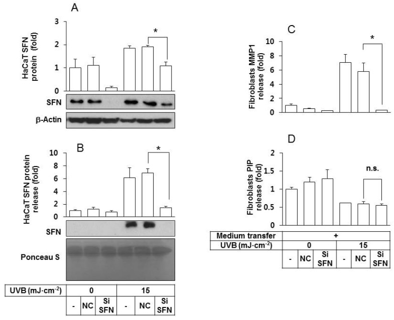
Fig. 3 An essential role for SFN derived from UVB-irradiated HaCaT keratinocytes in stimulating fibroblasts to express/release MMP1 protein. (A, B) HaCaT cells were transfected with a SFN siRNA or negative control siRNA for 24 h, followed by exposure to UVB at 15 mJ cm-2. After a 24 h incubation period, the intracellular (A) and released SFN protein levels (B) were determined by western blots of whole cell lysates and the conditioned media, respectively. For detection of total protein, transferred membrane was stained with Ponceau S. (C, D) Fibroblasts were cultured for 24 h in their own growth medium or the conditioned medium derived from HaCaT cells. The medium was then replaced by the fibroblasts growth medium without serum. After 24 h of incubation, MMP1 (C) and PIP (D) in the conditioned medium of fibroblasts were quantified using sandwich immunoassay kits. Data are presented as fold changes versus the control cells without medium transfer (Mean±SE, n=3). *p<0.05; n.s., not significant.
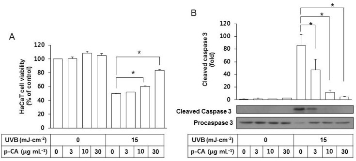
Fig. 4 Effects of p-CA on the UVB-induced cytotoxicity and caspase 3 activation in HaCaT cells. HaCaT cells were irradiated with UVB at 15 mJ cm-2 or not, in the absence or the presence of p-CA at the indicated concentrations and incubated for 24 h. (A) Cell viability was determined using an MTT assay. (B) Whole cell lysates of equal amounts of proteins were used for the western blot analysis of the active cleaved and inactive proforms of caspase 3. Data are presented as % of control or fold changes versus the control (Mean±SE, n=3). *p<0.05.
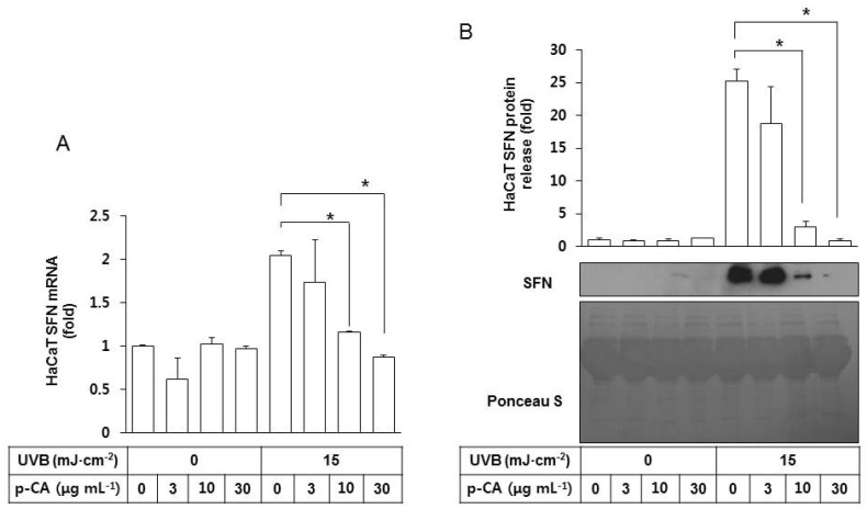
Fig. 5 Effects of p-CA on the UVB-induced SFN expression/release of HaCaT cells. HaCaT cells were irradiated with UVB at 15 mJ cm-2 or not, in the absence or the presence of p-CA at the indicated concentrations. The cells were then cultured in the fresh medium without p-CA for 24 h to harvest the cells and the conditioned medium. (A) SFN mRNA levels were determined by qRT-PCR analysis using GAPDH as a control. (B) The released SFN protein levels were determined by western blot analysis of the conditioned media derived from HaCaT cells. For detection of total protein, the transferred membrane was stained with Ponceau S. Data are presented as fold changes versus the control cells (mean±SE, n=3). *p<0.05.
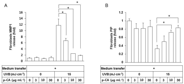
Fig. 6 Effects of the conditioned medium of HaCaT cells UVB-irradiated in the presence of p-CA on the releases of MMP1 and PIP from fibroblasts. HaCaT cells were irradiated with UVB at 15 mJ cm-2 or not, in the absence or the presence of p-CA at the indicated concentrations. The cells were then cultured in the fresh medium without p-CA for 24 h to harvest the conditioned medium. Fibroblasts were cultured for 24 h in the conditioned medium derived from HaCaT cells, and then the medium was replaced with fibroblast growth medium without serum. After incubating for 24 h, MMP1 (A) and PIP (B) in the fibroblasts conditioned medium were quantified using sandwich immunoassay kits. Data are presented as fold changes versus the control cells (mean±SE, n=3). *p<0.05.
Reference
-
1. Rabe JH, Mamelak AJ, McElgunn PJ, Morison WL, Sauder DN. Photoaging: mechanisms and repair. J Am Acad Dermatol. 2006; 55:1–19. PMID: 16781287.
Article2. Fisher GJ, Kang S, Varani J, Bata-Csorgo Z, Wan Y, Datta S, Voorhees JJ. Mechanisms of photoaging and chronological skin aging. Arch Dermatol. 2002; 138:1462–1470. PMID: 12437452.
Article3. Kähäri VM, Saarialho-Kere U. Matrix metalloproteinases in skin. Exp Dermatol. 1997; 6:199–213. PMID: 9450622.
Article4. Bode W, Fernandez-Catalan C, Tschesche H, Grams F, Nagase H, Maskos K. Structural properties of matrix metalloproteinases. Cell Mol Life Sci. 1999; 55:639–652. PMID: 10357232.
Article5. Brennan M, Bhatti H, Nerusu KC, Bhagavathula N, Kang S, Fisher GJ, Varani J, Voorhees JJ. Matrix metalloproteinase-1 is the major collagenolytic enzyme responsible for collagen damage in UV-irradiated human skin. Photochem Photobiol. 2003; 78:43–48. PMID: 12929747.
Article6. Scharffetter K, Wlaschek M, Hogg A, Bolsen K, Schothorst A, Goerz G, Krieg T, Plewig G. UVA irradiation induces collagenase in human dermal fibroblasts in vitro and in vivo. Arch Dermatol Res. 1991; 283:506–511. PMID: 1664713.
Article7. Brenneisen P, Oh J, Wlaschek M, Wenk J, Briviba K, Hommel C, Herrmann G, Sies H, Scharffetter-Kochanek K. Ultraviolet B wavelength dependence for the regulation of two major matrix-metalloproteinases and their inhibitor TIMP-1 in human dermal fibroblasts. Photochem Photobiol. 1996; 64:649–657. PMID: 8863471.
Article8. Ghahary A, Marcoux Y, Karimi-Busheri F, Li Y, Tredget EE, Kilani RT, Lam E, Weinfeld M. Differentiated keratinocyte-releasable stratifin (14-3-3 sigma) stimulates MMP-1 expression in dermal fibroblasts. J Invest Dermatol. 2005; 124:170–177. PMID: 15654971.
Article9. Lam E, Kilani RT, Li Y, Tredget EE, Ghahary A. Stratifin-induced matrix metalloproteinase-1 in fibroblast is mediated by c-fos and p38 mitogen-activated protein kinase activation. J Invest Dermatol. 2005; 125:230–238. PMID: 16098031.
Article10. Adachi H, Murakami Y, Tanaka H, Nakata S. Increase of stratifin triggered by ultraviolet irradiation is possibly related to premature aging of human skin. Exp Dermatol. 2014; 23(Suppl 1):32–36. PMID: 25234834.
Article11. An SM, Lee SI, Choi SW, Moon SW, Boo YC. p-Coumaric acid, a constituent of Sasa quelpaertensis Nakai, inhibits cellular melanogenesis stimulated by alpha-melanocyte stimulating hormone. Br J Dermatol. 2008; 159:292–299. PMID: 18544081.12. Kim M, Park J, Song K, Kim HG, Koh JS, Boo YC. Screening of plant extracts for human tyrosinase inhibiting effects. Int J Cosmet Sci. 2012; 34:202–208. PMID: 22220689.
Article13. Song K, Boo YC. UVB shielding effects of para-Coumaric acid. J Soc Cosmet Scientists Korea. 2012; 38:263–273.14. Lee SJ, Mun GI, An SM, Boo YC. Evidence for the association of peroxidases with the antioxidant effect of p-coumaric acid in endothelial cells exposed to high glucose plus arachidonic acid. BMB Rep. 2009; 42:561–567. PMID: 19788856.
Article15. Song K, An SM, Kim M, Koh JS, Boo YC. Comparison of the antimelanogenic effects of p-coumaric acid and its methyl ester and their skin permeabilities. J Dermatol Sci. 2011; 63:17–22. PMID: 21530181.
Article16. Zang LY, Cosma G, Gardner H, Shi X, Castranova V, Vallyathan V. Effect of antioxidant protection by p-coumaric acid on lowdensity lipoprotein cholesterol oxidation. Am J Physiol Cell Physiol. 2000; 279:C954–C960. PMID: 11003575.17. Lee SI, An SM, Mun GI, Lee SJ, Park KM, Park SH, Boo YC. Protective effect of Sasa quelpaertensis and p-coumaric acid on ethanol-induced hepatotoxicity in mice. J Appl Biol Chem. 2008; 51:148–154.
Article18. Park J, Seok JK, Suh HJ, Boo YC. Gardenia jasminoides extract attenuates the UVB-induced expressions of cytokines in keratinocytes and indirectly inhibits matrix metalloproteinase-1 expression in human dermal fibroblasts. Evid Based Complement Alternat Med. 2014; 2014:429246. PMID: 24711853.19. Canty-Laird EG, Lu Y, Kadler KE. Stepwise proteolytic activation of type I procollagen to collagen within the secretory pathway of tendon fibroblasts in situ. Biochem J. 2012; 441:707–717. PMID: 21967573.20. Sitailo LA, Tibudan SS, Denning MF. Activation of caspase-9 is required for UV-induced apoptosis of human keratinocytes. J Biol Chem. 2002; 277:19346–19352. PMID: 11919192.
Article21. Lee CH, Wu SB, Hong CH, Yu HS, Wei YH. Molecular mechanisms of UV-induced apoptosis and its effects on skin residential cells: the implication in UV-based phototherapy. Int J Mol Sci. 2013; 14:6414–6435. PMID: 23519108.
- Full Text Links
-
- Actions
-
Cited
- CITED
-
- Close
- Share
-
- Similar articles
-
- Low-dose UVB irradiation stimulates matrix metalloproteinase-1 expression via a BLT2-linked pathway in HaCaT cells
- The Effect of Supernatant from UVB - Irradiated Cultured Keratinocytes on the Growth , Melanin Content , and Tyrosinase Activity of Human Melanocyte
- Simvastatin as a Modulator of Tissue Remodeling through Inhibition of Matrix Metalloproteinase (MMP) Release from Human Lung Fibroblasts
- Oleanolic Acid Protects the Skin from Particulate Matter-Induced Aging
- Effects of Dexamethasone on Endothelin-1(ET-1) Production by Keratinocytes
