Inflammatory Myofibroblastic Tumor of the Kidney Misdiagnosed as Renal Cell Carcinoma
- Affiliations
-
- 1Department of Urology, Chonnam National University Medical School, Gwangju, Korea. ryusaint@naver.com
- 2Department of Urology, Chonbuk National University Medical School, Jeonju, Korea.
- 3Department of Pathology, Chonnam National University Medical School, Gwangju, Korea.
- KMID: 1779272
- DOI: http://doi.org/10.3346/jkms.2010.25.2.330
Abstract
- The inflammatory myofibroblastic tumor (IMT), also knowns as inflammatory pseuduotumor, is a soft tissue lesion of unknown etiology. In the urogenital tract, IMT mainly affects the urinary bladder or prostate, but rarely the kidney. It has been considered as a nonneoplastic reactive inflammatory lesion, but nowadays, it is regarded as a neoplasm due to its high recurrence rate and metastasis. We describe a case of a 61-yr-old woman that had originally been misdiagnosed as renal cell carcinoma, which was pathologically revealed to be an IMT.
MeSH Terms
Figure
-
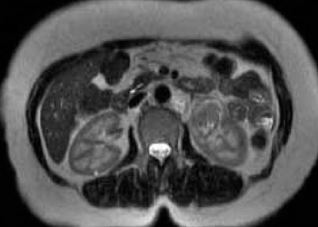
Fig. 1 Magnetic resonance image demonstrates a 3.0×2.5 cm size solid mass with mildly enhancement on left kidney lower pole with a central necrotic portion.

Fig. 2 Left kidney coronal opening specimen shows a well-circumscribed encapsulated mass measuring 2.7×2.8 cm size, involving the lower pole. The mass revealed areas of myxoid change necrosis, and cystic change.
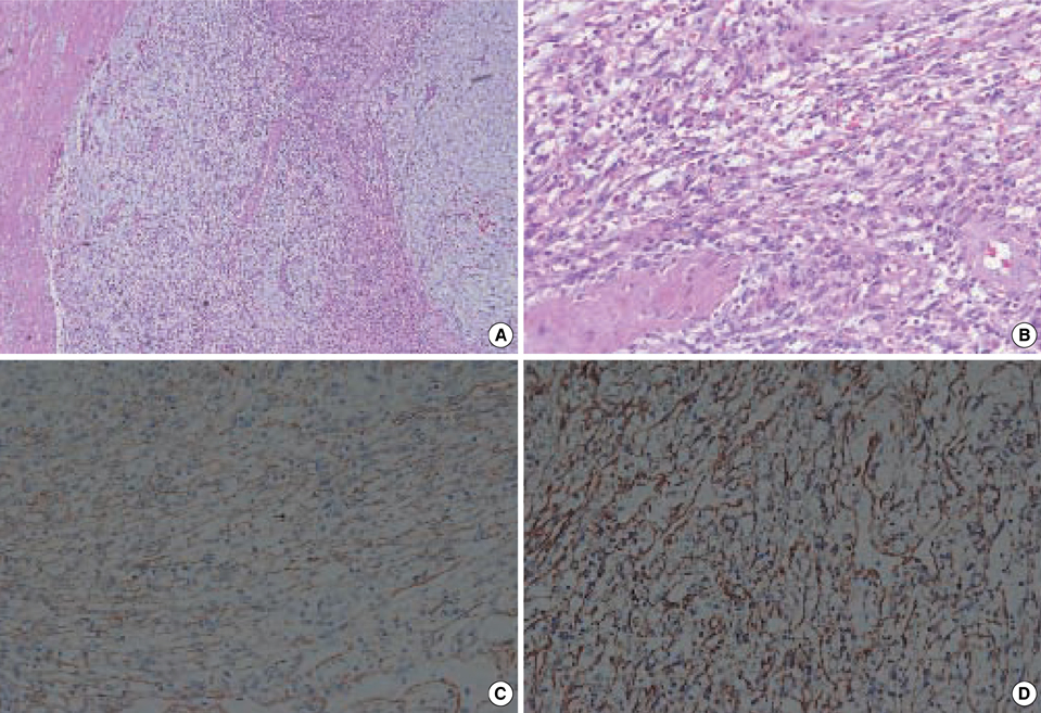
Fig. 3 Microscopic findings. (A) The low power appearance demonstrating a capsule of dense collagenous fibrous tissue and myxoid zone and inflammation with cellular zone consisting of spindle cells arranged in fascicles. (B) The area of myofibroblastic proliferation showing densely cellular fascicles. The tumor cells were potive for smooth muscle actin (C) and vimentin (D).
Reference
-
1. Brunn H. Two interesting benign lung tumors of contradictory histopathology; remarks on the necessity for maintaining chest tumor registry. J Thorac Surg. 1939. 9:119–131.2. Young RH, Eble JN. Non-neoplastic disorders of the urinary bladder. Urol Surg Pathol Mosby-Year Book. 1997. 166–214.
Article3. Larbcharoensub N, Chobpradit N, Kijvikai K, Chalermsanyakorn P. Primary renal inflammatory myofibroblastic tumor. Urol Int. 2006. 76:94–96.
Article4. Coffin CM, Watterson J, Priest JR, Dehner LP. Extrapulmonary inflammatory myofibroblastic tumor (inflammatory pseudotumor): a clinicopathologic and immunohistochemical study of 84 cases. Am J Surg Pathol. 1995. 19:859–872.
Article5. Kapusta LR, Weiss MA, Ramsay J, Lopez-Beltran A, Srigley JR. Inflammatory myofibroblastic tumors of the kidney: a clinicopathologic and immunohistochemical study of 12 cases. Am J Surg Pathol. 2003. 27:658–666.
Article6. Gwynn ES, Clark PE. Inflammatory myofibroblastic tumor associated with renal cell carcinoma. Urology. 2005. 66:880.
Article7. Donner LR, Trompler RA, White RR 4th. Progression of inflammatory myofibroblastic tumor (inflammatory pseudotumor) or soft tissue into sarcoma after several recurrences. Hum Pathol. 1996. 27:1095–1098.
Article8. Arber DA, Kamel OW, van de Rijn M, Davis RE, Medeiros LJ, Jaffe ES, Weiss LM. Frequent presence of the Epstein-Barr virus in inflammatory pseudotumor. Hum Pathol. 1995. 26:1093–1098.
Article9. Brittig F, Ajtay E, Jakso P, Kelenyi G. Follicular dendritic reticulum cell tumor mimicking inflammatory pseudotumor of the spleen. Pathol Oncol Res. 2004. 10:57–60.
Article
- Full Text Links
-
- Actions
-
Cited
- CITED
-
- Close
- Share
-
- Similar articles
-
- Inflammatory Myofibroblastic Tumor of Kidney
- Inflammatory Myofibroblastic Tumor of Nasal Septum after Septoplasty: A Case Report
- A Case of Renal Cell Carcinoma and Adult Wilms' Tumor in the Same Kidney
- Inflammatory Myofibroblastic Tumor in Posterior Mediastinum
- MR Findings of Inflammatory Myofibroblastic Tumor of the Thigh: A Case Report
