Atherosclerotic Progression Attenuates the Expression of Nogo-B in Autopsied Coronary Artery: Pathology and Virtual Histology Intravascular Ultrasound Analysis
- Affiliations
-
- 1Department of Internal Medicine, College of Medicine, Chung-Ang University, Seoul, Korea. wslee1227@dreamwiz.com
- 2Department of Pathology, College of Medicine, Chung-Ang University, Seoul, Korea.
- 3National Institute of Scientific Investigation, Seoul, Korea.
- KMID: 1779190
- DOI: http://doi.org/10.3346/jkms.2009.24.4.596
Abstract
- The relation of Nogo-B to atherosclerotic plaque progression is not well understood. Thus, the purpose of this study was to assess the expression of Nogo-B in fibroatheromas (FA) of different stages, classified using virtual histology intravascular ultrasound (VH-IVUS) analysis in 19 autopsied cases of non-sudden cardiac death. VH-IVUS imaging analysis was performed 30 mm from the ostium of each coronary artery. VH-IVUS revealed 11 early FAs (34.5+/-8.3 yr), 12 late FAs (42.6+/-16.6 yr), 8 thick-cap FAs (TkCFAs) (46.4+/-11.1 yr), and 6 thin-cap FAs (TCFAs) (51.8+/-6.8 yr). TkCFAs and TCFAs were defined as advanced FA. FA progression advanced with age (P=0.04). VH-IVUS analysis of small, early FAs showed smaller necrotic cores and relatively less calcium compared to more advanced FAs with large necrotic cores (P<0.001). Histopathology and immunohistochemical stains demonstrated that early or late FAs had smaller necrotic cores, less empty space of decalcification, and greater Nogo-B expression compared to advanced FAs (vs. early FA, P=0.013; vs. late FA, P=0.008, respectively). These findings suggest that FA progression is inversely associated with Nogo-B expression. Local reduction of Nogo-B may contribute to plaque formation and/or instability.
MeSH Terms
Figure
-
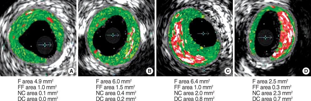
Fig. 1 Classification of fibroatheromas by virtual histology intravascular ultrasound. Early fibroatheromas (A) had less extensively necrotic cores (red color) and greater fibrous (green color) composition, while thick-cap fibroatheromas (C) and thin-cap fibroatheromas (D) had larger necrotic cores and more extensive calcium (white color) deposition. F, fibrous; FF, fibrofatty; NC, necrotic core; DC, dense calcium.
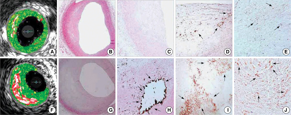
Fig. 2 Comparative VH-IVUS and microscopic findings between early fibroatheromas (FA) or late FAs and advanced FAs. Early FAs (A, B) had smaller necrotic cores compared to advanced FAs (F, G) with larger lipid pools. Late FAs (C) had few calcium components, while advanced FAs (H) had a large amount of calcium and decalcified empty spaces (arrows). The expression of CD68-positive macrophages (arrows) was strong in advanced FA (I) rather than in late FA (D). MMP-9 activity (arrows) was also prominent in advanced FA (J) as opposed to late FA (E). (B and G, H&E staining at ×40 magnification; C and H, von Kossa staining at ×40 and ×100 magnification, respectively; D and I, CD68 immunohistochemical staining at ×100 magnification; E and J, MMP-9 immunohistochemical staining at ×100 magnification).
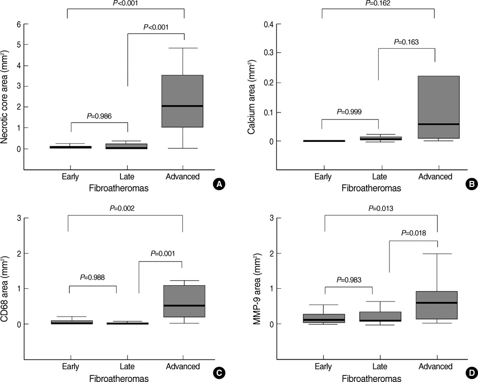
Fig. 3 Quantitative graphs of necrotic cores, calcium, CD68-positive macrophages, and MMP-9 activity in various stages of FA.
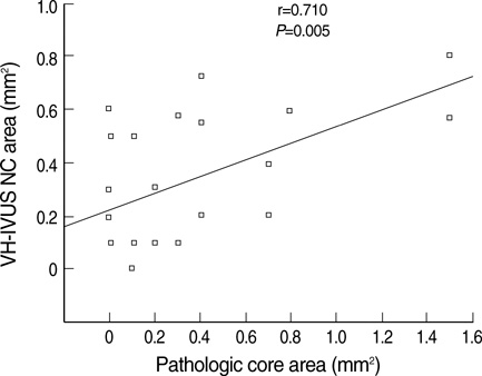
Fig. 4 The correlation of necrotic core area between VH-IVUS and pathology in human autopsied coronary arteries.
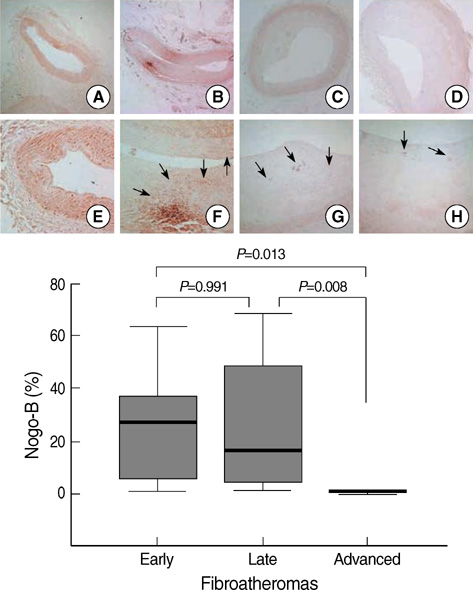
Fig. 5 Nogo-B expression with plaque growth. Early fibroatheromas (FA) (B and F) showed diffuse Nogo-B expression (arrows) comparable to that seen in controls (A and E). Late FAs (C and G) had weaker, focal expression (arrows) of Nogo-B compared to early FAs. Advanced FA (D and H) demonstrated much weaker focal Nogo-B activity (arrows) compared to that seen in early FA or late FA. I, quantitative graphs of Nogo-B expression in various FA stages. (Immunohistochemical staining for Nogo-B, A to D, ×40 magnification, E to H, ×100 magnification).
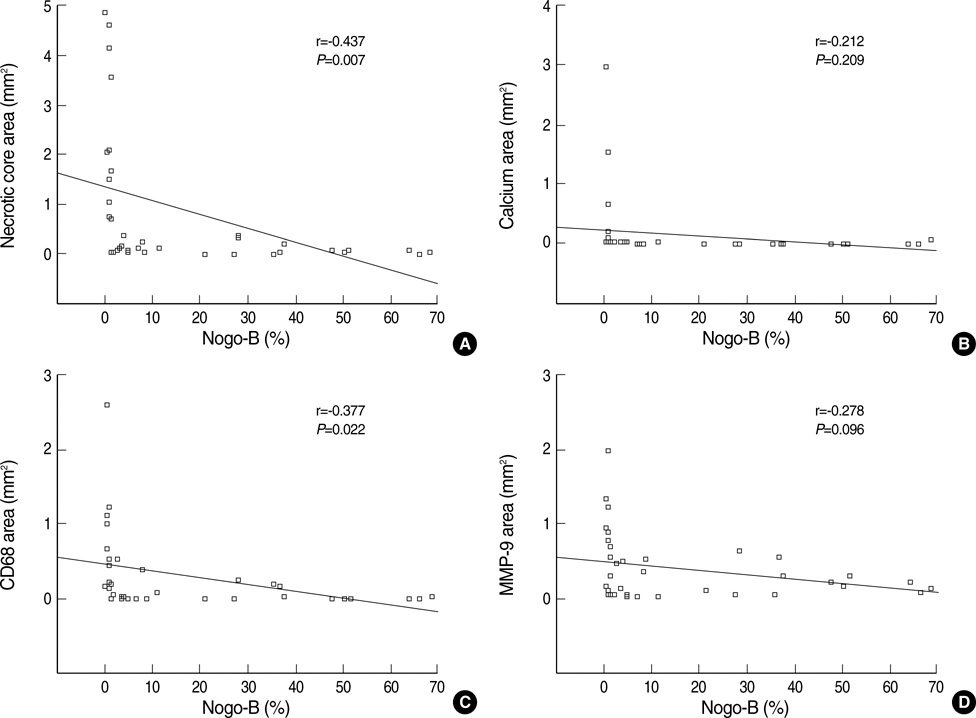
Fig. 6 The correlation between immunohistochemical Nogo-B activity, and plaque composition and CD68/MMP-9 expression. Nogo-B expression carried a significant negative correlation with core necrosis (A), but not with calcification (B). Nogo-B expression had a significant negative correlation with the number of CD68-positive cells (C), but not with MMP-9 expression (D).
Reference
-
1. Virmani R, Burke AP, Farb A, Kolodgie FD. Pathology of the vulnerable plaque. J Am Coll Cardiol. 2006. 47(8):Suppl. C13–C18.
Article2. Sen S, Hinderliter A, Sen PK, Simmons J, Beck J, Offenbacher S, Ohman EM, Oppenheimer SM. Aortic arch atheroma progression and recurrent vascular events in patients with stroke or transient ischemic attack. Circulation. 2007. 116:928–935.
Article3. Doonan AL, Karha J, Carrigan TP, Bavry AA, Begelman SM, Ellis SG, Yadav J, Bhatt DL. Presence of carotid and peripheral arterial disease in patients with left main disease. Am J Cardiol. 2007. 100:1087–1089.
Article4. Virmani R, Burke AP, Farb A. Sudden cardiac death. Cardiovasc Pathol. 2001. 10:275–282.
Article5. Kolodgie FD, Narula J, Burke AP, Haider N, Farb A, Hui-Liang Y, Smialek J, Virmani R. Localization of apoptotic macrophages at the site of plaque rupture in sudden coronary death. Am J Pathol. 2000. 157:1259–1268.
Article6. Schmermund A, Schwartz RS, Adamzik M, Sangiorgi G, Pfeifer EA, Rumberger JA, Burke AP, Farb A, Virmani R. Coronary atherosclerosis in unheralded sudden coronary death under age 50: histo-pathologic comparison with 'healthy' subjects dying out of hospital. Atherosclerosis. 2001. 155:499–508.
Article7. Tardif JC, Heinonen T, Orloff D, Libby P. Vascular biomarkers and surrogates in cardiovascular disease. Circulation. 2006. 113:2936–2942.
Article8. Koenig W, Khuseyinova N. Biomarkers of atherosclerotic plaque instability and rupture. Arterioscler Thromb Vasc Biol. 2007. 27:15–26.
Article9. Motoyama S, Kondo T, Sarai M, Sugiura A, Harigaya H, Sato T, Inoue K, Okumura M, Ishii J, Anno H, Virmani R, Ozaki Y, Hishida H, Narula J. Multislice computed tomographic characteristics of coronary lesions in acute coronary syndromes. J Am Coll Cardiol. 2007. 50:319–326.
Article10. Briley-Saebo KC, Mulder WJ, Mani V, Hyafil F, Amirbekian V, Aguinaldo JG, Fisher EA, Fayad ZA. Magnetic resonance imaging of vulnerable atherosclerotic plaques: current imaging strategies and molecular imaging probes. J Magn Reson Imaging. 2007. 26:460–479.
Article11. Riccio SA, House AA, Spence JD, Fenster A, Parraga G. Carotid ultrasound phenotypes in vulnerable populations. Cardiovasc Ultrasound. 2006. 4:44–52.
Article12. Hamdan A, Assali A, Fuchs S, Battler A, Kornowski R. Imaging of vulnerable coronary artery plaques. Catheter Cardiovasc Interv. 2007. 70:65–74.
Article13. Kawasaki M, Bouma BE, Bressner J, Houser SL, Nadkarni SK, Mac-Neill BD, Jang IK, Fujiwara H, Tearney GJ. Diagnostic accuracy of optical coherence tomography and integrated backscatter intravascular ultrasound images for tissue characterization of human coronary plaques. J Am Coll Cardiol. 2006. 48:81–88.
Article14. Sano K, Kawasaki M, Ishihara Y, Okubo M, Tsuchiya K, Nishigaki K, Zhou X, Minatoguchi S, Fujita H, Fujiwara H. Assessment of vulnerable plaques causing acute coronary syndrome using integrated backscatter intravascular ultrasound. J Am Coll Cardiol. 2006. 47:734–741.
Article15. DeMaria AN, Narula J, Mahmud E, Tsimikas S. Imaging vulnerable plaque by ultrasound. J Am Coll Cardiol. 2006. 47(8):Suppl. C32–C39.
Article16. Nasu K, Tsuchikane E, Katoh O, Vince DG, Virmani R, Surmely JF, Murata A, Takeda Y, Ito T, Ehara M, Matsubara T, Terashima M, Suzuki T. Accuracy of in vivo coronary plaque morphology assessment: a validation study of in vivo virtual histology compared with in vitro histopathology. J Am Coll Cardiol. 2006. 47:2405–2412.17. Nair A, Kuban BD, Tuzcu EM, Schoenhagen P, Nissen SE, Vince DG. Coronary plaque classification with intravascular ultrasound radiofrequency data analysis. Circulation. 2002. 106:2200–2206.
Article18. Dodd DA, Niederoest B, Bloechlinger S, Dupuis L, Loeffler JP, Schwab ME. Nogo-A, -B, and -C are found on the cell surface and interact together in many different cell types. J Biol Chem. 2005. 280:12494–12502.
Article19. Virmani R, Kolodgie FD, Burke AP, Finn AV, Gold HK, Tulenko TN, Wrenn SP, Narula J. Atherosclerotic plaque progression and vulnerability to rupture: angiogenesis as a source of intraplaque hemorrhage. Arterioscler Thromb Vasc Biol. 2005. 25:2054–2061.20. Rodriguez-Granillo GA, García-García HM, McFadden EP, Valgimigli M, Aoki J, de Feyter P, Serruys PW. In vivo intravascular ultrasound-derived thin-cap fibroatheroma detection using ultrasound radiofrequency data analysis. J Am Coll Cardiol. 2005. 46:2038–2042.
Article21. Rodriguez-Feo JA, Hellings WE, Verhoeven BA, Moll FL, de Kleijn DP, Prendergast J, Gao Y, van der Graaf Y, Tellides G, Sessa WC, Pasterkamp G. Low levels of Nogo-B in human carotid atherosclerotic plaques are associated with an atheromatous phenotype, restenosis, and stenosis severity. Arterioscler Thromb Vasc Biol. 2007. 27:1354–1360.
Article22. König A, Klauss V. Virtual histology. Heart. 2007. 93:977–982.
Article23. Granada JF, Wallace-Bradley D, Win HK, Alviar CL, Builes A, Lev EI, Barrios R, Schulz DG, Raizner AE, Kaluza GL. In vivo plaque characterization using intravascular ultrasound-virtual histology in a porcine model of complex coronary lesions. Arterioscler Thromb Vasc Biol. 2007. 27:387–393.
Article24. Ross R. Atherosclerosis-an inflammatory disease. N Engl J Med. 1999. 340:115–126.25. Sluijter JP, de Kleijn DP, Pasterkamp G. Vascular remodeling and protease inhibition-bench to bedside. Cardiovasc Res. 2006. 69:595–603.
Article26. Kondos GT, Hoff JA, Sevrukov A, Daviglus ML, Garside DB, Devries SS, Chomka EV, Liu K. Electron-beam tomography coronary artery calcium and cardiac events: a 37-month follow-up of 5635 initially asymptomatic low- to intermediate-risk adults. Circulation. 2003. 107:2571–2576.
Article27. Keelan PC, Bielak LF, Ashai K, Jamjoum LS, Denktas AE, Rumberger JA, Sheedy PF II, Peyser PA, Schwartz RS. Long-term prognostic value of coronary calcification detected by electron-beam computed tomography in patients undergoing coronary angiography. Circulation. 2001. 104:412–417.
Article28. Ostrom MP, Gopal A, Ahmadi N, Nasir K, Yang E, Kakadiaris I, Flores F, Mao SS, Budoff MJ. Mortality incidence and the severity of coronary atherosclerosis assessed by computed tomography angiography. J Am Coll Cardiol. 2008. 52:1335–1343.
Article29. Chatzizisis YS, Coskun AU, Jonas M, Edelman ER, Feldman CL, Stone PH. Role of endothelial shear stress in the natural history of coronary atherosclerosis and vascular remodeling: molecular, cellular, and vascular behavior. J Am Coll Cardiol. 2007. 49:2379–2393.30. Paszkowiak JJ, Maloney SP, Kudo FA, Muto A, Teso D, Rutland RC, Westvik TS, Pimiento JM, Tellides G, Sessa WC, Dardik A. Evidence supporting changes in Nogo-B levels as a marker of neointimal expansion but not adaptive arterial remodeling. Vascul Pharmacol. 2007. 46:293–301.
Article
- Full Text Links
-
- Actions
-
Cited
- CITED
-
- Close
- Share
-
- Similar articles
-
- Relationship between Neutrophil-to-Lymphocyte Ratio and Plaque Components in Patients with Coronary Artery Disease: Virtual Histology Intravascular Ultrasound Analysis
- A Case of Coronary Pseudostenosis, Diagnosed by Intravascular Ultrasound
- Practical Application of Coronary Imaging Devices in Cardiovascular Intervention
- Progression and Observational Frequency of Atheromatous Plaques in Autopsied Coronary Arteries
- Multimodal intravascular photoacoustic and ultrasound imaging
