Practical Application of Coronary Imaging Devices in Cardiovascular Intervention
- Affiliations
-
- 1Department of Internal Medicine, Keimyung University Dongsan Hospital, Daegu, Korea. shur@dsmc.or.kr
- KMID: 2297873
- DOI: http://doi.org/10.4070/kcj.2015.45.2.87
Abstract
- The significant morbidity and mortality associated with coronary artery disease has spurred the development of intravascular imaging devices to optimize the detection and assessment of coronary lesions and percutaneous coronary interventions. Intravascular ultrasound (IVUS) uses reflected ultrasound waves to quantitatively and qualitatively assess lesions; integrated backscatter and virtual histology IVUS more precisely characterizes plaque composition; angioscopy directly visualize thrombus and plaque; optical coherence tomography using near-infrared (NIR) light with very high spatial resolution provides more accurate images; and the recently introduced NIR spectroscopy identifies chemical components in coronary artery plaques based on differential light absorption in the NIR spectrum. This article reviews usefulness of these devices and hybrids thereof.
MeSH Terms
Figure
-
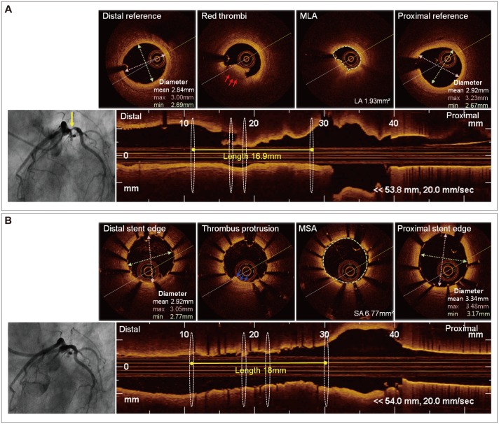
Fig. 1 OCT-guided PCI. A 63-year-old man was diagnosed with non-ST segment elevation myocardial infarction. A: baseline coronary angiogram showed significant stenosis of the proximal left circumflex artery. Pre-interventional OCT revealed a minimal lumen area of 1.93 mm2 and red thrombi (red arrow). The minimal lumen diameters at the proximal and distal reference segments were 2.69 mm and 2.67 mm, respectively, and the lesion length was 16.9 mm. B: a coronary angiogram after biolimus-eluting stent (2.75×18 mm) implantation. Post-interventional OCT showed A minimal stent area of 6.77 mm2 and a thrombic protrusion (blue arrow). OCT: optical coherence tomography, PCI: percutaneous coronary intervention, MLA: minimal lumen area, MSA: minimal stent area.
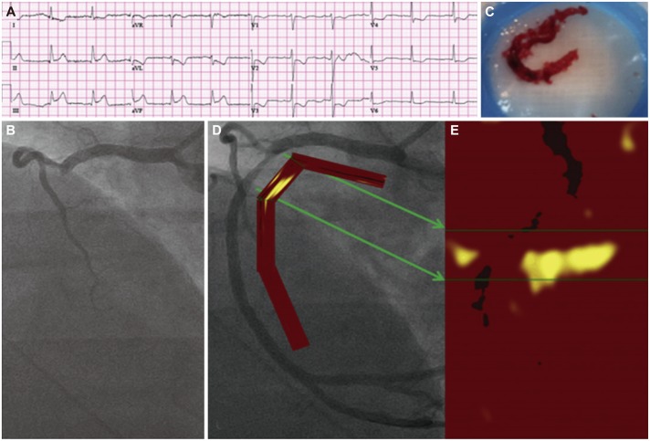
Fig. 2 Angiographic and NIRS findings in acute STEMI. A 56-year-old patient with acute chest pain and inferior-posterior injury was referred for primary PCI (A). Angiography of the right coronary artery revealed complete occlusion (B). Aspiration yielded a thrombus characteristic of STEMI (C) and resulted in a TIMI flow grade 3 (D). NIRS performed after the TIMI flow grade 3 was established revealed a prominent, nearly circumferential lipid core plaque concentrated at the culprit site (E). NIRS: near-infrared spectroscopy, STEMI: ST segment elevation myocardial infarction, PCI: percutaneous coronary intervention, TIMI: Thrombolysis in Myocardial Infarction. Adopted from Madder RD, Goldstein JA, Madden SP, et al. JACC Cardiovasc Interv 2013;6:838-46.74)
Reference
-
1. Lloyd-Jones D, Adams R, Carnethon M, et al. Heart disease and stroke statistics--2009 update: a report from the American Heart Association Statistics Committee and Stroke Statistics Subcommittee. Circulation. 2009; 119:480–486. PMID: 19171871.2. Arnett EN, Isner JM, Redwood DR, et al. Coronary artery narrowing in coronary heart disease: comparison of cineangiographic and necropsy findings. Ann Intern Med. 1979; 91:350–356. PMID: 475165.3. White CW, Wright CB, Doty DB, et al. Does visual interpretation of the coronary arteriogram predict the physiologic importance of a coronary stenosis? N Engl J Med. 1984; 310:819–824. PMID: 6700670.4. Hodgson JM, Graham SP, Savakus AD, et al. Clinical percutaneous imaging of coronary anatomy using an over-the-wire ultrasound catheter system. Int J Card Imaging. 1989; 4:187–193. PMID: 2527918.5. Lee SY, Mintz GS, Kim SY, et al. Attenuated plaque detected by intravascular ultrasound: clinical, angiographic, and morphologic features and post-percutaneous coronary intervention complications in patients with acute coronary syndromes. JACC Cardiovasc Interv. 2009; 2:65–72. PMID: 19463400.6. Potkin BN, Keren G, Mintz GS, et al. Arterial responses to balloon coronary angioplasty: an intravascular ultrasound study. J Am Coll Cardiol. 1992; 20:942–951. PMID: 1527306.7. Tang Z, Bai J, Su SP, et al. Cutting-balloon angioplasty before drug-eluting stent implantation for the treatment of severely calcified coronary lesions. J Geriatr Cardiol. 2014; 11:44–49. PMID: 24748881.8. Mintz GS, Pichard AD, Popma JJ, Kent KM, Satler LF, Leon MB. Preliminary experience with adjunct directional coronary atherectomy after high-speed rotational atherectomy in the treatment of calcific coronary artery disease. Am J Cardiol. 1993; 71:799–804. PMID: 8456757.9. Parise H, Maehara A, Stone GW, Leon MB, Mintz GS. Meta-analysis of randomized studies comparing intravascular ultrasound versus angiographic guidance of percutaneous coronary intervention in pre-drug-eluting stent era. Am J Cardiol. 2011; 107:374–382. PMID: 21257001.10. Ahn JM, Kang SJ, Yoon SH, et al. Meta-analysis of outcomes after intravascular ultrasound-guided versus angiography-guided drug-eluting stent implantation in 26,503 patients enrolled in three randomized trials and 14 observational studies. Am J Cardiol. 2014; 113:1338–1347. PMID: 24685326.11. Jang JS, Song YJ, Kang W, et al. Intravascular ultrasound-guided implantation of drug-eluting stents to improve outcome: a meta-analysis. JACC Cardiovasc Interv. 2014; 7:233–243. PMID: 24529934.12. de Jaegere P, Mudra H, Figulla H, et al. Intravascular ultrasound-guided optimized stent deployment. Immediate and 6 months clinical and angiographic results from the Multicenter Ultrasound Stenting in Coronaries Study (MUSIC Study). Eur Heart J. 1998; 19:1214–1223. PMID: 9740343.13. Zhang Y, Farooq V, Garcia-Garcia HM, et al. Comparison of intravascular ultrasound versus angiography-guided drug-eluting stent implantation: a meta-analysis of one randomised trial and ten observational studies involving 19,619 patients. EuroIntervention. 2012; 8:855–865. PMID: 23171805.14. Kawasaki M, Takatsu H, Noda T, et al. Noninvasive quantitative tissue characterization and two-dimensional color-coded map of human atherosclerotic lesions using ultrasound integrated backscatter: comparison between histology and integrated backscatter images. J Am Coll Cardiol. 2001; 38:486–492. PMID: 11499742.15. Kawasaki M, Takatsu H, Noda T, et al. In vivo quantitative tissue characterization of human coronary arterial plaques by use of integrated backscatter intravascular ultrasound and comparison with angioscopic findings. Circulation. 2002; 105:2487–2492. PMID: 12034654.16. Rodriguez-Granillo GA, Bruining N, Mc Fadden E, et al. Geometrical validation of intravascular ultrasound radiofrequency data analysis (Virtual Histology) acquired with a 30 MHz boston scientific corporation imaging catheter. Catheter Cardiovasc Interv. 2005; 66:514–518. PMID: 16281299.17. Nasu K, Tsuchikane E, Katoh O, et al. Impact of intramural thrombus in coronary arteries on the accuracy of tissue characterization by in vivo intravascular ultrasound radiofrequency data analysis. Am J Cardiol. 2008; 101:1079–1083. PMID: 18394436.18. Stone GW, Maehara A, Lansky AJ, et al. A prospective natural-history study of coronary atherosclerosis. N Engl J Med. 2011; 364:226–235. PMID: 21247313.19. Claessen BE, Maehara A, Fahy M, Xu K, Stone GW, Mintz GS. Plaque composition by intravascular ultrasound and distal embolization after percutaneous coronary intervention. JACC Cardiovasc Imaging. 2012; 5(3 Suppl):S111–S118. PMID: 22421225.20. Wu X, Maehara A, Mintz GS, et al. Virtual histology intravascular ultrasound analysis of non-culprit attenuated plaques detected by grayscale intravascular ultrasound in patients with acute coronary syndromes. Am J Cardiol. 2010; 105:48–53. PMID: 20102889.21. Rodriguez-Granillo GA, García-García HM, Mc Fadden EP, et al. In vivo intravascular ultrasound-derived thin-cap fibroatheroma detection using ultrasound radiofrequency data analysis. J Am Coll Cardiol. 2005; 46:2038–2042. PMID: 16325038.22. Uchida Y. Recent advances in coronary angioscopy. J Cardiol. 2011; 57:18–30. PMID: 21146367.23. Takano M, Mizuno K. Coronary angioscopic evaluation for serial changes of luminal appearance after pharmacological and catheter interventions. Circ J. 2010; 74:240–245. PMID: 20019412.24. Kotani J, Awata M, Nanto S, et al. Incomplete neointimal coverage of sirolimus-eluting stents: angioscopic findings. J Am Coll Cardiol. 2006; 47:2108–2111. PMID: 16697331.25. Alfonso F. The "vulnerable" stent why so dreadful? J Am Coll Cardiol. 2008; 51:2403–2406. PMID: 18565396.26. Yabushita H, Bouma BE, Houser SL, et al. Characterization of human atherosclerosis by optical coherence tomography. Circulation. 2002; 106:1640–1645. PMID: 12270856.27. Takarada S, Imanishi T, Liu Y, et al. Advantage of next-generation frequency-domain optical coherence tomography compared with conventional time-domain system in the assessment of coronary lesion. Catheter Cardiovasc Interv. 2010; 75:202–206. PMID: 19937788.28. Kubo T, Imanishi T, Takarada S, et al. Assessment of culprit lesion morphology in acute myocardial infarction: ability of optical coherence tomography compared with intravascular ultrasound and coronary angioscopy. J Am Coll Cardiol. 2007; 50:933–939. PMID: 17765119.29. Kubo T, Akasaka T, Shite J, et al. OCT compared with IVUS in a coronary lesion assessment: the OPUS-CLASS study. JACC Cardiovasc Imaging. 2013; 6:1095–1104. PMID: 24011777.30. Gonzalo N, Escaned J, Alfonso F, et al. Morphometric assessment of coronary stenosis relevance with optical coherence tomography: a comparison with fractional flow reserve and intravascular ultrasound. J Am Coll Cardiol. 2012; 59:1080–1089. PMID: 22421301.31. Kubo T, Imanishi T, Kitabata H, et al. Comparison of vascular response after sirolimus-eluting stent implantation between patients with unstable and stable angina pectoris: a serial optical coherence tomography study. JACC Cardiovasc Imaging. 2008; 1:475–484. PMID: 19356470.32. Gutiérrez-Chico JL, Wykrzykowska J, Nüesch E, et al. Vascular tissue reaction to acute malapposition in human coronary arteries: sequential assessment with optical coherence tomography. Circ Cardiovasc Interv. 2012; 5:20–29. S1–S8. PMID: 22319063.33. Im E, Kim BK, Ko YG, et al. Incidences, predictors, and clinical outcomes of acute and late stent malapposition detected by optical coherence tomography after drug-eluting stent implantation. Circ Cardiovasc Interv. 2014; 7:88–96. PMID: 24425586.34. Ino Y, Kubo T, Kitabata H, et al. Difference in neointimal appearance between early and late restenosis after sirolimus-eluting stent implantation assessed by optical coherence tomography. Coron Artery Dis. 2013; 24:95–101. PMID: 23363984.35. Kang SJ, Mintz GS, Akasaka T, et al. Optical coherence tomographic analysis of in-stent neoatherosclerosis after drug-eluting stent implantation. Circulation. 2011; 123:2954–2963. PMID: 21646494.36. Vergallo R, Yonetsu T, Uemura S, et al. Correlation between degree of neointimal hyperplasia and incidence and characteristics of neoatherosclerosis as assessed by optical coherence tomography. Am J Cardiol. 2013; 112:1315–1321. PMID: 23891431.37. Rath PC, Reddy K, Agarwal MK, Purohit BV, Deb T, Reddy AM. Optical coherence tomography guided PCI - initial experience at Apollo Health City, Jubilee Hills, Hyderabad. Indian Heart J. 2014; 66:31–37. PMID: 24581093.38. Prati F, Di Vito L, Biondi-Zoccai G, et al. Angiography alone versus angiography plus optical coherence tomography to guide decision-making during percutaneous coronary intervention: the Centro per la Lotta contro l'Infarto-Optimisation of Percutaneous Coronary Intervention (CLI-OPCI) study. EuroIntervention. 2012; 8:823–829. PMID: 23034247.39. Habara M, Nasu K, Terashima M, et al. Impact of frequency-domain optical coherence tomography guidance for optimal coronary stent implantation in comparison with intravascular ultrasound guidance. Circ Cardiovasc Interv. 2012; 5:193–201. PMID: 22456026.40. Kim IC, Hur SH, Cho YK, et al. Impact of optical coherence tomography-versus intravascular ultrasound-guided percutaneous coronary intervention on mid-term clinical outcomes. Eur Heart J. 2014; 35(suppl 1):1141.41. Cervinka P, Spaček R, Bystroň M, et al. Optical coherence tomography-guided primary percutaneous coronary intervention in ST-segment elevation myocardial infarction patients: a pilot study. Can J Cardiol. 2014; 30:420–427. PMID: 24680171.42. Moreno PR, Lodder RA, Purushothaman KR, Charash WE, O'Connor WN, Muller JE. Detection of lipid pool, thin fibrous cap, and inflammatory cells in human aortic atherosclerotic plaques by near-infrared spectroscopy. Circulation. 2002; 105:923–927. PMID: 11864919.43. Madder RD, Smith JL, Dixon SR, Goldstein JA. Composition of target lesions by near-infrared spectroscopy in patients with acute coronary syndrome versus stable angina. Circ Cardiovasc Interv. 2012; 5:55–61. PMID: 22253357.44. Waxman S, Dixon SR, L'Allier P, et al. In vivo validation of a catheter-based near-infrared spectroscopy system for detection of lipid core coronary plaques: initial results of the SPECTACL study. JACC Cardiovasc Imaging. 2009; 2:858–868. PMID: 19608137.45. Choi BJ, Prasad A, Gulati R, et al. Coronary endothelial dysfunction in patients with early coronary artery disease is associated with the increase in intravascular lipid core plaque. Eur Heart J. 2013; 34:2047–2054. PMID: 23569198.46. Dixon SR, Grines CL, Munir A, et al. Analysis of target lesion length before coronary artery stenting using angiography and near-infrared spectroscopy versus angiography alone. Am J Cardiol. 2012; 109:60–66. PMID: 21962996.47. Bourantas CV, Kourtis IC, Plissiti ME, et al. A method for 3D reconstruction of coronary arteries using biplane angiography and intravascular ultrasound images. Comput Med Imaging Graph. 2005; 29:597–606. PMID: 16278063.48. Pu J, Mintz GS, Brilakis ES, et al. In vivo characterization of coronary plaques: novel findings from comparing greyscale and virtual histology intravascular ultrasound and near-infrared spectroscopy. Eur Heart J. 2012; 33:372–383. PMID: 22019821.49. Schiele F, Meneveau N, Seronde MF, et al. Medical costs of intravascular ultrasound optimization of stent deployment. Results of the multicenter randomized ‘REStenosis after Intravascular ultrasound STenting' (RESIST) study. Int J Cardiovasc Intervent. 2000; 3:207–213. PMID: 12431345.50. Frey AW, Hodgson JM, Müller C, Bestehorn HP, Roskamm H. Ultrasound-guided strategy for provisional stenting with focal balloon combination catheter: results from the randomized Strategy for Intracoronary Ultrasound-guided PTCA and Stenting (SIPS) trial. Circulation. 2000; 102:2497–2502. PMID: 11076823.51. Mudra H, di Mario C, de Jaegere P, et al. Randomized comparison of coronary stent implantation under ultrasound or angiographic guidance to reduce stent restenosis (OPTICUS Study). Circulation. 2001; 104:1343–1349. PMID: 11560848.52. Oemrawsingh PV, Mintz GS, Schalij MJ, et al. Intravascular ultrasound guidance improves angiographic and clinical outcome of stent implantation for long coronary artery stenoses: final results of a randomized comparison with angiographic guidance (TULIP Study). Circulation. 2003; 107:62–67. PMID: 12515744.53. Gaster AL, Slothuus Skjoldborg U, Larsen J, et al. Continued improvement of clinical outcome and cost effectiveness following intravascular ultrasound guided PCI: insights from a prospective, randomised study. Heart. 2003; 89:1043–1049. PMID: 12923023.54. Gil RJ, Pawłowski T, Dudek D, et al. Comparison of angiographically guided direct stenting technique with direct stenting and optimal balloon angioplasty guided with intravascular ultrasound. The multicenter, randomized trial results. Am Heart J. 2007; 154:669–675. PMID: 17892989.55. Russo RJ, Silva PD, Teirstein PS, et al. A randomized controlled trial of angiography versus intravascular ultrasound-directed bare-metal coronary stent placement (the AVID Trial). Circ Cardiovasc Interv. 2009; 2:113–123. PMID: 20031704.56. Roy P, Steinberg DH, Sushinsky SJ, et al. The potential clinical utility of intravascular ultrasound guidance in patients undergoing percutaneous coronary intervention with drug-eluting stents. Eur Heart J. 2008; 29:1851–1857. PMID: 18550555.57. Park SJ, Kim YH, Park DW, et al. Impact of intravascular ultrasound guidance on long-term mortality in stenting for unprotected left main coronary artery stenosis. Circ Cardiovasc Interv. 2009; 2:167–177. PMID: 20031713.58. Jakabcin J, Spacek R, Bystron M, et al. Long-term health outcome and mortality evaluation after invasive coronary treatment using drug eluting stents with or without the IVUS guidance. Randomized control trial. HOME DES IVUS. Catheter Cardiovasc Interv. 2010; 75:578–583. PMID: 19902491.59. Claessen BE, Mehran R, Mintz GS, et al. Impact of intravascular ultrasound imaging on early and late clinical outcomes following percutaneous coronary intervention with drug-eluting stents. JACC Cardiovasc Interv. 2011; 4:974–981. PMID: 21939937.60. Kim JS, Hong MK, Ko YG, et al. Impact of intravascular ultrasound guidance on long-term clinical outcomes in patients treated with drug-eluting stent for bifurcation lesions: data from a Korean multicenter bifurcation registry. Am Heart J. 2011; 161:180–187. PMID: 21167352.61. Youn YJ, Yoon J, Lee JW, et al. Intravascular ultrasound-guided primary percutaneous coronary intervention with drug-eluting stent implantation in patients with ST-segment elevation myocardial infarction. Clin Cardiol. 2011; 34:706–713. PMID: 22057856.62. Park KW, Kang SH, Yang HM, et al. Impact of intravascular ultrasound guidance in routine percutaneous coronary intervention for conventional lesions: data from the EXCELLENT trial. Int J Cardiol. 2013; 167:721–726. PMID: 22481046.63. Ahn SG, Yoon J, Sung JK, et al. Intravascular ultrasound-guided percutaneous coronary intervention improves the clinical outcome in patients undergoing multiple overlapping drug-eluting stents implantation. Korean Circ J. 2013; 43:231–238. PMID: 23682282.64. Ahn JM, Han S, Park YK, et al. Differential prognostic effect of intravascular ultrasound use according to implanted stent length. Am J Cardiol. 2013; 111:829–835. PMID: 23273529.65. Chen SL, Ye F, Zhang JJ, et al. Intravascular ultrasound-guided systematic two-stent techniques for coronary bifurcation lesions and reduced late stent thrombosis. Catheter Cardiovasc Interv. 2013; 81:456–463. PMID: 22899562.66. Chieffo A, Latib A, Caussin C, et al. A prospective, randomized trial of intravascular-ultrasound guided compared to angiography guided stent implantation in complex coronary lesions: the AVIO trial. Am Heart J. 2013; 165:65–72. PMID: 23237135.67. Hur SH, Kang SJ, Kim YH, et al. Impact of intravascular ultrasound-guided percutaneous coronary intervention on long-term clinical outcomes in a real world population. Catheter Cardiovasc Interv. 2013; 81:407–416. PMID: 21805605.68. Kim JS, Kang TS, Mintz GS, et al. Randomized comparison of clinical outcomes between intravascular ultrasound and angiography-guided drug-eluting stent implantation for long coronary artery stenoses. JACC Cardiovasc Interv. 2013; 6:369–376. PMID: 23523455.69. Witzenbichler B, Maehara A, Weisz G, et al. Relationship between intravascular ultrasound guidance and clinical outcomes after drug-eluting stents: the assessment of dual antiplatelet therapy with drug-eluting stents (ADAPT-DES) study. Circulation. 2014; 129:463–470. PMID: 24281330.70. Shiono Y, Kitabata H, Kubo T, et al. Optical coherence tomography-derived anatomical criteria for functionally significant coronary stenosis assessed by fractional flow reserve. Circ J. 2012; 76:2218–2225. PMID: 22785153.71. Pyxaras SA, Tu S, Barbato E, et al. Quantitative angiography and optical coherence tomography for the functional assessment of nonobstructive coronary stenoses: comparison with fractional flow reserve. Am Heart J. 2013; 166:1010–1018. PMID: 24268215.72. Pawlowski T, Prati F, Kulawik T, Ficarra E, Bil J, Gil R. Optical coherence tomography criteria for defining functional severity of intermediate lesions: a comparative study with FFR. Int J Cardiovasc Imaging. 2013; 29:1685–1691. PMID: 23999603.73. Reith S, Battermann S, Jaskolka A, et al. Relationship between optical coherence tomography derived intraluminal and intramural criteria and haemodynamic relevance as determined by fractional flow reserve in intermediate coronary stenoses of patients with type 2 diabetes. Heart. 2013; 99:700–707. PMID: 23543283.74. Madder RD, Goldstein JA, Madden SP, et al. Detection by near-infrared spectroscopy of large lipid core plaques at culprit sites in patients with acute ST-segment elevation myocardial infarction. JACC Cardiovasc Interv. 2013; 6:838–846. PMID: 23871513.
- Full Text Links
-
- Actions
-
Cited
- CITED
-
- Close
- Share
-
- Similar articles
-
- Cardiac CT in Detecting a Dislodged Stent in Right Coronary Artery after Primary Percutaneous Coronary Intervention
- Recent Advances in Percutaneous Coronary Intervention in Coronary Artery Disease
- Functional Magnetic Resonance Imaging of the Brain: Principle and Practical Application
- Angiographical Changes Induced by Tiny Coronary Fistula
- Application of Artificial Intelligence to Cardiovascular Computed Tomography
