Advanced Surface Reconstruction Technique to Build Detailed Surface Models of the Liver and Neighboring Structures from the Visible Korean Human
- Affiliations
-
- 1Department of Anatomy, Ajou University School of Medicine, Suwon, Korea. dissect@ajou.ac.kr
- 2Department of Anatomy, Dongguk University College of Medicine, Gyeongju, Korea.
- 3Department of Film, TV and Multimedia, Sungkyunkwan University, Seoul, Korea.
- 4Korea Institute of Science and Technology Information, Daejeon, Korea.
- KMID: 1779151
- DOI: http://doi.org/10.3346/jkms.2009.24.3.375
Abstract
- Unlike volume models, surface models, which are empty three-dimensional images, have small file size, so that they can be displayed, rotated, and modified in a real time. For the reason, the surface models of liver and neighboring structures can be effectively applied to virtual hepatic segmentectomy, virtual laparoscopic cholecystectomy, and so on. The purpose of this research is to present surface models of detailed structures inside and outside the liver, which promote medical simulation systems. Forty-seven chosen structures were liver structures such as portal triad, hepatic vein, and neighboring structures such as the stomach, duodenum, muscles, bones, and skin. The structures were outlined in the serially sectioned images from the Visible Korean Human to prepare segmented images. From the segmented images, serial outlines of each structure were stacked; on the popular commercial software, advanced surface reconstruction technique was applied to build surface model of the structure. A surface model of the liver was divided into eight models of hepatic segments according to distribution of the portal vein. The surface models will be distributed to encourage researchers to develop the various kinds of medical simulation of the abdomen.
Keyword
MeSH Terms
Figure
-
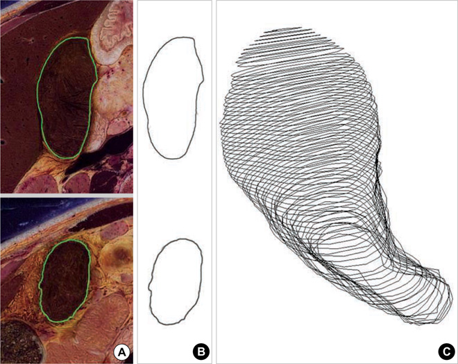
Fig. 1 Drawing and stacking of gallbladder's outlines. Segmented images on the serially sectioned images (A). Vectorized segmented images (B). Outlines lines model (C).
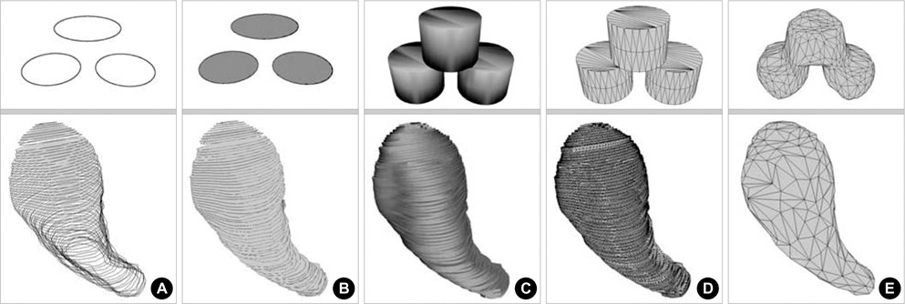
Fig. 2 Surface reconstruction of a schematic structure with three outlines (top row) and that of the gallbladder (bottom row), where neighboring outlines are overlapped. Outlines model (A). Packed outlines model (B). Columns volume model (C). Columns surface model (D). Smoothed surface model (E).

Fig. 3 Surface reconstruction of a schematic structure with four outlines (top row) and that of the hepatic artery (bottom row), where neighboring outlines are not-overlapped. Outlines model (A). Disassembled surface models (B). Assembled surface model (C). Smoothed surface model (D).

Fig. 4 Smoothed surface model of the gallbladder, superimposed on the outlines model (A). That superimposed on the serially sectioned image (B).
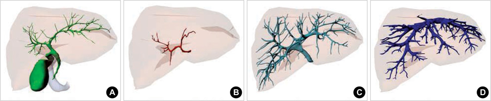
Fig. 5 Surface models of the portal triad and hepatic vein. Hepatic duct (A). Hepatic artery (B). Portal vein (C). Hepatic vein (D).
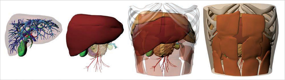
Fig. 6 Combined surface models of the liver and neighboring structures, which are differently selected to display.

Fig. 7 Surface model of the portal vein, which is differently colored according to the segmental branches (A). Surface model of the liver, which is divided into models of the hepatic segments (B).

Fig. 8 Volume model of the liver, which is rotated.
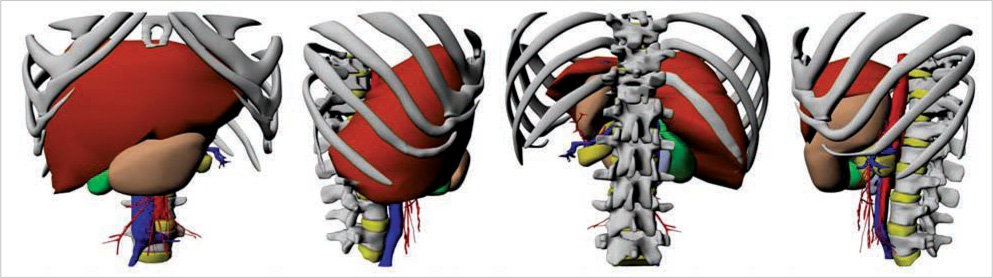
Fig. 9 Combined surface models of the liver and neighboring structures, which are rotated.
Reference
-
1. Muller MA, Marincek B, Frauenfelder T. State of the art 3D imaging of abdominal organs. JBR-BTR. 2007. 90:467–474.2. Jastrow H, Vollrath L. Anatomy online: presentation of a detailed WWW atlas of human gross anatomy. Reference for medical education. Clin Anat. 2002. 15:402–408.3. Jastrow H, Vollrath L. Teaching and learning gross anatomy by using modern electronic media based on the Visible Human Project. Clin Anat. 2003. 16:44–54.4. Doherty M, Bordes N, Hugh T, Pailthorpe B. 3D visualization of tumours and blood vessels in human liver. 2002. 22:In : Pan-Sydney Area Workshop on Visual Information Processing (VIP2002); 27–31.5. Spitzer VM, Ackerman MJ, Scherzinger AL, Whitlock DG. The visible human male. A technical report. J Am Med Inform Assoc. 1996. 3:118–130.
Article6. Park JS, Chung MS, Hwang SB, Lee YS, Har DH, Park HS. Visible Korean Human. Improved serially sectioned images of the entire body. IEEE Trans Med Imaging. 2005. 24:352–360.7. Park JS, Chung MS, Hwang SB, Shin BS, Park HS. Visible Korean Human. Its techniques and applications. Clin Anat. 2006. 19:216–224.
Article8. Park JS, Shin DS, Chung MS, Hwang SB, Chung J. Technique of semiautomatic surface reconstruction of the Visible Korean Human data by using commercial software. Clin Anat. 2007. 20:871–879.9. Uhl JF, Park JS, Chung MS, Delmas V. Three-dimensional reconstruction of urogenital tract from Visible Korean Human. Anat Rec Part A Discov Mol Cell Evol Biol. 2006. 288:893–899.
Article10. Park JS, Chung MS, Hwang SB, Lee YS, Har DH. Technical report on semiautomatic segmentation by using the Adobe Photoshop. J Digit Imaging. 2005. 18:333–343.11. Wilkins MR, Kazmier C. MEL Scripting for Maya Animators. 2005. 2nd ed. San Francisco: Morgan Kaufmann.12. FCAT (Federative Committee on Anatomical Terminology). Terminologia Anatomica, International Anatomical Terminology. 1998. Stuttgart, New York: Thieme.13. Moore KL, Dalley AF. Clinically Oriented Anatomy. 2006. 5th ed. Philadelphia: Lippincott Williams and Wilkins;293–295.14. Lehmann KS, Ritz JP, Valdeig S, Schenk A, Holmer C, Peitgen HO, Buhr HJ, Frericks BB. Portal vein segmentation of a 3D-planning system for liver surgery--in vivo evaluation in a porcine model. Ann Surg Oncol. 2008. 15:1899–1907.15. Sainz M, Susin A, Bagherzadeh N. MTMesh. Image based mesh reconstruction and rendering. 2003. In : IAESTED Conference in Visualization, Imaging and Image Processing (VIIP'03); 785–790.16. Haas A, Fischer MS. Three-dimensional reconstruction of histological sections using modern product-design software. Anat Rec. 1997. 249:510–516.
Article
- Full Text Links
-
- Actions
-
Cited
- CITED
-
- Close
- Share
-
- Similar articles
-
- Surface models of the male urogenital organs built from the Visible Korean using popular software
- Three types of the serial segmented images suitable for surface reconstruction
- The Visible Korean: Movable Surface Models of the Foot
- Dawn of the Visible Monkey: Segmentation of the Rhesus Monkey for 2D and 3D Applications
- Three Dimensional Automatic Surface Reconstruction Software
