Three types of the serial segmented images suitable for surface reconstruction
- Affiliations
-
- 1Department of Anatomy, Ajou University School of Medicine, Suwon, Korea. dissect@ajou.ac.kr
- 2Department of Anatomy, Dongguk University College of Medicine, Gyeongju, Korea.
- KMID: 2005876
- DOI: http://doi.org/10.5115/acb.2012.45.2.128
Abstract
- Stereoscopic surface models of human organs can be manipulated in real time. This is a significant feature of an interactive simulation system used for clinical practice. Objective surface models are obtainable from the accumulation of each structure's serial outlines, followed by surface reconstruction. The segmented images including the outlines can be divided into outlined images, white-filled images, and color-filled images. The purpose of this study was to report the benefits of the three types of segmented images for surface reconstruction. For the raw data, sectioned images of a male cadaver head were used. In the sectioned images, 91 structures were delineated for the preparation of 234 serial outlined images. The outlined images were converted into white-filled and color-filled images; the reverse conversion was also possible. The outlined images, including the original sectioned images, could be the source not only of surface models but also of volume models. The white-filled images, with a minimal file size, were preferred for separate surface reconstruction of the individual structures. The color-filled images, which allowed for recognition of the entire outlined structures simultaneously, were regarded as a good choice for the construction of several surface models. For the process, we employed a variety of software packages including those for animation, where the images were compatible. This information can be used by other investigators to build their own three-dimensional models. In addition, the surface models of detailed structures in the head, accompanied by the corresponding sectioned and segmented images, will hopefully contribute to various simulations that can be useful to clinicians.
Keyword
MeSH Terms
Figure
-
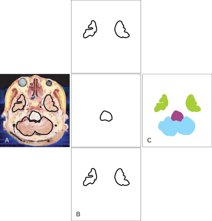
Fig. 1 Three types of segmented images of the cerebrum, cerebellum, and brainstem. (A) Outlined image drawn and displayed on the sectioned image. (B) White-filled images of individual structures. (C) Color-filled image.

Fig. 2 Schemes of demonstrating the conversion between three sorts of segmented images (1-6) and the surface reconstruction from them (7-9).

Fig. 3 Timeline panel of After Effects revealing the distribution of color-filled outlines of selected head structures.
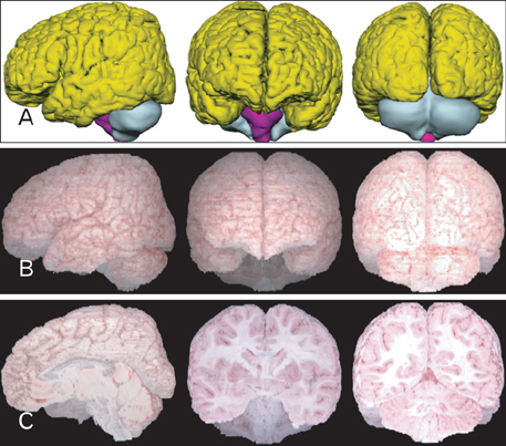
Fig. 4 Surface and volume models of the brain. (A) Surface models of the cerebrum (yellow), cerebellum (cyan), and brainstem (magenta). (B) Corresponding volume models. (C) Sectioned volume models.
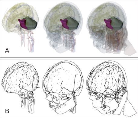
Fig. 5 Surface models of the head structures including brain. They are displayed either (A) semitransparently like a glass specimen or (B) opaquely like a sketch on paper.
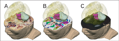
Fig. 6 Surface models of the brain, displayed with several types of two-dimensional images, such as (A) sectioned image, (B) color-filled image, and (C) magnetic resonance imaging.
Reference
-
1. Mahmoudi SE, Akhondi-Asl A, Rahmani R, Faghih-Roohi S, Taimouri V, Sabouri A, Soltanian-Zadeh H. Web-based interactive 2D/3D medical image processing and visualization software. Comput Methods Programs Biomed. 2010. 98:172–182.2. Park JS, Chung MS, Shin DS, Har DH, Cho ZH, Kim YB, Han JY, Chi JG. Sectioned images of the cadaver head including the brain and correspondences with ultrahigh field 7.0 T MRIs. Proc IEEE. 2009. 97:1988–1996.3. Park JS, Chung MS, Hwang SB, Lee YS, Har DH. Technical report on semiautomatic segmentation using the Adobe Photoshop. J Digit Imaging. 2005. 18:333–343.4. Park JS, Shin DS, Chung MS, Hwang SB, Chung J. Technique of semiautomatic surface reconstruction of the Visible Korean Human data using commercial software. Clin Anat. 2007. 20:871–879.5. Shin DS, Chung MS, Lee JW, Park JS, Chung J, Lee SB, Lee SH. Advanced surface reconstruction technique to build detailed surface models of the liver and neighboring structures from the Visible Korean Human. J Korean Med Sci. 2009. 24:375–383.6. Shin DS, Park JS, Shin BS, Chung MS. Surface models of the male urogenital organs built from the Visible Korean using popular software. Anat Cell Biol. 2011. 44:151–159.7. Jang HG, Chung MS, Shin DS, Park SK, Cheon KS, Park HS, Park JS. Segmentation and surface reconstruction of the detailed ear structures, identified in sectioned images. Anat Rec (Hoboken). 2011. 294:559–564.8. Moore KL, Dalley AF, Agur AM. Clinically oriented anatomy. 2010. 6th ed. Baltimore/Philadelphia: Wolters Kluwer/Lippincott Williams & Wilkins;820–980.9. Shin DS, Park JS, Park HS, Hwang SB, Chung MS. Outlining of the detailed structures in sectioned images from Visible Korean. Surg Radiol Anat. 2012. 34:235–247.10. Shin DS, Chung MS, Park HS, Park JS, Hwang SB. Browsing software of the Visible Korean data used for teaching sectional anatomy. Anat Sci Educ. 2011. 4:327–332.11. Au AG, Palathinkal D, Liggins AB, Raso VJ, Carey J, Lambert RG, Amirfazli A. A NURBS-based technique for subject-specific construction of knee bone geometry. Comput Methods Programs Biomed. 2008. 92:20–34.12. Park JS, Jung YW, Lee JW, Shin DS, Chung MS, Riemer M, Handels H. Generating useful images for medical applications from the Visible Korean Human. Comput Methods Programs Biomed. 2008. 92:257–266.13. Shin DS, Chung MS, Park JS, Park HS, Lee SB, Lee SH, Choi HN, Riemer M, Handels H, Lee JE, Jung W. Three-dimensional surface models of detailed lumbosacral structures reconstructed from the Visible Korean. Ann Anat. 2011. 193:64–70.14. Pommert A, Höhne KH, Pflesser B, Richter E, Riemer M, Schiemann T, Schubert R, Schumacher U, Tiede U. Creating a high-resolution spatial/symbolic model of the inner organs based on the Visible Human. Med Image Anal. 2001. 5:221–228.15. Shin DS, Park JS, Lee SB, Lee SH, Chung J, Chung MS. Surface model of the gastrointestinal tract constructed from the Visible Korean. Clin Anat. 2009. 22:601–609.16. Shin DS, Chung MS, Park JS. Systematized methods of surface reconstruction from the serial sectioned images of a cadaver head. J Craniofac Surg. 2012. 23:190–194.17. Viceconti M, Taddei F, Montanari L, Testi D, Leardini A, Clapworthy G, Van Sint Jan S. Multimod data manager: a tool for data fusion. Comput Methods Programs Biomed. 2007. 87:148–159.18. Brenton H, Hernandez J, Bello F, Strutton P, Purkayastha S, Firth T, Darzi A. Using multimedia and Web3D to enhance anatomy teaching. Comput Educ. 2007. 49:32–53.19. Ravichandiran K, Ravichandiran M, Oliver ML, Singh KS, McKee NH, Agur AM. Determining physiological cross-sectional area of extensor carpi radialis longus and brevis as a whole and by regions using 3D computer muscle models created from digitized fiber bundle data. Comput Methods Programs Biomed. 2009. 95:203–212.20. Park JS, Chung MS, Hwang SB, Lee YS, Har DH, Park HS. Visible Korean Human: improved serially sectioned images of the entire body. IEEE Trans Med Imaging. 2005. 24:352–360.21. Filippi S, Motyl B, Bandera C. Analysis of existing methods for 3D modelling of femurs starting from two orthogonal images and development of a script for a commercial software package. Comput Methods Programs Biomed. 2008. 89:76–82.22. Färber M, Hummel F, Gerloff C, Handels H. Virtual reality simula tor for the training of lumbar punctures. Methods Inf Med. 2009. 48:493–501.
- Full Text Links
-
- Actions
-
Cited
- CITED
-
- Close
- Share
-
- Similar articles
-
- Serial Slice Images and Segmented Images of the Brainstem for Recognizing the Stereoscopic Morphology of its Nuclei and Tracts
- Three Dimensional Automatic Surface Reconstruction Software
- Dawn of the Visible Monkey: Segmentation of the Rhesus Monkey for 2D and 3D Applications
- Manufacture of the Serially Sectioned Images of the Whole Body (Fifth Report: Methods for Manufacture of the Three Dimensional Images and Virtual Dissection Software)
- Serially Sectioned and Segmented Images of the Mouse for Learning Mouse Anatomy
