Gastric Metastasis from Breast Cancer
- Affiliations
-
- 1Department of Internal Medicine, Ewha Womans University School of Medicine, Seoul, Korea. shimkn@ewha.ac.kr
- KMID: 1775789
- DOI: http://doi.org/10.4166/kjg.2013.61.1.54
Abstract
- No abstract available.
MeSH Terms
-
Adenocarcinoma/*diagnosis/radiography/secondary
Adult
Antineoplastic Agents/therapeutic use
Breast Neoplasms/*diagnosis/drug therapy/pathology
Carrier Proteins/metabolism
Doxorubicin/therapeutic use
Drug Therapy, Combination
Endoscopy, Digestive System
Female
Glycoproteins/metabolism
Humans
Mastectomy, Modified Radical
Positron-Emission Tomography and Computed Tomography
Stomach Neoplasms/*diagnosis/radiography/secondary
Taxoids/therapeutic use
Tomography, X-Ray Computed
Antineoplastic Agents
Carrier Proteins
Glycoproteins
Taxoids
Doxorubicin
Figure
-
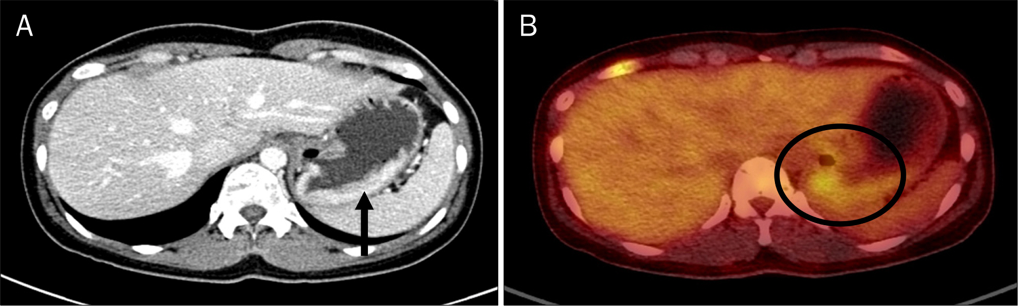
Fig. 1. Abdominal CT and PET-CT findings. (A) Diffuse wall thickening and enhancement (black arrow) were seen on the fundus. (B) It showed fluorodeoxyglucose uptake in the stomach and fundus (black circle), maximum standardized uptake value of 5.2.
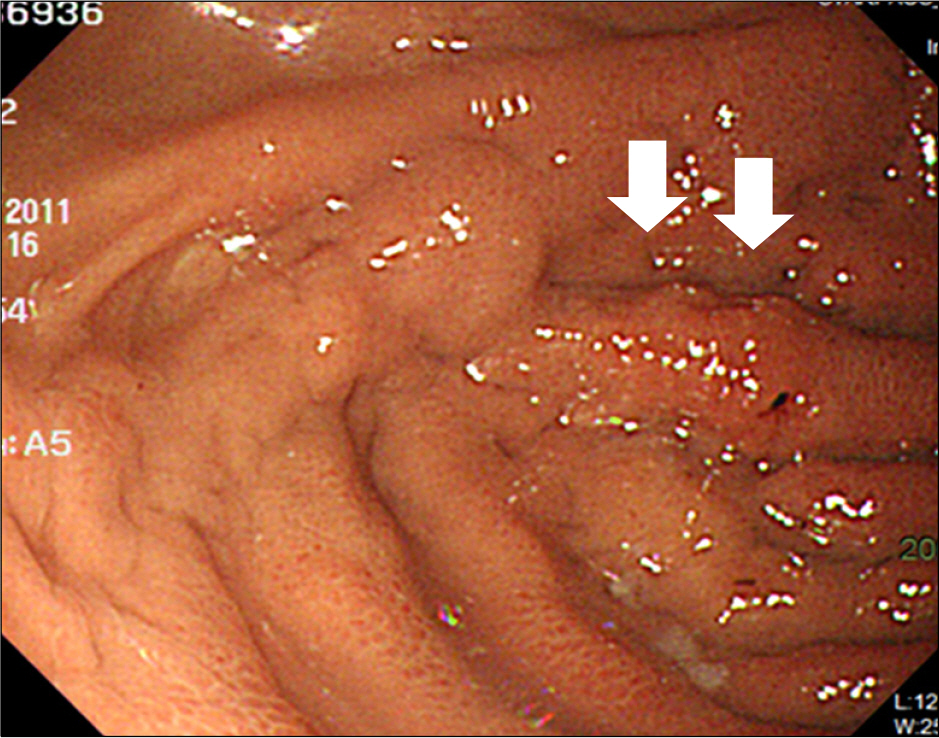
Fig. 2. Initial esophagogastroduodenoscopic finding. Congestive nodularity with hemorrhagic spot (white arrows) was seen on the fundus.
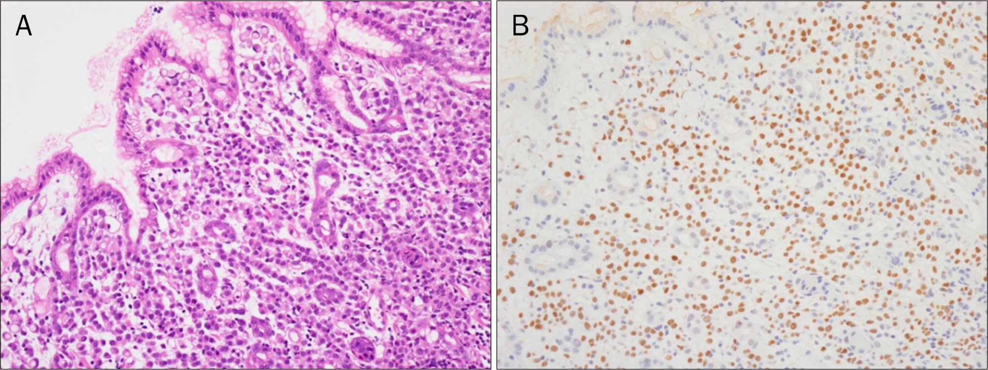
Fig. 3. Microscopic findings of the gastric biopsy. (A) It showed diffuse infiltrated adenocarcinoma with signet ring cell feature, consistent with metastatic lobular carcinoma with signet ring cell feature from the breast (H&E, ×200). (B) In additional estrogen receptor immunohistochemical stain, it revealed positive finding (Estrogen receptor, ×200).
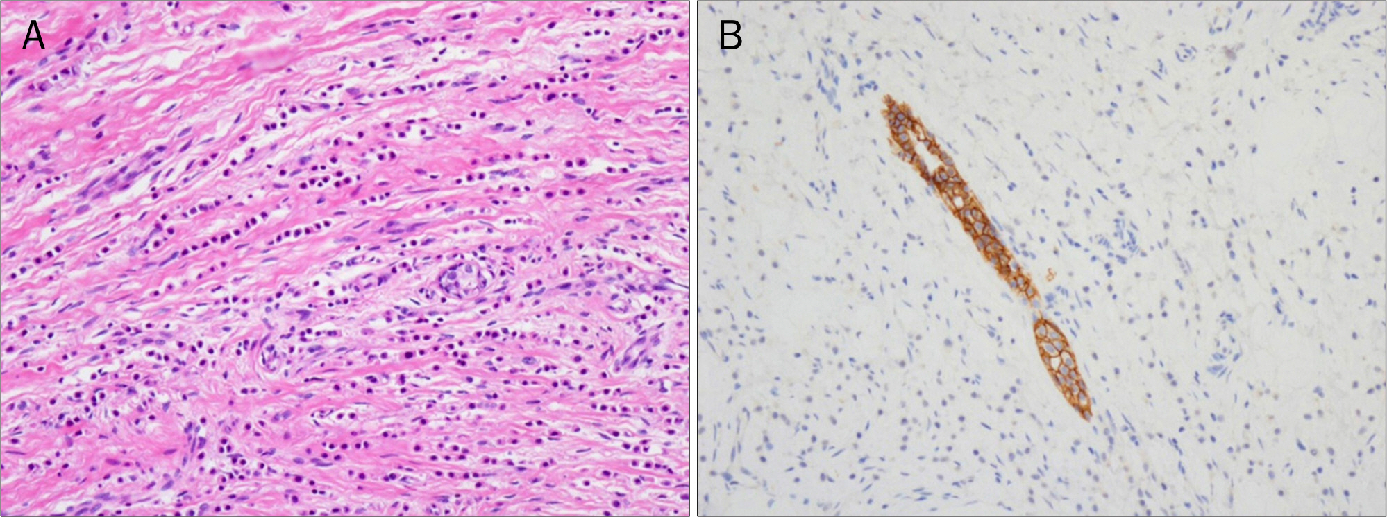
Fig. 4. Microscopic findings of the breast biopsy. (A) It was composed of non-cohesive cells individually dispersed in single-file linear patterns,‘Indian files', in fibrous stroma (H&E, ×200). (B) There was no detection of E-cadherin expression, and it was compatible with invasive lobular carcinoma (E-cadherin, ×200).
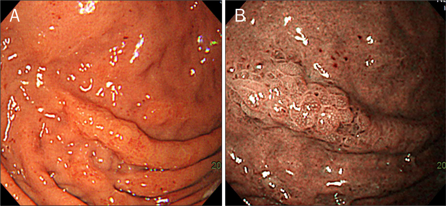
Fig. 5. One year follow-up esophagogastroduodenoscopic findings. (A) At the fundus, nodular elevated lesion with enlarged thichkened folds was seen without significant interval change. (B) It showed irregular nodularity in narrow band image.
Cited by 1 articles
-
Gastric Metastasis from Ovarian Cancer Presenting as a Submucosal Tumor: A Case Report
Eun Young Kim, Cho Hyun Park, Eun Sun Jung, Kyo Young Song
J Gastric Cancer. 2014;14(2):138-141. doi: 10.5230/jgc.2014.14.2.138.
Reference
-
References
1. McLemore EC, Pockaj BA, Reynolds C, et al. Breast cancer: presentation and intervention in women with gastrointestinal metastasis and carcinomatosis. Ann Surg Oncol. 2005; 12:886–894.
Article2. Jones GE, Strauss DC, Forshaw MJ, Deere H, Mahedeva U, Mason RC. Breast cancer metastasis to the stomach may mimic primary gastric cancer: report of two cases and review of literature. World J Surg Oncol. 2007; 5:75.
Article3. Koike K, Kitahara K, Higaki M, Urata M, Yamazaki F, Noshiro H. Clinicopathological features of gastric metastasis from breast cancer in three cases. Breast Cancer. 2011. [Epub ahead of print].
Article4. Taal BG, den Hartog Jager FC, Steinmetz R, Peterse H. The spectrum of gastrointestinal metastases of breast carcinoma: I. Stomach. Gastrointest Endosc. 1992; 38:130–135.
Article5. Jeon SH, Lee YS, Kwon TK, et al. A case of gastric metastasis from breast carcinoma manifested by upper gastrointestinal bleeding. Korean J Gastrointest Endosc. 2002; 24:220–224.6. Almubarak MM, Laé M, Cacheux W, et al. Gastric metastasis of breast cancer: a single centre retrospective study. Dig Liver Dis. 2011; 43:823–827.
Article7. Cheoi KS, Lee WY, Eum YO, et al. A case of stomach metastasis from breast cancer. Korean J Med. 2006; 71:567–572.8. Taal BG, Peterse H, Boot H. Clinical presentation, endoscopic features, and treatment of gastric metastases from breast carcinoma. Cancer. 2000; 89:2214–2221.
Article9. Cormier WJ, Gaffey TA, Welch JM, Welch JS, Edmonson JH. Linitis plastica caused by metastatic lobular carcinoma of the breast. Mayo Clin Proc. 1980; 55:747–753.10. Dumoulin FL, Sen Gupta R. Breast cancer metastasis to the stomach resembling small benign gastric polyps. Gastrointest Endosc. 2009; 69:174–175.
Article11. Yamamoto D, Yoshida H, Sumida K, et al. Gastric tumor from metastasis of breast cancer. Anticancer Res. 2010; 30:3705–3708.12. Kojima O, Takahashi T, Kawakami S, Uehara Y, Matsui M. Localization of estrogen receptors in gastric cancer using immunohistochemical staining of monoclonal antibody. Cancer. 1991; 67:2401–2406.
Article13. Wick MR, Lillemoe TJ, Copland GT, Swanson PE, Manivel JC, Kiang DT. Gross cystic disease fluid protein-15 as a marker for breast cancer: immunohistochemical analysis of 690 human neoplasms and comparison with alpha-lactalbumin. Hum Pathol. 1989; 20:281–287.
Article14. Park KW, Im YH, Lee J, et al. Use of GCDFP-15 (BRST-2) as a specific immunocytochemical marker for diagnosis of gastric metastasis of breast carcinoma. Cancer Res Treat. 2003; 35:460–464.
Article
- Full Text Links
-
- Actions
-
Cited
- CITED
-
- Close
- Share
-
- Similar articles
-
- A case of stomach metastasis from breast cancer
- An Unusual Case of Gastric Cancer Presenting with Breast Metastasis with Pleomorphic Microcalcifications
- Occult Invasive Lobular Carcinoma of Breast Detected by Stomach Metastasis: A Case Report
- Breast Cancer Metastasis to the Stomach Resembling Early Gastric Cancer
- Stomach and Colon Metastasis from Breast Cancer
