An Unusual Case of Gastric Cancer Presenting with Breast Metastasis with Pleomorphic Microcalcifications
- Affiliations
-
- 1Department of Radiology, Pamela Youde Nethersole Eastern Hospital, Chai Wan, Hong Kong, China. lys177@ha.org.hk
- 2Department of Pathology, Pamela Youde Nethersole Eastern Hospital, Chai Wan, Hong Kong, China.
- KMID: 2286463
- DOI: http://doi.org/10.4048/jbc.2012.15.3.356
Abstract
- Breast metastasis from gastric carcinoma is rare. We present a case of right breast mass with microcalcification in which the diagnosis of poorly differentiated adenocarcinoma from the stomach was made after a biopsy. Pleomorphic microcalcification was noted in the ill-defined breast mass, which is a rare feature in breast metastasis. Since breast metastasis usually signifies advanced metastatic disease, differentiating primary breast cancer from metastasis is important for appropriate treatment.
Figure
-
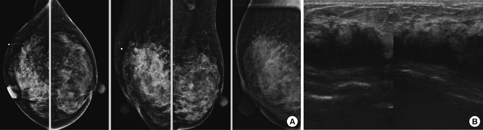
Figure 1 Mammographic findings. (A) Bilateral craniocaudal and medial-oblique views mammography of both breasts, cone-compression medialoblique view of the right breast mammography showing a large area of pleomorphic microcalcifications with architectural distortion in the upper outer quadrant of the right breast. (B) Ultrasonography revealed an ill-defined hypoechoic mass in the upper outer quadrant of the right breast.

Figure 2 Microscopic findings. (A, B) Immunohistochemical stain that showed cells from the right breast mass were negative for estrogen receptor and c-erbB-2 staining (Immunoperoxidase stain, ×200). (C, E) The malignant cells showed positivity in cytokeratin AE1/AE3, PAS-D, and E-cadherin staining (Immunoperoxidase stain, ×200). (D) (PAS stain, ×200). (F, G) A biopsy of the right breast mass showing infiltration of the stroma by malignant cells, most of which show signet ring features, compatible with metastasis from the stomach (H&E stain, ×200, ×400). (H) Pathological examination of bilateral ovarian lesions showing metastatic poorly differentiated adenocarcinoma with signet ring features (H&E stain, ×100). (I, J) Immunohistochemical examination of the ovarian lesions showed that the malignant cells were diffusely positive for cytokeratin 7 and focally positive of cytokeratin 20 (Immunoperoxidase stain, ×100).
Reference
-
1. Feder JM, de Paredes ES, Hogge JP, Wilken JJ. Unusual breast lesions: radiologic-pathologic correlation. Radiographics. 1999. 19(Spec No):S11–S26.
Article2. Chung SY, Oh KK. Imaging findings of metastatic disease to the breast. Yonsei Med J. 2001. 42:497–502.
Article3. Qureshi SS, Shrikhande SV, Tanuja S, Shukla PJ. Breast metastases of gastric signet ring cell carcinoma: a differential diagnosis with primary breast signet ring cell carcinoma. J Postgrad Med. 2005. 51:125–127.4. Boutis AL, Andreadis C, Patakiouta F, Mouratidou D. Gastric signetring adenocarcinoma presenting with breast metastasis. World J Gastroenterol. 2006. 12:2958–2961.
Article5. Parrell Soler C, Palacios Marqués A, Saco López L, Bermejo De Las Heras R, Pertusa Martínez S. Breast metastatic localization of signet-ring cell gastric carcinoma. ISRN Obstet Gynecol. 2011. 2011:426150.6. Madan AK, Ternovits C, Huber SA, Pei LA, Jaffe BM. Gastrointestinal metastasis to the breast. Surgery. 2002. 132:889–893.
Article7. Kwak JY, Kim EK, Oh KK. Radiologic findings of metastatic signet ring cell carcinoma to the breast from stomach. Yonsei Med J. 2000. 41:669–672.
Article8. Murphy WA, DeSchryver-Kecskemeti K. Isolated clustered microcalcifications in the breast: radiologic-pathologic correlation. Radiology. 1978. 127:335–341.
Article9. Yang WT, Suen M, Ahuja A, Metreweli C. In vivo demonstration of microcalcification in breast cancer using high resolution ultrasound. Br J Radiol. 1997. 70:685–690.
Article10. Dinkel HP, Gassel AM, Tschammler A. Is the appearance of microcalcifications on mammography useful in predicting histological grade of malignancy in ductal cancer in situ? Br J Radiol. 2000. 73:938–944.
Article11. Sato T, Muto I, Fushiki M, Hasegawa M, Sakai T, Sekiya M. Metastatic breast cancer from gastric and ovarian cancer, mimicking inflammatory breast cancer: report of two cases. Breast Cancer. 2008. 15:315–320.
Article12. Lee SK, Kim WW, Kim SH, Hur SM, Kim S, Choi JH, et al. Characteristics of metastasis in the breast from extramammary malignancies. J Surg Oncol. 2010. 101:137–140.
Article13. Çil T, Altintaş A, Paşa S, Işikdoğan A. Gastric ring cell carcinoma metastasis to the breast: two case reports. Turk J Cancer. 2009. 39:62–65.
- Full Text Links
-
- Actions
-
Cited
- CITED
-
- Close
- Share
-
- Similar articles
-
- A case of stomach metastasis from breast cancer
- Ultrasonographic Features and the Diagnostic Role of Core Needle Biopsy at Metastatic Breast Cancer in the Thyroid gland: A Case Report
- Increased Malignant Microcalcifications after Neoadjuvant Chemotherapy in Advanced Breast Cancer
- The Biologic Singificance of Mammographic Calcification
- Mucinous Breast Carcinoma Presenting as a Coarse and Densely Calcified Mass on Mammography: A Case Report
