J Korean Surg Soc.
2012 Dec;83(6):388-392. 10.4174/jkss.2012.83.6.388.
Primary cutaneous malignant melanoma of the breast
- Affiliations
-
- 1Department of Surgery, Chonbuk National University Medical School, Jeonju, Korea. yhj0903@jbnu.ac.kr
- 2Research Institute of Clinical Medicine of Chonbuk National University, Jeonju, Korea.
- 3Biomedical Research Institute of Chonbuk National University Hospital, Jeonju, Korea.
- 4Department of Pathology, Chonbuk National University Medical School, Jeonju, Korea.
- KMID: 1437538
- DOI: http://doi.org/10.4174/jkss.2012.83.6.388
Abstract
- Cutaneous malignant melanoma of the breast can be divided into two categories: primary and metastatic lesions. Cutaneous malignant melanoma of the breast is a rare tumor, accounting for less than 5% of all malignant melanomas. Clinical features and diagnostic methods of primary cutaneous malignant melanoma of the breast are similar to those arising from other cutaneous areas. Treatment of choice is wide local excision with adequate resection margin according to tumor thickness. Sentinel lymph node biopsy should be performed because the presence of lymph node metastasis is the most important prognostic factor. There have been only limited reports involving primary cutaneous malignant melanoma of the breast. Thus, we report a case of primary cutaneous malignant melanoma in a 59-year-old woman with a review of the recent literature.
MeSH Terms
Figure
-
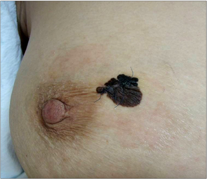
Fig. 1 A 59-year-old woman with a 2.0 × 1.5 cm sized and an ulcerative pigmented skin lesion in the right breast.

Fig. 2 Gross finding (A) and cut surface (B) of the right breast lesion after wide local excision.

Fig. 3 Microscopic findings of the right breast lesion. (A) Low power view reveals many tumor cell nests located in superficial dermis just beneath the epidermis (arrows). Tumor cell nests are arranged in laterally spreading pattern and some of them are brown-black pigmented (H&E, ×40). (B) High power view reveals tumor cells with large nuclei, prominent nucleoli and abundant cytoplasm with brown-black pigments (H&E, ×400).
Reference
-
1. Kurul S, Tas F, Buyukbabani N, Mudun A, Baykal C, Camlica H. Different manifestations of malignant melanoma in the breast: a report of 12 cases and a review of the literature. Jpn J Clin Oncol. 2005. 35:202–206.2. Alzaraa A, Sharma N. Primary cutaneous melanoma of the breast: a case report. Cases J. 2008. 1:212.3. Papachristou DN, Kinne DW, Rosen PP, Ashikari R, Fortner JG. Cutaneous melanoma of the breast. Surgery. 1979. 85:322–328.4. Biswas A, Goyal S, Jain A, Suri V, Mathur S, Julka PK, et al. Primary amelanotic melanoma of the breast: combating a rare cancer. Breast Cancer. 2010. 10. 27. [Epub]. http://dx.doi.org/10.1007/s12282-010-0231-8.5. Bassi F, Gatti G, Mauri E, Ballardini B, De Pas T, Luini A. Breast metastases from cutaneous malignant melanoma. Breast. 2004. 13:533–535.6. Balch CM, Gershenwald JE, Atkins MB, Buzaid AC, Cascinelli N, Cochran AJ, et al. Edge SB, Byrd DR, Compton CC, Fritz AG, Greene FL, Trotti A, editors. Melanoma of the skin. AJCC cancer staging manual. 2010. 7th ed. New York: Springer;325–344.7. Lee YT, Sparks FC, Morton DL. Primary melanoma of skin of the breast region. Ann Surg. 1977. 185:17–22.8. English DR, Armstrong BK, Kricker A, Fleming C. Sunlight and cancer. Cancer Causes Control. 1997. 8:271–283.9. Bono A, Baldi M, Maurichi A, Tomatis S. Distribution of melanoma on breast surface suggests its etiology. Int J Cancer. 2003. 105:434.10. Mitchell S, Lachica R, Randall MB, Beech DJ. Paget's disease of the breast areola mimicking cutaneous melanoma. Breast J. 2006. 12:233–236.
- Full Text Links
-
- Actions
-
Cited
- CITED
-
- Close
- Share
-
- Similar articles
-
- Cutaneous Malignant Melanoma Associated with Papillary Thyroid Cancer
- A Case of Malignant Melanoma Presenting as a Breast Mass
- Primary malignant melanoma arising in a cystic teratoma
- Second Primary Cancers in Patients with Cutaneous Malignant Melanoma
- Three Cases of Malignant Melanoma Possibly Arising in a Long Standing Melanocytic Nevus
