Cystic Changes in Intraabdominal Extrahepatic Metastases from Gastrointestinal Stromal Tumors Treated with Imatinib
- Affiliations
-
- 1Department of Radiology, Seoul National University College of Medicine, Institute of Radiation Medicine, SNUMRC, and Clinical Research Institute, Seoul National University Hospital, Korea. leejm@radcom.snu.ac.kr
- 2Department of Radiology, Kangwon National University College of Medicine, Korea.
- 3Radiation Medicine Branch, National Cancer Center.
- KMID: 1118820
- DOI: http://doi.org/10.3348/kjr.2004.5.3.157
Abstract
OBJECTIVE
This study was undertaken for the purpose of describing the CT features of intra-abdominal extra-hepatic metastases from gastrointestinal stromal tumors in patients who were treated with imatinib. MATERIALS AND METHODS: Eleven patients with intra-abdominal extra-hepatic metastases from gastrointestinal stromal tumors, who were treated with imatinib between May 2001 and December 2003, were included in this study. The clinical findings and CT scans were retrospectively reviewed. The metastatic lesions were assessed according to the location, size (greatest diameter), attenuation, and the enhancing pattern before and after imatinib treatment. RESULTS: Prior to the treatment, the sizes and attenuation values of the metastatic lesions ranged from 5 to 20 cm and from 63 to 131 H, respectively. The metastatic lesions showed a heterogeneous enhancement pattern on the contrast-enhanced CT scans. After the treatment, the metastatic lesions became smaller in all 11 patients, and the corresponding attenuation value ranged from 15 to 51 H. The metastatic lesions became homogeneous and cystic in appearance on the follow-up CT scans, mimicking ascites. CONCLUSION: Intra-abdominal extra-hepatic metastases of patients with gastrointestinal stromal tumors treated with imatinib may appear as well-circumscribed cystic lesions on contrast-enhanced CT. These metastases are likely to become smaller and resemble ascites, but may persist indefinitely on the follow-up CT.
MeSH Terms
-
Adult
Aged
Antineoplastic Agents/*therapeutic use
Contrast Media
Gastrointestinal Stromal Tumors/*pathology/surgery
Humans
Iohexol/*analogs & derivatives/diagnostic use
Liver Neoplasms/drug therapy/*radiography/secondary
Male
Middle Aged
Peritoneal Neoplasms/drug therapy/*radiography/secondary
Piperazines/*therapeutic use
Protein-Tyrosine Kinase/antagonists & inhibitors
Pyrimidines/*therapeutic use
Research Support, Non-U.S. Gov't
Retrospective Studies
Tomography, X-Ray Computed/methods
Figure
-
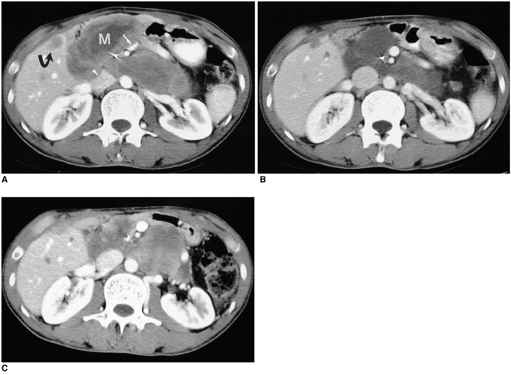
Fig. 1 A 34-year-old man with peritoneal seeding and liver metastases after resection of gastrointestinal stromal tumor of the duodenum. A. CT scan before treatment shows multiple heterogeneous masses (82 H) (M) compressing inferior vena cava (arrowhead) and superior mesenteric vein (arrow). Metastatic lesion in liver (curved arrow). B. CT scan obtained after 8 weeks of treatment with imatinib shows peritoneal and hepatic metastases that have decreased in size and density (35 H). Note the decompressed inferior vena cava and superior mesenteric vein and shrunk metastatic lesions in the liver. C. CT scan obtained 2 months after stopping imatinib therapy shows increased size and density of peritoneal and hepatic metastases. After resumption of imatinib therapy, the metastatic lesions showed cystic changes again (now shown).

Fig. 2 A 68-year-old man with peritoneal seeding after resection of gastrointestinal stromal tumor of the stomach. A. CT scan before treatment shows 15×13 cm heterogeneous metastatic lesion (81 H) (G) in left subphrenic space. Note small metastatic nodule (arrow) in right subphrenic space and ascites (15 H). B. CT scan obtained after 8 weeks of treatment with imatinib shows metastatic lesion (G) that has decreased in size to 8×6 cm and is cyst-like in appearance lesion (28 H) around spleen (S). Note the disappearance of the ascites. C. CT scan obtained after 13 months of treatment with imatinib shows 5×3.5 cm cystic lesion (20 H).
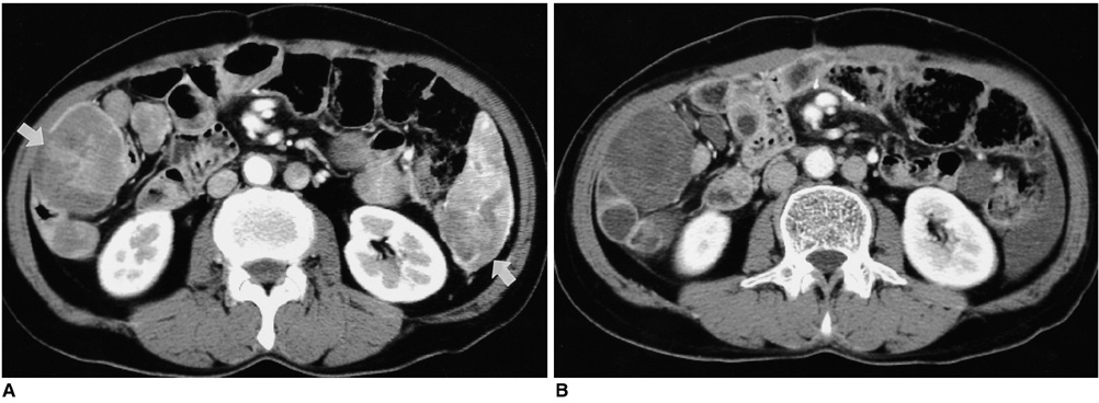
Fig. 3 A 52-year-old man with peritoneal seeding after resection of gastrointestinal stromal tumor of the mesentery. A. CT scan before treatment shows multiple peritoneal implants (arrows) in both paracolic gutters. B. CT scan obtained after 4 weeks of treatment with imatinib shows that the metastatic lesions in the right paracolic gutter have some solid components, while that in the left paracolic gutter resembles ascites.
Reference
-
1. Levy AD, Remotti HE, Thompson WM, Sobin LH, Miettinen M. Gastrointestinal stromal tumors: radiologic features with pathologic correlation. RadioGraphics. 2003. 23:283–304.2. Hirota S, Isozaki K, Moriyama Y, et al. Gain-of-function mutations of c-kit in human gastrointestinal stromal tumors. Science. 1998. 279:577–580.3. Demetri GD, von Mehren M, Blanke CD, et al. Efficacy and safety of imatinib mesylate in advanced gastrointestinal stromal tumors. N Engl J Med. 2002. 347:472–480.4. Burkill GJ, Badran M, Al-Muderis O, et al. Malignant gastrointestinal stromal tumor: distribution, imaging features, and pattern of metastatic spread. Radiology. 2003. 226:527–532.5. Levy AD, Remotti HE, Thompson WM, Sobin LH, Miettinen M. Anorectal gastrointestinal stromal tumors: CT and MR imaging features with clinical and pathologic correlation. AJR Am J Roentgenol. 2003. 180:1607–1612.6. Nishida T, Kumano S, Sugiura T, et al. Multidetector CT of high-risk patients with occult gastrointestinal stromal tumors. AJR Am J Roentgenol. 2003. 180:185–189.7. Kim H-C, Lee JM, Kim SH, et al. Primary gastrointestinal stromal tumors in the omentum and mesentery: CT findings and pathologic correlations. AJR Am J Roentgenol. 2004. 182:1463–1467.8. Kim H-C, Lee JM, Son KR, et al. Gastrointestinal stromal tumors of the duodenum: CT and barium study findings. AJR Am J Roentgenol. 2004. 183:415–419.9. Megibow AJ, Balthazar EJ, Hulnick DH, Naidich DP, Bosniak MA. CT evaluation of gastrointestinal leiomyomas and leiomyosarcomas. AJR Am J Roentgenol. 1985. 144:727–731.10. Joensuu H, Roberts PJ, Sarlomo-Rikala M, et al. Effect of the tyrosine kinase inhibitor STI571 in a patient with a metastatic gastrointestinal stromal tumor. N Engl J Med. 2001. 344:1052–1056.11. Chen MY, Bechtold RE, Savage PD. Cystic changes in hepatic metastases from gastrointestinal stromal tumors (GISTs) treated with Gleevec (imatinib mesylate). AJR Am J Roentgenol. 2002. 179:1059–1062.12. Bechtold RE, Chen MY, Stanton CA, Savage PD, Levine EA. Cystic changes in hepatic and peritoneal metastases from gastrointestinal stromal tumors treated with Gleevec. Abdom Imaging. 2003. 28:808–814.13. Bumming P, Andersson J, Meis-Kindblom JM, et al. Neoadjuvant, adjuvant and palliative treatment of gastrointestinal stromal tumours (GIST) with imatinib: a centre-based study of 17 patients. Br J Cancer. 2003. 89:460–464.14. Gayed I, Vu T, Iyer R, et al. The role of 18F-FDG PET in staging and early prediction of response to therapy of recurrent gastrointestinal stromal tumors. J Nucl Med. 2004. 45:17–21.
- Full Text Links
-
- Actions
-
Cited
- CITED
-
- Close
- Share
-
- Similar articles
-
- Metastatic Cutaneous Duodenal Gastrointestinal Stromal Tumor: A Possible Clue to Multiple Metastases
- Imatinib-induced DRESS Syndrome in Gastrointestinal Stromal Tumor
- Imatinib-induced hepatitis treated by corticosteroids in a patient with metastatic gastrointestinal stromal tumor
- A rare case of osteonecrosis of the jaw related to imatinib
- Two different KIT mutations may lead to different responses to imatinib in metastatic gastrointestinal stromal tumor
