Comparison of bare metal stent and paclitaxel-eluting stent using a novel rat aorta stent model
- Affiliations
-
- 1Department of Cardiology, Chonnam National University Hospital, Gwanju 501-757, Korea. cecilyk@chonnam.ac.kr
- 2Department of Cardiology, Chungbuk National University Hospital, Cheongju 361-711, Korea. kdwoon@chungbuk.ac.kr
- KMID: 1106556
- DOI: http://doi.org/10.4142/jvs.2011.12.2.143
Abstract
- The purpose of our study was to create a novel rat aorta stent implantation model. Stainless steel bare metal stents (BMS) or paclitaxel-eluting stents (PES) were implanted in male Sprague-Dawley rats (BW 400 +/- 20 g). Two and four weeks after stent implantation, the aorta were collected, fixed with 2% glutaraldehyde, and cut into two segments. One segment was used for scanning electron microscopy analysis to evaluate re-endothelialization, and the other segment was used to calculate the neointimal area. At 2 weeks after stenting, the appearance of neointimal hyperplasia was less in the PES group than in the BMS group. At 4 weeks after stenting, no significant difference in neointimal hyperplasia was observed between two groups. On the other hand, the PES group showed more thrombus formation and less re-endothelialization compared to the BMS group. This study demonstrated the ability of a novel rat model of aorta stenting via a common carotid artery to measure the efficacy and safety of commercially available drug-eluting stents.
Keyword
MeSH Terms
Figure
-
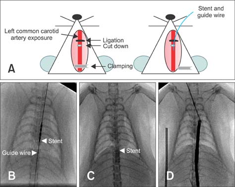
Fig. 1 Establishment of a novel aorta stent implantation procedure in rats. Representative illustrations and fluoroscopic angiographs of the implantation of a bare metal stent or paclitaxel-eluting stent in the thoracic aorta. (A) Operation procedure. (B) and (C) Operation procedure involved a guide wire that was advanced in the thoracic aorta after excision of the left common carotid artery and a stent inserted into the thoracic aorta. (D) Follow-up thoracic angiography was performed with a catheter in the right common carotid artery.
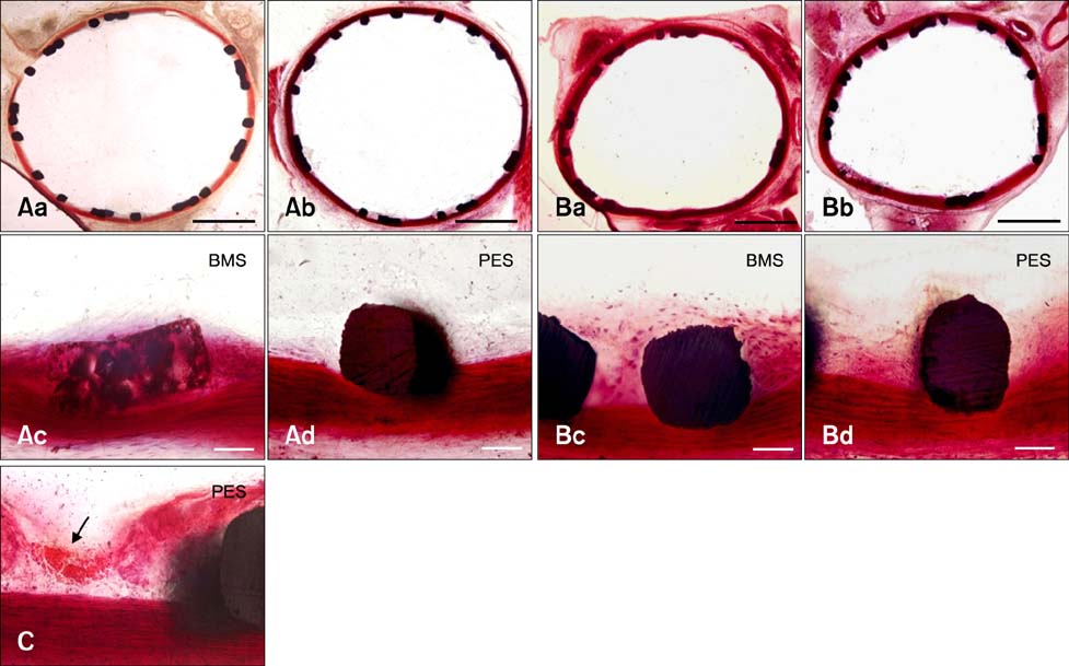
Fig. 2 (A) 2 weeks (wk) and (B) 4 wk after implantation of the bare metal stent (BMS) or paclitaxel-eluting stent (PES). (C) Thrombus in the outer and inner area of the neointima at 4 wk. Scale bars = Aa and b, Ba and b: 1 mm; Ac and d, Bc and d, C: 50 µm. H&E stain.

Fig. 3 Histomorphometric results of BMS or PES implantation. (A) Neointimal area. (B) Representation of neointimal area, media area (green line) and inner luminal area (square line). (C) Uncovered strut ratio. (D) Focal thrombus ratio. BMS 2 wk vs. PES 2 wk (*p < 0.05). BMS 4 wk vs. PES 4 wk (**p < 0.05), BMS 2 wk vs. BMS 4 wk (#p < 0.05), PES 2 wk vs. PES 4 wk (##p < 0.05).
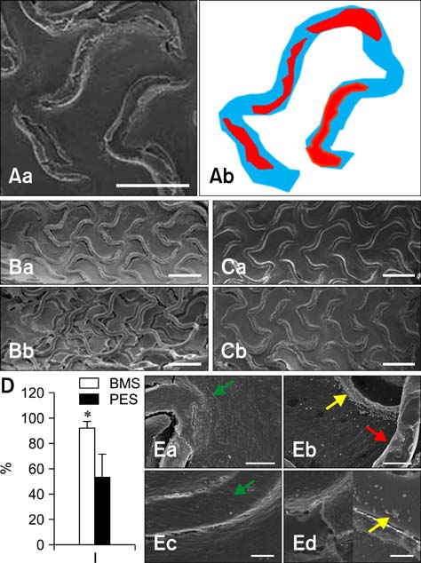
Fig. 4 Scanning electron microscopy photographs of BMS and PES in rat aorta. (A) Representation of strut area (blue) and uncovered area (red). (B) BMS and PES at 2 wk and (C) at 4 wk. (D) The covered stent ratio at 4 wk after stent implantation. (E) High magnification. Endothelial cells (green arrow), inflammatory cells (yellow arrow), and stent strut (red arrow). *p < 0.05. Ba and Ea: BMS 2 wk, Bb and Eb: PES 2 wk, Ca and Ec: BMS 4 wk, Cb and Ed: PES 4 wk. Scale bars = Aa: 500 µm, B and C: 1 mm, Ea and b: 100 µm, Ec and d: 50 µm.
Reference
-
1. Alhan C, Karabulut H, Senay S, Cagil H, Toraman F. Endovascular treatment of occlusive abdominal aortic thrombosis. Heart Vessels. 2010. 25:70–72.
Article2. Busseuil D, Collin B, Rioufol G, Korandji C, Zeller M, Maingon P, Briot F, Cottin Y, Rochette L. Combining sirolimus-eluting stents and external irradiation in cholesterol-fed rabbits increased incomplete stent apposition and decreased re-endothelialization. J Cardiovasc Pharmacol. 2009. 53:318–324.
Article3. Curfman GD, Morrissey S, Jarcho JA, Drazen JM. Drug-eluting coronary stents - Promise and uncertainty. N Engl J Med. 2007. 356:1059–1060.
Article4. Drachman DE, Edelman ER, Seifert P, Groothuis AR, Bornstein DA, Kamath KR, Palasis M, Yang D, Nott SH, Rogers C. Neointimal thickening after stent delivery of paclitaxel: change in composition and arrest of growth over six months. J Am Coll Cardiol. 2000. 36:2325–2332.
Article5. Ertaş G, van Beusekom HM, van Giessen WJ. Late stent thrombosis, endothelialisation and drug-eluting stents. Neth Heart J. 2009. 17:177–180.
Article6. Farb A, Boam AB. Stent thrombosis redux - The FDA perspective. N Engl J Med. 2007. 356:984–987.
Article7. Finn AV, Gold HK, Tang A, Weber DK, Wight TN, Clermont A, Virmani R, Kolodgie FD. A novel rat model of carotid artery stenting for the understanding of restenosis in metabolic diseases. J Vasc Res. 2002. 39:414–425.
Article8. Finn AV, Kolodgie FD, Harnek J, Guerrero LJ, Acampado E, Tefera K, Skorija K, Weber DK, Gold HK, Virmani R. Differential response of delayed healing and persistent inflammation at sites of overlapping sirolimus- or paclitaxel-eluting stents. Circulation. 2005. 112:270–278.
Article9. Finn AV, Nakazawa G, Joner M, Kolodgie FD, Mont EK, Gold HK, Virmani R. Vascular responses to drug eluting stents: importance of delayed healing. Arterioscler Thromb Vasc Biol. 2007. 27:1500–1510.10. Finn AV, Joner M, Nakazawa G, Kolodgie F, Newell J, John MC, Gold HK, Virmani R. Pathological correlates of late drug-eluting stent thrombosis: strut coverage as a marker of endothelialization. Circulation. 2007. 115:2435–2441.
Article11. Gallo R, Padurean A, Jayaraman T, Marx S, Roque M, Adelman S, Chesebro J, Fallon J, Fuster V, Marks A, Badimon JJ. Inhibition of intimal thickening after balloon angioplasty in porcine coronary arteries by targeting regulators of the cell cycle. Circulation. 1999. 99:2164–2170.
Article12. Indolfi C, Esposito G, Stabile E, Cavuto L, Pisani A, Coppola C, Torella D, Perrino C, Di Lorenzo E, Curcio A, Palombini L, Chiariello M. A new rat model of small vessel stenting. Basic Res Cardiol. 2000. 95:179–185.
Article13. Jang HS, Nam HY, Kim JM, Hahm DH, Nam SH, Kim KL, Joo JR, Suh W, Park JS, Kim DK, Gwon HC. Effects of curcumin for preventing restenosis in a hypercholesterolemic rabbit iliac artery stent model. Catheter Cardiovasc Interv. 2009. 74:881–888.
Article14. Johnson GJ, Griggs TR, Badimon L. The utility of animal models in the preclinical study of interventions to prevent human coronary artery restenosis: analysis and recommendations. On behalf of the Subcommittee on Animal, Cellular and Molecular Models of Thrombosis and Haemostasis of the Scientific and Standardization Committee of the International Society on Thrombosis and Haemostasis. Thromb Haemost. 1999. 81:835–843.15. Kastrati A, Mehilli J, Pache J, Kaiser C, Valgimigli M, Kelbæk H, Menichelli M, Sabaté M, Suttorp MJ, Baumgart D, Seyfarth M, Pfisterer ME, Schömig A. Analysis of 14 trials comparing sirolimus-eluting stents with bare-metal stents. N Engl J Med. 2007. 356:1030–1039.
Article16. Kotani J, Awata M, Nanto S, Uematsu M, Oshima F, Minamiguchi H, Mintz GS, Nagata S. Incomplete neointimal coverage of sirolimus-eluting stents: angioscopic findings. J Am Coll Cardiol. 2006. 47:2108–2111.17. Lagerqvist B, James SK, Stenestrand U, Lindbäck J, Nilsson T, Wallentin L. the SCAAR Study Group. Long-term outcomes with drug-Eluting stents versus Bare-Metal stents in Sweden. N Engl J Med. 2007. 356:1009–1019.
Article18. Langeveld B, Roks AJM, Tio RA, van Boven AJ, van der Want JJL, Henning RH, van Beusekom HMM, van der Giessen WJ, Zijlstra F, van Gilst WH. Rat abdominal aorta stenting: a new and reliable small animal model for in-stent restenosis. J Vasc Res. 2004. 41:377–386.
Article19. Li J, Jabara R, Pendyala L, Otsuka Y, Shinke T, Hou D, Robinson K, Chronos N. Abnormal vasomotor function of porcine coronary arteries distal to sirolimus-eluting stents. JACC Cardiovasc Interv. 2008. 1:279–285.
Article20. Lim SY, Jeong MH, Hong SJ, Lim DS, Moon JY, Hong YJ, Kim JH, Ahn Y, Kang JC. Inflammation and delayed endothelization with overlapping drug-eluting stents in a porcine model of in-stent restenosis. Circ J. 2008. 72:463–468.
Article21. Maisel WH. Unanswered questions - Drug-eluting stents and the risk of late thrombosis. N Engl J Med. 2007. 356:981–984.
Article22. Mauri L, Hsieh WH, Massaro JM, Ho KKL, D'Agostino R, Cutlip DE. Stent thrombosis in randomized clinical trials of drug-Eluting stents. N Engl J Med. 2007. 356:1020–1029.
Article23. Park SJ, Shim WH, Ho DS, Raizner AE, Park SW, Hong MK, Lee CW, Choi D, Jang Y, Lam R, Weissman NJ, Mintz GS. A paclitaxel-eluting stent for the prevention of coronary restenosis. N Engl J Med. 2003. 348:1537–1545.
Article24. Suzuki T, Kopia G, Hayashi S, Bailey LR, Llanos G, Wilensky R, Klugherz BD, Papandreou G, Narayan P, Leon MB, Yeung AC, Tio F, Tsao PS, Falotico R, Carter AJ. Stent-based delivery of sirolimus reduces neointimal formation in a porcine coronary model. Circulation. 2001. 104:1188–1193.
Article25. Spaulding C, Daemen J, Boersma E, Cutlip DE, Serruys PW. A pooled analysis of data comparing sirolimus-eluting stents with bare-metal stents. N Engl J Med. 2007. 356:989–997.
Article26. Stone GW, Moses JW, Ellis SG, Schofer J, Dawkins KD, Morice MC, Colombo A, Schampaert E, Grube E, Kirtane AJ, Cutlip DE, Fahy M, Pocock SJ, Mehran R, Leon MB. Safety and efficacy of sirolimus- and paclitaxel-eluting coronary stents. N Engl J Med. 2007. 356:998–1008.
Article27. Suzuki Y, Ikeno F, Koizumi T, Tio F, Yeung AC, Yock PG, Fitzgerald PJ, Fearon WF. In vivo comparison between optical coherence tomography and intravascular ultrasound for detecting small degrees of in-stent neointima after stent implantation. JACC Cardiovasc Interv. 2008. 1:168–173.
Article28. Virmani R, Kolodgie FD, Farb A, Lafont A. Drug eluting stents: are human and animal studies comparable? Heart. 2003. 89:133–138.
Article
- Full Text Links
-
- Actions
-
Cited
- CITED
-
- Close
- Share
-
- Similar articles
-
- A Case of Neointimal Calcification in a Drug-Eluting Stent
- Late Stent Thrombosis Associated with Late Stent Malapposition after Drug-Eluting Stenting: A Case Report
- A Case of Stent Strut Fracture of a Paclitaxel-Eluting Stent at the Time of Stent Implantation in a Complex Coronary Lesion
- A Case of Stent Thrombosis Occurred at 5 Years after Sirolimus-Eluting Stent Implantation
- Stent Thrombosis in the Era of the Drug-Eluting Stent
