Immunohistochemical localization of calcium binding proteins and some neurotransmitters in myenteric plexus of goat stomach
- Affiliations
-
- 1Department of Anatomy and Cell Biology, College of Veterinary Medicine, Seoul National University, Seoul 151-742, Korea. leehss@snu.ac.kr
- KMID: 1089476
- DOI: http://doi.org/10.4142/jvs.2006.7.4.315
Abstract
- To understand the neurochemical properties of the gastric myenteric plexus of ruminants, the expression patterns of calbindin D-28k (CB), calretinin (CR), substance P (SP) and calcitonin gene-related peptide (CGRP) were explored in the Korean native goat. In gastric myenteric plexus, CB and SP immunoreactivity were observed in round- or ovalshaped neurons. CR and CGRP immunoreactivity were detected only in the nerve fibers. This immunohistochemical localization of CB, CR, CGRP and SP in the myenteric plexus of the goat stomach exhibited species-specific patterns. These findings suggest that these substances may be directly or indirectly related to the gastric functions of the goat stomach.
Keyword
MeSH Terms
Figure
-
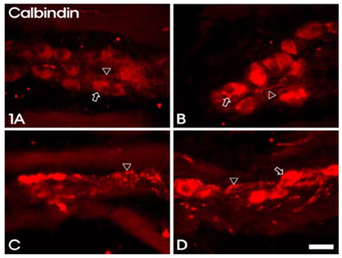
Fig. 1 CB immunoreactivity in the stomach myenteric plexus of Korean native goat. 1A: rumen, B: reticulum, C: omasum, D: abomasum. CB immunoreactivity is observed in both the nerve cell bodies (open arrows) and the fibers (arrowheads) (A, B and D), although only CB-IR fibers are observed in the omasum (C). bar = 50 µm.
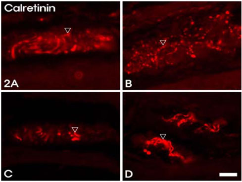
Fig. 2 CR immunoreactivity in the stomach myenteric plexus of Korean native goat. 2A: rumen, B: reticulum, C: omasum, D: abomasum. CR immunoreactivity is observed only in the fibers (arrowheads) (A-D), and varicosities of CR-IR fibers are dominant in the reticulum (B). bar = 50 µm.
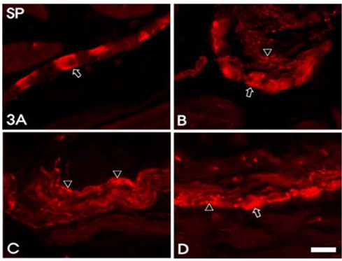
Fig. 3 SP immunoreactivity in the stomach myenteric plexus of Korean native goat. 3A: rumen, B: reticulum, C: omasum, D: abomasum. SP immunoreactivity is observed in both the nerve cell bodies (open arrows) and the fibers (arrowheads) (A, B and D), although only SP-IR fibers are observed in the omasum (C). bar = 50 µm.
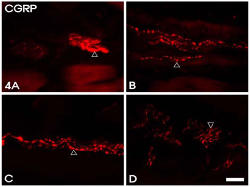
Fig. 4 CGRP immunoreactivity in the stomach myenteric plexus of Korean native goat. 4A: rumen, B: reticulum, C: omasum, D: abomasum. CGRP immunoreactivity is only observed in the fibers (arrowheads) (A-D). bar = 50 µm.
Reference
-
1. El-Salhy M, Spangeus A. Substance P in the gastrointestinal tract of non-obese diabetic mice. Scand J Gastroenterol. 1998. 33:394–400.
Article2. Furness JB. Types of neurons in the enteric nervous system. J Auton Nerv Syst. 2000. 81:87–96.
Article3. Furness JB, Lloyd KCK, Sternini C, Walsh JH. Projections of substance P, vasoactive intestinal peptide and tyrosine hydroxylase immunoreactive nerve fibres in the canine intestine, with special reference to the innervation of the circular muscle. Arch Histol Cytol. 1990. 53:129–140.
Article4. Furness JB, Trussell DC, Pompolo S, Bornstein JC, Smith TK. Calbindin neurons of the guinea-pig small intestine: quantitative analysis of their numbers and projections. Cell Tissue Res. 1990. 260:261–272.
Article5. Furness JB, Trussell DC, Pompolo S, Bornstein JC, Maley BE, Storm-Mathisen J. Shapes and projections of neurons with immunoreactivity for gamma-aminobutyric acid in the guinea-pig small intestine. Cell Tissue Res. 1989. 256:293–301.
Article6. Goyal RK, Hirano I. The enteric nervous system. N Engl J Med. 1996. 334:1106–1115.
Article7. Gregory PC. Forestomach motility in the chronically vagotomized sheep. J Physiol. 1982. 328:431–447.
Article8. Hens J, Schrodl F, Brehmer A, Adriaensen D, Neuhuber W, Scheuermann DW, Schemann M, Timmermans JP. Mucosal projections of enteric neurons in the porcine small intestine. J Comp Neurol. 2000. 421:429–436.
Article9. Hansen MB. The enteric nervous system I: organization and classification. Pharmacol Toxicol. 2003. 92:105–113.10. Lees GM, MacKenzie GM, Pearson GT. Complex correlations between the morphology, electrophysiology and peptide immunohistochemistry of guinea-pig enteric neurones. Eur J Morphol. 1992. 30:123–136.11. National Research Council. Guide for the care and use of laboratory animals. 1996. Reissue ed. Washington DC: National Academy Press;21–48. 56–65.12. Pearson GT. Structural organization and neuropeptide distributions in the equine enteric nervous system: an immunohistochemical study using whole-mount preparations from the small intestine. Cell Tissue Res. 1994. 276:523–534.
Article13. Pfannkuche H, Schemann M, Gäbel G. Ruminal muscle of sheep is innervated by non-polarized pathways of cholinergic and nitrergic myenteric neurones. Cell Tissue Res. 2002. 309:347–354.
Article14. Resibois A, Vienne G, Pochet R. Calbindin D-28K and the peptidergic neuroendocrine system in rat gut: an immunohistochemical study. Biol Cell. 1988. 63:67–75.
Article15. Rukebusch Y. Schultz SG, Wood JD, Rauner BB, editors. Gastrointestinal motor functions in ruminants. Handbook of Physiology. The Gastrointestinal System. 1989. Vol. 1. New York: Oxford University Press;1225–1283.16. Sang Q, Young HM. Chemical coding of neurons in the myenteric plexus and external muscle of the small and large intestine of the mouse. Cell Tissue Res. 1996. 284:39–53.
Article17. Scheuermann DW, Stach W, De Groodt-Lasseel MH, Timmermans JP. Calcitonin gene-related peptide in morphologically well-defined type II neurons of the enteric nervous system in the porcine small intestine. Acta Anat (Basel). 1987. 129:325–328.
Article18. Seo JH, Cho SS, Lee IS, Lee HS. Anatomical and neuropeptidergic properties of the duodenal neurons projecting to the gallbladder in the golden hamster. Arch Histol Cytol. 2002. 65:317–321.
Article19. Song ZM, Brookes SJ, Costa M. All calbindin-immunoreactive myenteric neurons project to the mucosa of the guinea-pig small intestine. Neurosci Lett. 1994. 180:219–222.
Article20. Timmermans JP, Adriaensen D, Cornelissen W, Scheuermann DW. Structural organization and neuropeptide distribution in the mammalian enteric nervous system, with special attention to those components involved in mucosal relaxes. Comp Biochem Physiol A Physiol. 1997. 118:331–340.
Article21. Wasserman RH, Taylor AN. Vitamin D3-induced calcium-binding protein in chick intestinal mucosa. Science. 1966. 152:791–793.
Article22. Wathuta EM. The distribution of vasoactive intestinal polypeptide-like, substance P-like and bombesin-like immunoreactivity in the digestive system of the sheep. Q J Exp Physiol. 1986. 71:615–631.
Article
- Full Text Links
-
- Actions
-
Cited
- CITED
-
- Close
- Share
-
- Similar articles
-
- Changes of Immunoreactivities of Calcium Channel alpha(1B)Subunit in Myenteric Plexus of Capsaicin Treated Adult Rats
- Impact of Myenteric Plexus Alterations on Diabetes Related Gastrointestinal Dysmotility
- Unusual Histology of Eosinophilic Myenteric Ganglionitis: A Case Report
- Immunohistochemical Analysis of Calretinin and Parvalbumin in the Goat Main Olfactory Bulb
- Double Immunohistochemical Studies on Distribution and Coexistence with Putative Neurotransmitters and Calretinin in Trigeminal Ganglion of Korean Native Goat
