J Pathol Transl Med.
2017 May;51(3):320-324. 10.4132/jptm.2016.09.07.
Unusual Histology of Eosinophilic Myenteric Ganglionitis: A Case Report
- Affiliations
-
- 1Department of Pathology, Eulji University School of Medicine, Daejeon, Korea.
- 2Department of Radiology, Eulji University School of Medicine, Daejeon, Korea.
- 3Department of Surgery, Eulji University School of Medicine, Daejeon, Korea. jhjang@eulji.ac.kr
- KMID: 2392599
- DOI: http://doi.org/10.4132/jptm.2016.09.07
Abstract
- Eosinophilic myenteric ganglionitis is a disorder characterized by infiltration of the Auerbach myenteric plexus by eosinophils. As a cause of chronic intestinal pseudo-obstruction (CIPO), eosinophilic myenteric ganglionitis has been rarely reported and the majority of the reported cases in the literature were children. We experienced a case of eosinophilic myenteric ganglionitis associated with CIPO in a 53-year-old female patient. Histologic examination of the resected descending colon showed moderate eosinophilic infiltrates with hypogangliosis in the myenteric plexus. Immunohistochemical study revealed increased number of CD4-positive lymphocytes and stronger but scantier glial fibillary acid protein expression in the inflamed myenteric plexus.
Keyword
MeSH Terms
Figure
-
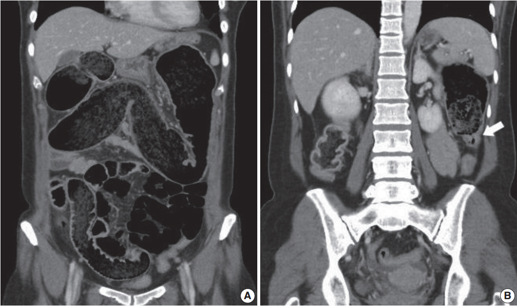
Fig. 1. Abdominopelvic computed tomography shows markedly dilated colon filled with feces (A). Transition point between the dilated proximal and collapsed distal colon is noted (B, arrow).

Fig. 2. Approximately half of the resected segment of the descending colon was dilated.
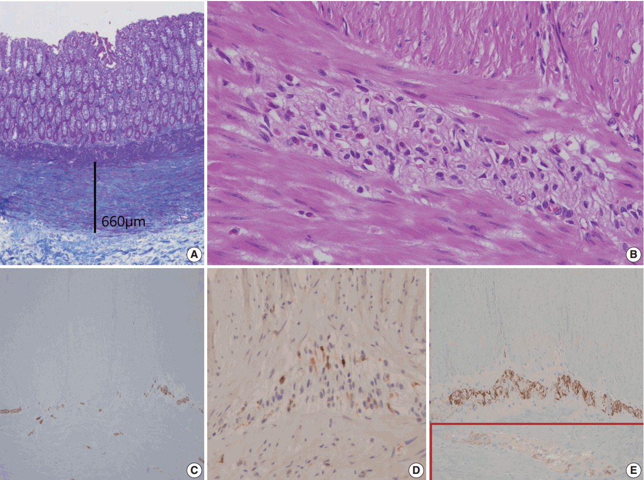
Fig. 3. Summary of histologic findings: thickened muscularis mucosa in the dilated colon (A), many eosinophils in Auerbach’s myenteric plexus (B), hypogangliosis in synaptophysin immunostain (C), scattered CD4-positive lymphocytes in the affected myenteric plexus (D) and stonger but scantier glial fibrillary acidic protein positivity in our patient (E) in contrast to the control patient (in box).
Reference
-
1. De Giorgio R, Sarnelli G, Corinaldesi R, Stanghellini V. Advances in our understanding of the pathology of chronic intestinal pseudo-obstruction. Gut. 2004; 53:1549–52.
Article2. Lee BH, Kim N, Kang SB, et al. Two cases of chronic idiopathic intestinal pseudo-obstruction with different clinical features. J Neurogastroenterol Motil. 2010; 16:83–9.
Article3. Schäppi MG, Smith VV, Milla PJ, Lindley KJ. Eosinophilic myenteric ganglionitis is associated with functional intestinal obstruction. Gut. 2003; 52:752–5.4. Chander B, Fiedler P, Jain D. Eosinophilic myenteric ganglionitis: a case of intestinal pseudo-obstruction in a 93-year-old female. J Clin Gastroenterol. 2011; 45:314–6.5. De Giorgio R, Camilleri M. Human enteric neuropathies: morphology and molecular pathology. Neurogastroenterol Motil. 2004; 16:515–31.
Article6. De Giorgio R, Bovara M, Barbara G, et al. Anti-HuD-induced neuronal apoptosis underlying paraneoplastic gut dysmotility. Gastroenterology. 2003; 125:70–9.
Article7. Lakhan SE, Kirchgessner A. Neuroinflammation in inflammatory bowel disease. J Neuroinflammation. 2010; 7:37.
Article8. Rosenbaum C, Schick MA, Wollborn J, et al. Activation of myenteric glia during acute inflammation in vitro and in vivo. PLoS One. 2016; 11:e0151335.
Article9. De Giorgio R, Guerrini S, Barbara G, et al. Inflammatory neuropathies of the enteric nervous system. Gastroenterology. 2004; 126:1872–83.
Article10. De Winter BY, van den Wijngaard RM, de Jonge WJ. Intestinal mast cells in gut inflammation and motility disturbances. Biochim Biophys Acta. 2012; 1822:66–73.
Article
- Full Text Links
-
- Actions
-
Cited
- CITED
-
- Close
- Share
-
- Similar articles
-
- Four Cases of Unusual Eosinophilic Pustular Folliculits
- A Case of Eosinophilic Cholecystitis associated with Eosinophilic Cholangitis and Pancreatitis
- Enterobiliary Fistula as a Complication of Eosinophilic Gastroenteritis: a Case Report
- Impact of Myenteric Plexus Alterations on Diabetes Related Gastrointestinal Dysmotility
- A case of subserosal type of eosinophilic gastroenteritis with ascites
