Usefulness of Multidetector-row CT in the Evaluation of Reperfused Myocardial Infarction in a Rabbit Model
- Affiliations
-
- 1Department of Radiology and Center for Imaging Science, Samsung Medical Center, Sungkyunkwan University School of Medicine. yhchoe@smc.samsung.co.kr
- KMID: 1066241
- DOI: http://doi.org/10.3348/kjr.2004.5.1.19
Abstract
OBJECTIVE
To evaluate the usefulness of multidetector-row computed tomography (CT) in the evaluation of reperfused myocardial infarction. MATERIALS AND METHODS: Eleven rabbits were subjected to 90-min occlusion of the left anterior descending coronary artery followed by reperfusion. Multidetector-row CT was performed 31 hours+/-21 after the procedure and preand post-contrast multiphase helical CT images were obtained up to 10 min after contrast injection. The animals were sacrificed after 30 days and histochemical staining of the resected specimens was perfomed with 2'3'5-triphenyl tetrazolium chloride (TTC). RESULTS: In all 11 cases, the areas of myocardial infarction demonstrated with TTC-staining were identified on the CT images and the lesions showed hypoenhancement on the early phases up to 62 sec and hyperenhancement on the delayed phases of 5 min and 10 min compared with normal myocardial enhancement. The percentage area of the lesion with respect to the left ventricle wall on CT was significantly correlated with that of the TTC-staining results (p < 0.001 for both early and delayed phase CT) according to the generalized linear model analysis. The areas showing hypoenhancement on early CT were significantly smaller than those with hyperenhancement on delayed CT (p < 0.0001). CONCLUSION: Multidetector-row CT may be useful in the detection and sizing of reperfused myocardial infarction.
Keyword
MeSH Terms
Figure
-
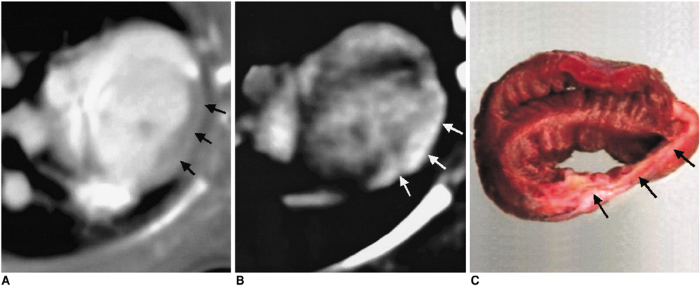
Fig. 1 Extensive myocardial infarction. Early-phase CT obtained at 27 sec following administration of contrast material (A) showed hypoenhancement (arrows) and 10-min-delayed image (B) showed hyperenhancement (arrows) in the anterior wall compared with normal myocardium. 2'3'5-triphenyl tetrazolium chloride-stained specimen obtained at four weeks after coronary artery occlusion and reperfusion (C) shows damaged myocardium (arrows) as a 2'3'5-triphenyl tetrazolium chloride-unstained area with wall thinning.

Fig. 2 Line graph of pooled data from all 11 rabbits shows enhancement patterns of normal myocardium and infarct myocardium on CT images. Infarct areas showed hypoenhancement at early phases and hyperenhancement at late phases compared with normal myocardium.
Cited by 2 articles
-
Myocardial Contractility, Perfusion, and Viability Analysis Using Multidetector CT in Patients with Ischemic Heart Disease
Sung Min Ko
J Korean Med Assoc. 2007;50(2):143-150. doi: 10.5124/jkma.2007.50.2.143.Time Efficiency and Diagnostic Accuracy of New Automated Myocardial Perfusion Analysis Software in 320-Row CT Cardiac Imaging
Matthias Rief, Fabian Stenzel, Anisha Kranz, Peter Schlattmann, Marc Dewey
Korean J Radiol. 2013;14(1):21-29. doi: 10.3348/kjr.2013.14.1.21.
Reference
-
1. McNarama MT, Tscholakoff D, Revel D, et al. Differentiation of reversible and irreversible myocardial injury by MR imaging with and without gadolinium-DTPA. Radiology. 1986. 158:765–769.2. Saeed M, Wendland MF, Takehara Y, Higgins CB. Reversible and irreversible injury in the reperfused myocardium: differentiation with contrast material-enhanced MR imaging. Radiology. 1990. 175:633–637.3. Pereira RS, Prato FS, Wisenberg G, Sykes J. The determination of myocardial viability using Gd-DTPA in a canine model of acute myocardial ischemia and reperfusion. Magn Reson Med. 1996. 36:684–693.4. Gray WR Jr, Parkey RW, Buja LM, et al. Computed tomography: in vitro evaluation of myocardial infarction. Radiology. 1977. 122:511–513.5. Hessel SJ, Adams DF, Judy PF, Fishbein MC, Abrams HL. Detection of myocardial ischemia in vitro by computed tomography. Radiology. 1978. 127:413–418.6. Higgins CB, Siemers PT, Schmidt W, et al. Evaluation of myocardial ischemic damage of various ages by computerized transmission tomography. Time-dependent effects of contrast material. Circulation. 1979. 60:284–291.7. Cipriano PR, Nassi M, Ricci MT, Reitz BA, Brody WR. Acute myocardial ischemia detected in vivo by computed tomography. Radiology. 1981. 140:727–731.8. Mochizuki T, Murase K, Higashino H, Koyama Y, Azemoto S, Ikezoe J. Demonstration of acute myocardial infarction by subsecond spiral computed tomography: early defect and delayed enhancement. Circulation. 1999. 99:2058–2059.9. Hilfiker PR, Weishaupt D, Marincek B. Multislice spiral computed tomography of subacute myocardial infarction. Circulation. 2001. 104:1083.10. Fishbein MC, Meerbaum S, Rit J, et al. Early phase acute myocardial infarct size quantification: validation of the triphenyl tetrazolium chloride tissue enzyme staining technique. Am Heart J. 1981. 101:593–600.11. Sicari R, Picano E, Landi P, et al. Prognostic value of dobutamine-atropine stress echocardiography early after acute myocardial infarction. Echo Dobutamine International Cooperative (EDIC) Study. J Am Coll Cardiol. 1997. 29:254–260.12. Williams MJ, Odabashian J, Lauer MS, Thomas JD, Marwick TH. Prognostic value of dobutamine echocardiography in patients with left ventricular dysfunction. J Am Coll Cardiol. 1996. 27:132–139.13. Phelps ME, Hoffman EJ, Selin C, et al. Investigation of [18F]2-deoxyglucose for the measure of myocardial glucose metabolism. J Nucl Med. 1978. 19:1311–1319.14. Burt RW, Perkins OW, Oppenheim BE, et al. Direct comparison of fluorine-18-FDG SPECT, fluorine-18-FDG PET and rest thallium-201 SPECT for detection of myocardial viability. J Nucl Med. 1995. 36:176–179.15. Kuijper AF, Vliegen HW, van der Wall EE, et al. The clinical impact of thallium-201 reinjection scintigraphy for detection of myocardial viability. Eur J Nucl Med. 1992. 19:783–789.16. Wesbey G, Higgins CB, Lanzer P, Botvinick E, Lipton MJ. Imaging and characterization of acute myocardial infarction in vivo by gated nuclear magnetic resonance. Circulation. 1984. 69:125–130.17. Ratner RV, Okada RD, Newell JB, Pohost GM. The relationship between proton nuclear magnetic resonance relaxation parameters and myocardial perfusion with acute coronary arterial occlusion and reperfusion. Circulation. 1985. 71:823–828.18. Tscholakoff D, Higgins CB, McNamara MT, Derugin N. Early-phase myocardial infarction: evaluation by MR imaging. Radiology. 1986. 159:667–672.19. McNamara MT, Tscholakoff D, Revel D, et al. Differentiation of reversible and irreversible myocardial injury by MR imaging with and without gadolinium-DTPA. Radiology. 1986. 158:765–769.20. Peshock RM, Malloy CR, Buja LM, Nunnally RL, Parkey RW, Willerson JT. Magnetic resonance imaging of acute myocardial infarction: gadolinium diethylenetriamine pentaacetic acid as a marker of reperfusion. Circulation. 1986. 74:1434–1440.21. McNamara MT, Higgins CB, Ehman RL, Revel D, Sievers R, Brasch RC. Acute myocardial ischemia: magnetic resonance contrast enhancement with gadolinium-DTPA. Radiology. 1984. 153:157–163.22. Tscholakoff D, Higgins CB, Sechtem U, McNamara MT. Occlusive and reperfused myocardial infarcts: effects of Gd-DTPA on ECG-gated MR imaging. Radiology. 1986. 160:515–519.23. Wesbey GE, Higgins CB, McNarama MT, et al. Effect of gadolinium-DTPA on the magnetic relaxation times of normal and infarcted myocardium. Radiology. 1984. 153:165–169.24. Kim RJ, Fieno DS, Parrish TB, et al. Relationship of MRI delayed contrast enhancement to irreversible injury, infarct age, and contractile function. Circulation. 1999. 100:1992–2002.25. Choi SI, Jiang CZ, Lim KH, et al. Application of breath-hold T2-weighted, first-pass perfusion and gadolinium-enhanced T1-weighted MR imaging for assessment of myocardial viability in a pig model. J Magn Reson Imaging. 2000. 11:476–480.26. Saeed M, Bremerich J, Wendland MF, Wyttenbach R, Weinmann HJ, Higgins CB. Reperfused myocardial infarction as seen with use of necrosis-specific versus standard extracellular MR contrast media in rats. Radiology. 1999. 213:247–257.27. Arheden H, Saeed M, Higgins CB, et al. Reperfused rat myocardium subjected to various durations of ischemia: estimation of the distribution volume of contrast material with echo-planar MR imaging. Radiology. 2000. 215:520–528.28. Schaefer S, Malloy CR, Katz J, et al. Gadolinium-DTPA-enhanced nuclear magnetic resonance imaging of reperfused myocardium: identification of the myocardial bed at risk. J Am Coll Cardiol. 1988. 12:1064–1072.29. Saeed M, Lund G, Wendland MF, Bremerich J, Weinmann H, Higgins CB. Magnetic resonance characterization of the periinfarction zone of reperfused myocardial infarction with necrosis-specific and extracellular nonspecific contrast media. Circulation. 2001. 103:871–876.30. Rogers WJ Jr, Kramer CM, Geskin G, et al. Early contrast-enhanced MRI predicts late functional recovery after reperfused myocardial infarction. Circulation. 1999. 99:744–750.31. Willmann JK, Szente-Varga M, Roos JE, Hilfiker PR, Weishaupt D. Three-dimensional images of extra-anatomic arterial bypass graft using multidetector row spiral computed tomography data with volume rendering. Circulation. 2001. 104:E154–E155.32. Budoff MJ, Achenbach S, Duerinckx A. Clinical utility of computed tomography and magnetic resonance techniques for noninvasive coronary angiography. J Am Coll Cardiol. 2003. 42:1867–1878.
- Full Text Links
-
- Actions
-
Cited
- CITED
-
- Close
- Share
-
- Similar articles
-
- Sacroiliitis in Ankylosing Spondylitis: Comparison with Multidetector Row CT and Plain Radiography
- Early and Delayed Myocardial Enhancement in Myocardial Infarction Using Two-Phase Contrast-Enhanced Multidetector-Row CT
- Multidetector-row CT in Ischemic Heart Disease
- Reverse Redistribution in Myocardial Perfusion Imaging: Revisited with 64-slice MDCT
- Evaluation of Reperfused Myocardial Infarction by Low-Dose Multidetector Computed Tomography Using Prospective Electrocardiography (ECG)-Triggering: Comparison with Magnetic Resonance Imaging
