Comparison of Transaxial Source Images and 3-Plane, Thin-Slab Maximal Intensity Projection Images for the Diagnosis of Coronary Artery Stenosis with Using ECG-Gated Cardiac CT
- Affiliations
-
- 1Department of Radiology, Research Institute of Radiology, University of Ulsan College of Medicine, Asan Medical Center, Seoul, Korea. seojb@amc.seoul.kr
- 2Department of Radiology, Seoul National University College of Medicine, Seoul, Korea.
- KMID: 753912
- DOI: http://doi.org/10.3348/kjr.2006.7.1.20
Abstract
OBJECTIVE
We wanted to compare the transaxial source images with the optimized three plane, thin-slab maximum intensity projection (MIP) images from electrocardiographic (ECG)-gated cardiac CT for their ability to detect hemodynamically significant stenosis (HSS), and we did this by means of performing a receiver operating characteristic (ROC) analysis. MATERIALS AND METHODS: Twenty-eight patients with a heart rate less than 66 beats per minute and who were undergoing both retrospective ECG-gated cardiac CT and conventional coronary angiography were included in this study. The contrast-enhanced CT scans were obtained with a collimation of 16x0.75-mm and a rotation time of 420 msec. The transaxial images were reconstructed at the mid-diastolic phase with a 1-mm slice thickness and a 0.5-mm increment. Using the transaxial images, the slab MIP images were created with a 4-mm thickness and a 2-mm increment, and they covered the entire heart in the horizontal long axis (4 chamber view), in the vertical long axis (2 chamber view) and in the short axis. The transaxial images and MIP images were independently evaluated for their ability to detect HSS. Conventional coronary angiograms of the same study group served as the standard of reference. Four radiologists were requested to rank each image with using a five-point scale (1 = definitely negative, 2 = probably negative, 3 = indeterminate, 4 = probably positive, and 5 = definitely positive) for the presence of HSS; the data were then interpreted using ROC analysis. RESULTS: There was no statistical difference in the area under the ROC curve between transaxial images and MIP images for the detection of HSS (0.8375 and 0.8708, respectively; p > 0.05). The mean reading time for the transaxial source images and the MIP images was 116 and 126.5 minutes, respectively. CONCLUSION: The diagnostic performance of the MIP images for detecting HSS of the coronary arteries is acceptable and this technique's ability to detect HSS is comparable to that of the transaxial source images.
Keyword
MeSH Terms
Figure
-
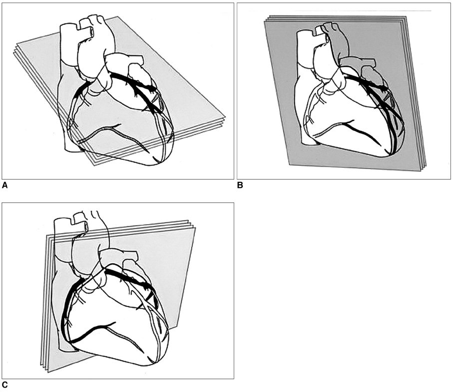
Fig. 1 Schematic diagram of the range of parallel thin-slab maximum intensity projections arranged in a horizontal long axis (4 chamber view, A), vertical long axis (2 chamber view, B), and short axis of the heart (C).
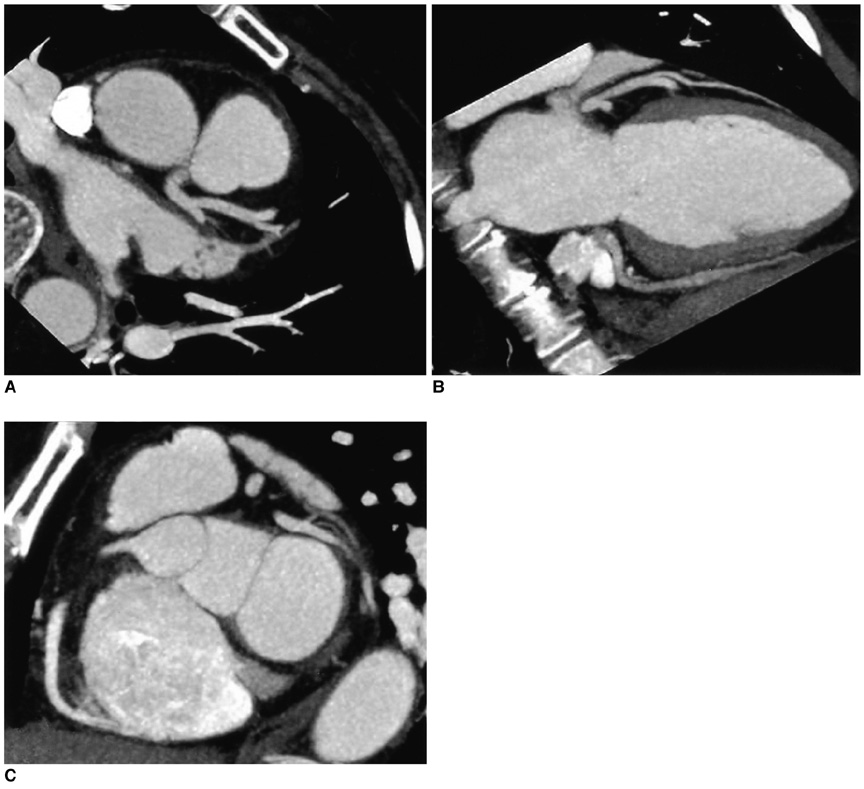
Fig. 2 Representative maximal intensity projection images of the coronary arteries using the standardized 3-plane, thin-slab maximum intensity projection technique. A. Horizontal long axis (4 chamber) view, B. Vertical long axis (2 chamber) view, C. Short axis view.
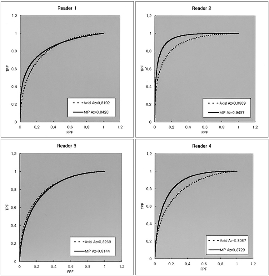
Fig. 3 Receiver operating characteristic curves for the transaxial images and the MIP images from each reader. The Az values of the MIP images were larger than those of the transaxial images except for reader 3. However, the difference was statistically significant only for reader 2. Axial = Transaxial image, TPF = true positive fraction, FPF = false positive fraction.
Reference
-
1. Rumberger JA. Noninvasive coronary angiography using computed tomography: ready to kick it up another notch [editorial]. Circulation. 2002. 106:2036–2038.2. Nieman K, Oudkerk M, Rensing BJ, van Ooijen P, Munne A, van Geuns RJ, et al. Coronary angiography with multislice computed tomography. Lancet. 2001. 357:599–603.3. Nieman K, Cademartiri F, Lemos PA, Raaijmakers R, Pattynama PM, de Feyter PJ. Reliable noninvasive coronary angiography with fast submillimeter multislice spiral computed tomography. Circulation. 2002. 106:2051–2054.4. Kopp AF, Schroeder S, Kuettner A, Baumbach A, Georg C, Ohnesorge B, et al. Non-invasive coronary angiography with high resolution mutidetector-row computed tomography: results in 102 patients. Eur Heart J. 2002. 23:1714–1725.5. Kopp AF, Schroeder S, Kuettner A, Heuchmid M, Georg C, Ohnesorge B, et al. Coronary arteries: retrospectively ECG-gated multi-detector row CT angiography with selective optimization of the image reconstruction window. Radiology. 2001. 221:683–688.6. Vogl TJ, Adolmaali ND, Diebold T, Engelmann K, Ay M, Dogan S, et al. Techniques for the detection of coronary atherosclerosis: multi-detector row CT coronary angiography. Radiology. 2002. 223:212–220.7. Gerber TC, Kuzo RS, Lane GE, O'Brien PC, Karstaedt N, Morin RL, et al. Image quality in a standardized algorithm for minimally invasive coronary angiography with multislice spiral computed tomography. J Comput Assist Tomogr. 2003. 27:62–69.8. Mahnken AH, Wildberger JE, Sinha AM, Dedden K, Stanzel S, Hoffmann R, et al. Value of 3D-volume rendering in the assessment of coronary arteries with retrospectively ECG-gated multislice spiral CT. Acta Radiol. 2003. 44:302–309.9. Addis KA, Hopper KD, Lyriboz TA, Liu Y, Wise SW, Kasales CJ, et al. CT angiography: in vitro comparison of five reconstruction methods. AJR Am J Roentgenol. 2001. 177:1171–1176.10. Hirai T, Korogi Y, Ono K, Murata Y, Takahashi M, Suginohara K, et al. Maximum stenosis of extracranial internal carotid artery: effect of luminal morphology on stenosis measurement by using CT angiography and conventional DSA. Radiology. 2001. 221:802–807.11. Rubin G, Dake MD, Napel S, Jeffrey RB, McDonnell CH, Sommer FG, et al. Spiral CT of renal artery stenosis: comparison of three-dimensional rendering techniques. Radiology. 1994. 190:181–189.12. Berg MH, Manninen HI, Vanninen RL, Vainio RA, Soimakallio S. Assessment of renal artery stenosis with CT angiography: usefulness of multiplanar reformation, quantitative stenosis measurement, and densitometric analysis of renal parenchymal enhancement as adjuncts to MIP film rendering. J Comput Assist Tomogr. 1998. 22:533–540.13. Heath DG, Soyer PA, Kuszyk BS, Bliss DF, Calhoun PS, Bluemke DA, et al. Three-dimensional spiral CT during arterial portography: comparison of three rendering techniques. RadioGraphics. 1995. 15:1001–1011.14. Lu Bin, Dai RP, Jiang SL, Bai H, He S, Zhuang N, et al. Effects of window and threshold levels on the accuracy of three-dimensional rendering techniques in coronary artery electron-beam CT angiography. Acad Radiol. 2001. 8:754–761.15. Rensing BJ, Bongaerts A, van Geuns RJ, van Ooijen P, Oudkerk M, de Feyter PJ. Intravenous coronary angiography using electron beam computed tomography. Prog Cardiovasc Dis. 1999. 42:139–148.16. Peter MA, van Ooijen P, Ho KY, Dorgelo J, Oudkerk M. Coronary artery imaging with multidetector CT: visualization issues. RadioGraphics. 2003. 08. Available from: URL: http://radiographics.rsnajnls.org/cgi/content/full/e16v1.17. Pannu HK, Flohr TG, Corl FM, Fishman EK. Current concepts in multi-detector row CT evaluation of the coronary arteries: principles, techniques, and anatomy. RadioGraphics. 2003. 23:s111–s125.18. Soto JA, Munera F, Cardoso N, Guarin O, Medina S. Diagnostic performance of helical CT angiography in trauma to large arteries of the extremities. J Comput Assist Tomogr. 1999. 23:188–196.19. Sugahara T, Korogi Y, Hirai T, Hamatake S, Komohara Y, Okuda T, et al. CT angiography in vascular intervention for steno-occlusive diseases: role of multiplanar reconstruction and source images. Br J Radiol. 1998. 71:601–611.20. Regenfus M, Ropers D, Achenback S, Schlundt C, Kessler W, Laub G, et al. Diagnostic value of maximum intensity projections versus source images for assessment of contrast-enhanced three-dimensional breath-hold magnetic resonance coronary angiography. Invest Radiol. 2003. 38:200–206.21. Bae KT, Whiting BR. CT data storage reduction by means of compressing projection data instead of images: feasibility study. Radiology. 2003. 219:850–855.
- Full Text Links
-
- Actions
-
Cited
- CITED
-
- Close
- Share
-
- Similar articles
-
- Diagnosis of Vertebral Artery Ostial Stenosis on Contrast-Enhanced MR Angiography: Usefulness of a Thin-Slab MIP Technique
- Prospectively Electrocardiogram-Gated High-Pitch Spiral Acquisition Mode Dual-Source CT Coronary Angiography in Patients with High Heart Rates: Comparison with Retrospective Electrocardiogram-Gated Spiral Acquisition Mode
- Clinical Value of Cardiovascular Calcifications on Non-Enhanced, Non-ECG-Gated Chest CT
- Evaluation of Reperfused Myocardial Infarction by Low-Dose Multidetector Computed Tomography Using Prospective Electrocardiography (ECG)-Triggering: Comparison with Magnetic Resonance Imaging
- Technical Aspect of Coronary CT Angiography: Imaging Tips and Safety Issues
