Portal cavernography during endoscopic retrograde cholangiopancreatography: from bilhemia to hemobilia
- Affiliations
-
- 1Department of Gastroenterology, Hepatopancreatology, and Digestive Oncology, Erasme University Hospital, Université Libre de Bruxelles, Brussels, Belgium
- 2Department of Gastroenterology and Digestive Oncology, Meuse and Sambre Regional Hospital Center, Namur, Belgium
- KMID: 2544575
- DOI: http://doi.org/10.5946/ce.2021.276
Abstract
- Portobiliary fistulas are rare but may lead to life-threatening complications. Biliary plastic stent-induced portobiliary fistulas during endoscopic retrograde cholangiopancreatography have been described. Herein, we present a case of portal cavernography and recurrent hemobilia after endoscopic retrograde cholangiopancreatography in which a portobiliary fistula was detected in a patient with portal biliopathy. This likely indicates a change in clinical presentation (from bilhemia to hemobilia) after biliary drainage that was successfully treated by placement of a fully covered, self-expandable metallic stent.
Keyword
Figure
-

Fig. 1. (A) T2-weighted image (axial plane) depicting compression of the common bile duct between two venous structures (arrows). (B) T2-weighted image (coronal plane) showing a vessel as a low signal tubular structure (arrow) parallel to the common bile duct.
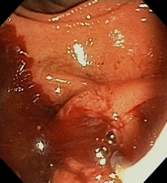
Fig. 2. Endoscopic image showing active bleeding from the papillary orifice (hemobilia).

Fig. 3. (A, B) Cholangiogram showing opacification of a venous structure compatible with the known cavernoma (black arrows) and contrast dye in a venous branch parallel to the bile duct (long white arrow) with possible orifice of portobiliary fistula (short white arrow).
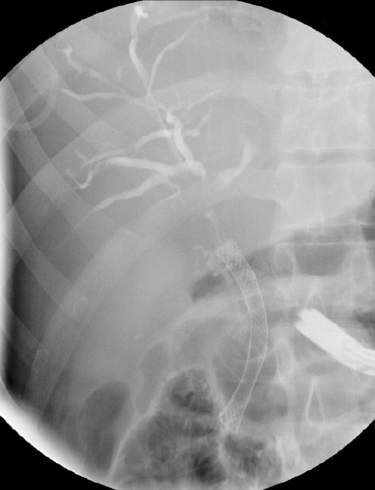
Fig. 4. Radiography showing the fully covered self-expandable metal stent deployed in the main bile duct.

Fig. 5. (A) Radiography showing opacification of the common bile duct with no residual fistula during the endoscopic retrograde cholangiopancreatography performed to replace the initially placed fully covered self-expandable metal stent. (B) Radiography showing opacification of the common bile duct with no residual fistula or stenosis after definitive removal of the fully covered self-expandable metal stent.
Reference
-
1. Yamashita H, Chijiiwa K, Ogawa Y, et al. The internal biliary fistula: reappraisal of incidence, type, diagnosis and management of 33 consecutive cases. HPB Surg. 1997; 10:143–147.2. DiCocco JM, Fabian TC. Biliovenous fistula. In : Vincent JL, Hall JB, editors. Encyclopedia of intensive care medicine. Berlin: Springer;2012. p. 303–305.3. Huibregtse K, Gish R, Tytgat GN. A frightening event during endoscopic papillotomy. Gastrointest Endosc. 1988; 34:67–68.4. Ricci E, Mortilla MG, Conigliaro R, et al. Portal vein filling: a rare complication associated with ERCP for endoscopic biliary stent placement. Gastrointest Endosc. 1992; 38:524–525.5. Tighe M, Jacobson I. Bleeding from bile duct varices: an unexpected hazard during therapeutic ERCP. Gastrointest Endosc. 1996; 43:250–252.6. Kennedy C, Larvin M, Linsell J. Fatal hepatic air embolism following ERCP. Gastrointest Endosc. 1997; 45:187–188.7. Mutignani M, Shah SK, Bruni A, et al. Endoscopic treatment of extrahepatic bile duct strictures in patients with portal biliopathy carries a high risk of haemobilia: report of 3 cases. Dig Liver Dis. 2002; 34:587–591.8. Espinel J, Pinedo ME, Calleja JL. Portal vein filling: an unusual complication of needle-knife sphincterotomy. Endoscopy. 2007; 39 Suppl 1:E245.9. Layec S, D’Halluin PN, Pagenault M, et al. Massive hemobilia during extraction of a covered self-expandable metal stent in a patient with portal hypertensive biliopathy. Gastrointest Endosc. 2009; 70:555–556.10. Furuzono M, Hirata N, Saitou J, et al. A rare complication during ERCP and sphincterotomy: placement of an endoscopic nasobiliary drainage tube in the portal vein. Gastrointest Endosc. 2009; 70:588–590.11. Kawakami H, Kuwatani M, Kudo T, et al. Portobiliary fistula: unusual complication of wire-guided cannulation during endoscopic retrograde cholangiopancreatography. Endoscopy. 2011; 43 Suppl 2 UCTN:E98–E99.12. Kalaitzakis E, Stern N, Sturgess R. Portal vein cannulation: an uncommon complication of endoscopic retrograde cholangiopancreatography. World J Gastroenterol. 2011; 17:5131–5132.13. Dawwas MF, Oppong KW, John SK, et al. Endoscopic ultrasound diagnosis of an ERCP-related portobiliary fistula. Endoscopy. 2013; 45 Suppl 2 UCTN:E214–E216.14. So H, Song TJ, Lee D, et al. Endoscopic treatment of recurrent bleeding from a portobiliary fistula with a fully covered self-expandable metal stent. Endoscopy. 2015; 47 Suppl 1:E616–E617.
- Full Text Links
-
- Actions
-
Cited
- CITED
-
- Close
- Share
-
- Similar articles
-
- Delayed Severe Hemobilia after Endoscopic Biliary Plastic Stent Insertion
- Massive Hemobilia due to Hepatic Arteriobiliary Fistula during Endoscopic Retrograde Cholangiopancreatography: An Extremely Rare Guidewire-Related Complication
- Delayed Hemobilia Caused by Penetration of Biliary Plastic Stent into Portal Vein
- Pancreaticoduodenal artery pseudoaneurysm-induced hemobilia caused by a plastic biliary stent
- Two Cases of Xanthogranulomatous Cholecystitis and Gallbladder Cancer with Hemobilia
