Congestive myelopathy due to spinal dural arteriovenous fistula mimicking CNS demyelinating disease
- Affiliations
-
- 1Department of Neurology, College of Medicine, Hanyang University, Seoul, Korea
- KMID: 2537326
- DOI: http://doi.org/10.7461/jcen.2022.E2021.11.003
Abstract
- Spinal dural arteriovenous fistula (SDAVF) is known for its ambiguous and various clinical presentations. Among these presentations, congestive myelopathy is one of the most common, yet it is challenging to correctly diagnose SDAVF at initial presentation. Several diseases present as myelopathy, including demyelinating diseases. Herein, we present two cases of congestive myelopathy due to SDAVF presenting to the emergency room (ER) with progressive quadriparesis. Even though the patients had a proper magnetic resonance imaging (MRI) examination from the initial presentation, there was a delay in making a final diagnosis. Both patients’ clinical presentation and MRI mimicked central nervous system (CNS) demyelinating disease initially, and a more thorough examination revealed SDAVF. Such a delay in diagnosis can result in more neurological deterioration and may result in more sequelae. Hence, SDAVF should always be considered as a differential diagnosis when examining patients with myelopathy.
Keyword
Figure
-
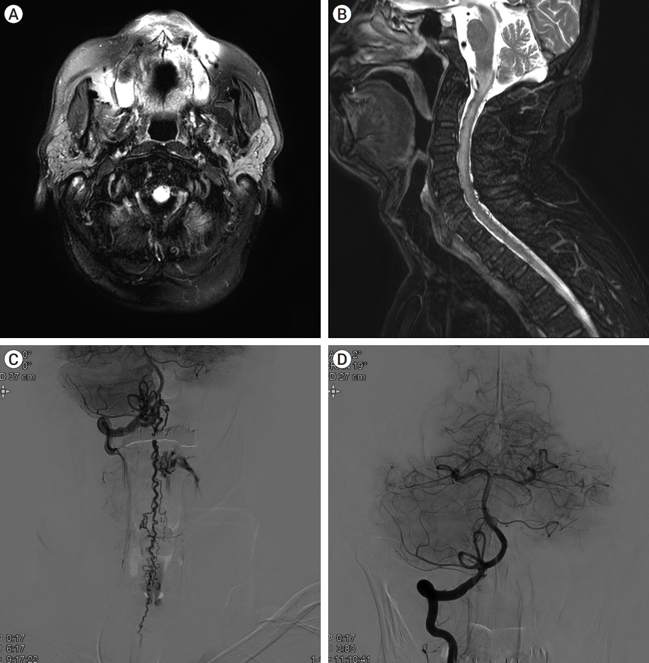
Fig. 1. MRI and DSA of Case 1.Initial brain MRI showing increased signal intensity at the medulla in fluid-attenuated inversion recovery (FLAIR) imaging (A). C-spine MRI shows multiple small abnormal vessel flow voids on the dorsal pial surface from the cerebellum to the T-spine in T2 weighted image, and intramedullary high signal and a peripheral low signal from the cervicomedullary junction to T1 with cord swelling (B). DSA image confirms AV shunt at the C1 level with prominent perimedullary draining vein along the anterior and posterior surface of the spinal cord (C). Shunt feeder from the right. The V3-V4 junction was embolized, and the final angiogram shows the disappearance of the AV shunt (D). MRI, magnetic resonance imaging; DSA, Digital subtraction angiography; AV, arteriovenous
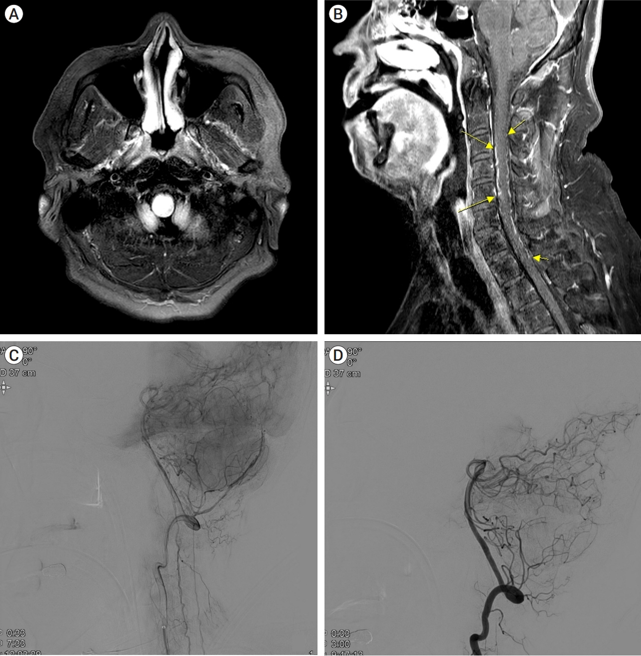
Fig. 2. MRI and DSA of Case 2.Initial brain MRI shows T2 high signal intensity in the medulla and cervical cord without diffusion restriction (A). Along with the high signal intensity at the medulla, cervical T1 enhanced image demonstrates abnormal enhancing serpentine vessels in the ventral and dorsal surfaces of the spinal cord from the C3 to C7 level (B, yellow arrows). DSA reveals dural AVF at posterior Rt, meningeal angiography (C). Embolization of the Rt. The posterior meningeal artery was performed, and the final angiogram shows occlusion of the dural AVF (D). MRI, magnetic resonance imaging; DSA, Digital subtraction angiography; AVF, arteriovenous fistula
Reference
-
1. Fox S, Hnenny L, Ahmed U, Meguro K, Kelly ME. Spinal dural arteriovenous fistula: a case series and review of imaging findings. Spinal Cord Ser Cases. 2017; Jul. 3:17024.
Article2. Howard RS. Spinal vascular disease: a neglected cause of myelopathy. Pract Neurol. 2019; Jun. 19(3):184–6.
Article3. Koch C, Zeumer H, Westphal M. Spinal vascular malformations: therapeutic options. In : Lanzer P, Topol EJ, editors. Pan Vascular Medicine. Berlin, Heidelberg: Springer;2002. p. 1253–64.4. Li C, Yu J, Li K, Hou K, Yu J. Dural arteriovenous fistula of the lateral foramen magnum region: a review. Interv Neuroradiol. 2018; Aug. 24(4):425–34.
Article5. Maimon S, Luckman Y, Strauss I. Spinal dural arteriovenous fistula: a review. Adv Tech Stand Neurosurg. 2016; (43):111–37.
Article6. Rain S, Udding J, Broere D. Acute clinical worsening after steroid administration in cervical myelitis may reveal a subdural arteriovenous fistula. Case Rep Neurol. 2016; Nov. 8(3):234–42.
Article7. Ronald AA, Yao B, Winkelman RD, Piraino D, Masaryk TJ, Krishnaney AA. Spinal dural arteriovenous fistula: diagnosis, outcomes, and prognostic factors. World Neurosurg. 2020; Dec. 144:e306–15.
Article
- Full Text Links
-
- Actions
-
Cited
- CITED
-
- Close
- Share
-
- Similar articles
-
- Myelopathy due to Spinal Dural Arteriovenous Fistula: A Case Report
- Myelopathy Caused by Spinal Dural Arterio-Venous Fistula after First Lumbar Vertebral Body Fracture: A Case Report
- Syringomyelia Associated with Spinal Dural Arteriovenous Fistula: Clinical and Radiological Improvement after Embolization
- Intracranial Dural Arteriovenous Fistula Draining into Spinal Perimedullary Veins: A Rare Cause of Myelopathy
- A Case of Myelopathy due to Spinal Dural Arteriovenous Fistula Supplied by Branches of the Internal Iliac Arteries
