Myelopathy Caused by Spinal Dural Arterio-Venous Fistula after First Lumbar Vertebral Body Fracture: A Case Report
- Affiliations
-
- 1Department of Rehabilitation Medicine, Gangneung Asan Medical Center, University of Ulsan College of Medicine, Gangneung 210-711, Korea. mdjhkoo@gnah.co.kr
- KMID: 2267237
- DOI: http://doi.org/10.5535/arm.2011.35.5.729
Abstract
- Spinal dural arteriovenous fistula is a rare vascular lesion of the spinal cord associated with progressive myelopathy. Symptoms include progressive gait dysfunction, weakness, sensory loss, and bowel and bladder dysfunction. Because these symptoms overlap with other common causes of myelopathy and the disease is rare, spinal dural arteriovenous fistula is often not suspected and the time to diagnosis is long. We report the case of a 60-year-old woman who presented with progressive lower limb weakness and gait disturbance diagnosed as spinal dural arteriovenous fistula involving a fractured L1 vertebral body.
MeSH Terms
Figure
-
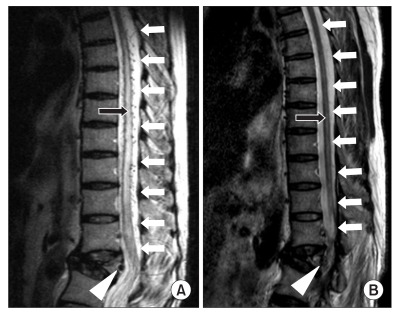
Fig. 1 (A) Sagittal T2-weighted magnetic resonance image of the thoracolumbar spine showing edema of the thoracic cord and conus medullaris (black arrow) and regional dilated perimedullary vessels (white arrows) suggestive of a spinal dural arteriovenous fistula (white arrow head) in the fractured vertebral body at L1 level. (B) After glue embolization, sagittal T2-weighted magnetic resonance image of the thoracolumbar spine showing normal finding of the thoracic cord and conus medullaris (black arrow) and disappearance of dilated perimedullary veins (white arrows) and a spinal dural arteriovenous fistula (white arrow head).
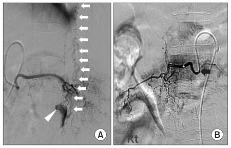
Fig. 2 (A) Selective spinal angiogram of the right L1 segmental radicular artery showing a fistulous connection (white arrow head) between the feeding artery and the perimedullary venous plexus (white arrows) (The lateral view). (B) Right spinal angiograms following microcatheter placement for glue (*) embolization in the right L1 and T12 radicular artery from above and across to below the fistula (Black arrow). After embolization, there was complete cessation of flow through the fistula (The anteropostetrior view).
Reference
-
1. Gilbertson JR, Miller GM, Goldman MS, Marsh WR. Spinal dural arteriovenous fistulas; MR and myelographic findings. AJNR Am J Neuroradiol. 1995; 16:2049–2057. PMID: 8585493.2. Atkinson JL, Miller GM, Krauss WE, Marsh WR, Piepgras DG, Atkinson PP, Brown RD Jr, Lane JI. Clinical and radiographic features of dural arteriovenous fistula, a treatable cause of myelopathy. Mayo Clin Proc. 2001; 76:1120–1130. PMID: 11702900.
Article3. Isu T, Iwasaki Y, Akino M, Koyanagi I, Abe H. Magnetic resonance imaging in cases of spinal dural arteriovenous malformation. Neurosurgery. 1989; 24:919–923. PMID: 2747870.
Article4. Heuer GG, Gabel BC, Bhowmick DA, Stiefel MF, Hurst RW, Schuster JM. Symptomatic high-flow arteriovenous fistula after a C2 fracture: case report. J Neurosurg Spine. 2008; 8:381–384. PMID: 18377324.5. Chul Suh D, Choi GC, Sung KB, Kim KK, Rhim SC. Spinal osseous epidural arteriovenous fistula with multiple small arterial feeders converging to a round fistular nidus as a target of venous approach. AJNR Am J Neuroradiol. 2004; 25:69–73. PMID: 14729531.6. Leape LL, Palacios E. Acute traumatic vertebral arteriovenous fistula. Ann Surg. 1971; 174:908–910. PMID: 5132435.
Article7. Hui F, Trosselo MP, Meisel HJ, Alvarez H, Sequeira E, Lasjaunias P. Paraspinal arteriovenous shunts in children. Neuroradiology. 1994; 36:69–73. PMID: 8108004.
Article8. Jones BV, Ernst RJ, Tomsick TA, Tew J Jr. Spinal dural arteriovenous fistulas; recognizing the spectrum of magnetic resonance imaging findings. J Spinal Cord Med. 1997; 20:43–48. PMID: 9097255.
Article
- Full Text Links
-
- Actions
-
Cited
- CITED
-
- Close
- Share
-
- Similar articles
-
- A Case of Dural Arterio-venous Malformation Involving the Cavernous Sinus
- Intracranial Dural Arteriovenous Fistula Draining into Spinal Perimedullary Veins: A Rare Cause of Myelopathy
- Endovascular Treatment of Spinal Dural and Epidural Arteriovenous Fistula as Complication of Lumbar Surgery
- Myelopathy due to Spinal Dural Arteriovenous Fistula: A Case Report
- Syringomyelia Associated with Spinal Dural Arteriovenous Fistula: Clinical and Radiological Improvement after Embolization
