Syringomyelia Associated with Spinal Dural Arteriovenous Fistula: Clinical and Radiological Improvement after Embolization
- Affiliations
-
- 1Department of Neurology, Bombay Hospital and Medical Research Centre, Mumbai, India
- 2Department of Neurology, Fortis Hospital, Kalyan, Kalyan, India
- KMID: 2508066
- DOI: http://doi.org/10.5469/neuroint.2020.00192
Abstract
- Spinal dural arteriovenous fistulae (AVF) are rare and can result in spinal cord dysfunction. We present one such case wherein the patient presented with a venous congestive myelopathy. Magnetic resonance imaging showed a syrinx formation, spinal cord edema, and flow voids. Digital subtraction angiography confirmed the dural AVF, which was treated with embolization. The syrinx disappeared, other spinal cord changes improved, and the patient had remarkable clinical improvement. The case is presented to draw attention to the rare formation of a syrinx in a spinal dural arteriovenous fistula and its disappearance after successful embolization.
Keyword
Figure
-
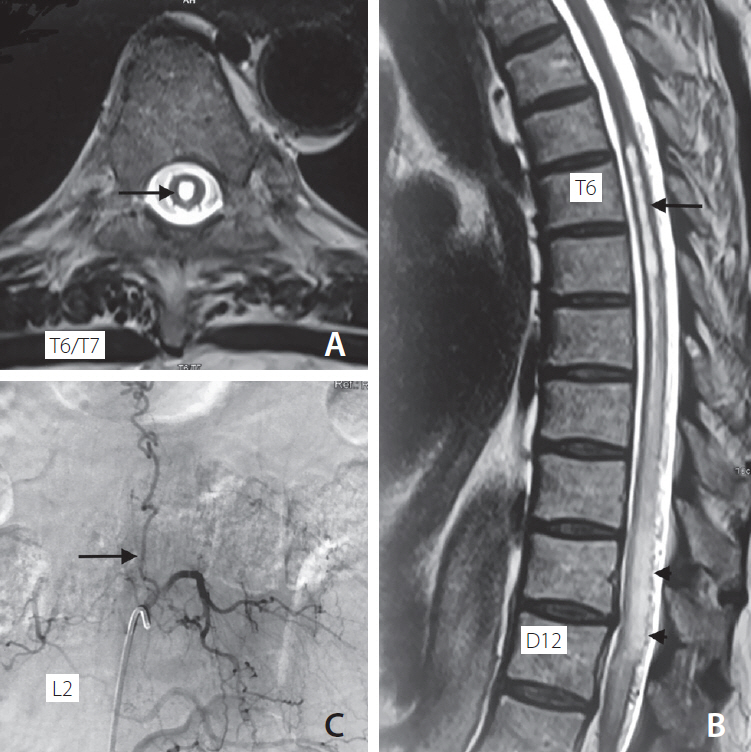
Fig. 1. Pre embolization status showing (A) T2-weighted axial section of cord at T6/T7 level showing syrinx (arrow), (B) T2 weighted sagittal section of spinal cord showing syrinx from upper border of T6 to T8 (arrow) and hyperintense signal from T7 to L1 with flow-voids from T10 to T12 (arrowheads), (C) DSA at L2 level showing dural AV fistula (arrow) with draining vein reaching up to thoracic level. DSA, digital subtraction angiography; AV, arteriovenous.
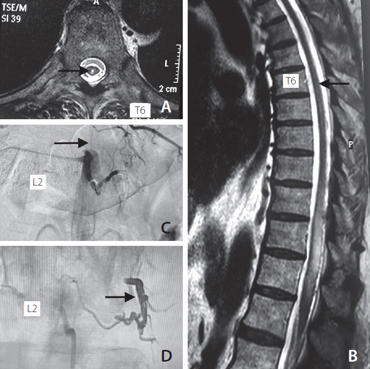
Fig. 2. Post embolization status showing (A) T2 weighted axial image of cord at T6 level showing reduction in syrinx size (arrow), (B) T2 weighted sagittal cord image showing reduction in hyperintense signal, syrinx size (arrow), and disappearance of flow voids, (C) DSA at L2 level showing successful embolization of dural AV fistula (arrow), (D) Onyx cast post embolization of fistula (arrow). DSA, digital subtraction angiography; AV, arteriovenous.
Reference
-
1. Kramer CL. Vascular disorders of the spinal cord. Continuum (Minneap Minn). 2018; 24:407–426.
Article2. Suh DC, Song Y, Park D, et al. New grading system for the clinical evaluation of patients with spinal vascular lesions. Neuroradiology. 2018; 60:1035–1041.
Article3. Hunt R, Roberts RM, Mortimer AM. Spinal dural arteriovenous fistula: delay to radiological diagnosis and sources of radiological error. Clin Radiol. 2018; 73:835.e11–835.e16.
Article4. Gardner WJ. Hydrodynamic mechanism of syringomyelia: its relationship to myelocele. J Neurol Neurosurg Psychiatry. 1965; 28:247–259.
Article5. Williams B. Progress in syringomyelia. Neurol Res. 1986; 8:130–145.
Article6. Oldfield EH, Muraszko K, Shawker TH, Patronas NJ. Pathophysiology of syringomyelia associated with Chiari I malformation of the cerebellar tonsils. Implications for diagnosis and treatment. J Neurosurg. 1994; 80:3–15.7. Heiss JD, Snyder K, Peterson MM, et al. Pathophysiology of primary spinal syringomyelia. J Neurosurg Spine. 2012; 17:367–380.
Article8. Brinjikji W, Nasr DM, Morris JM, Rabinstein AA, Lanzino G. Clinical outcomes of patients with delayed diagnosis of spinal dural arteriovenous fistulas. AJNR Am J Neuroradiol. 2016; 37:380–386.
Article9. Finsterer J, Bavinzski G, Ungersböck K. Spinal dural arteriovenous fistula associated with syringomyelia. J Neuroradiol. 2000; 27:211–214.10. Zaed I, Pinto MV, Mauermann ML, Lanzino G. Teaching neuroimages: spinal cord syrinx secondary to a spinal dural arteriovenous fistula. Neurology. 2018; 91:e295–e296.
Article
- Full Text Links
-
- Actions
-
Cited
- CITED
-
- Close
- Share
-
- Similar articles
-
- Endovascular Treatment of Spinal Dural and Epidural Arteriovenous Fistula as Complication of Lumbar Surgery
- Stereotactic radiosurgery for dural arteriovenous fistula
- Intracranial Dural Arteriovenous Fistula Draining into Spinal Perimedullary Veins: A Rare Cause of Myelopathy
- Novalis Stereotactic Radiosurgery for Spinal Dural Arteriovenous Fistula
- Dural Arteriovenous Fistula Involving Transverse Sinus: Successful Embolization Using Onyx(R)
