Radiation safety for pain physicians: principles and recommendations
- Affiliations
-
- 1Department of Anesthesiology and Pain Medicine, Konkuk University School of Medicine, Seoul, Korea
- KMID: 2527764
- DOI: http://doi.org/10.3344/kjp.2022.35.2.129
Abstract
- C-arm fluoroscopy is a useful tool for interventional pain management. However, with the increasing use of C-arm fluoroscopy, the risk of accumulated radiation exposure is a significant concern for pain physicians. Therefore, efforts are needed to reduce radiation exposure. There are three types of radiation exposure sources: (1) the primary X-ray beam, (2) scattered radiation, and (3) leakage from the X-ray tube. The major radiation exposure risk for most medical staff members is scattered radiation, the amount of which is affected by many factors. Pain physicians can reduce their radiation exposure by use of several effective methods, which utilize the following main principles: reducing the exposure time, increasing the distance from the radiation source, and radiation shielding. Some methods reduce not only the pain physician’s but also the patient’s radiation exposure. Taking images with collimation and minimal use of magnification are ways to reduce the intensity of the primary X-ray beam and the amount of scattered radiation. It is also important to carefully select the C-arm fluoroscopy mode, such as pulsed mode or low-dose mode, for ensuring the physician’s and patient’s radiation safety. Pain physicians should practice these principles and also be aware of the annual permissible radiation dose as well as checking their radiation exposure. This article aimed to review the literature on radiation safety in relation to C-arm fluoroscopy and provide recommendations to pain physicians during C-arm fluoroscopy-guided interventional pain management.
Keyword
Figure
-

Fig. 1 Three major causes of radiation exposure: primary X-ray beams (yellow), scattered X-rays (red), and leakage X-rays (blue).
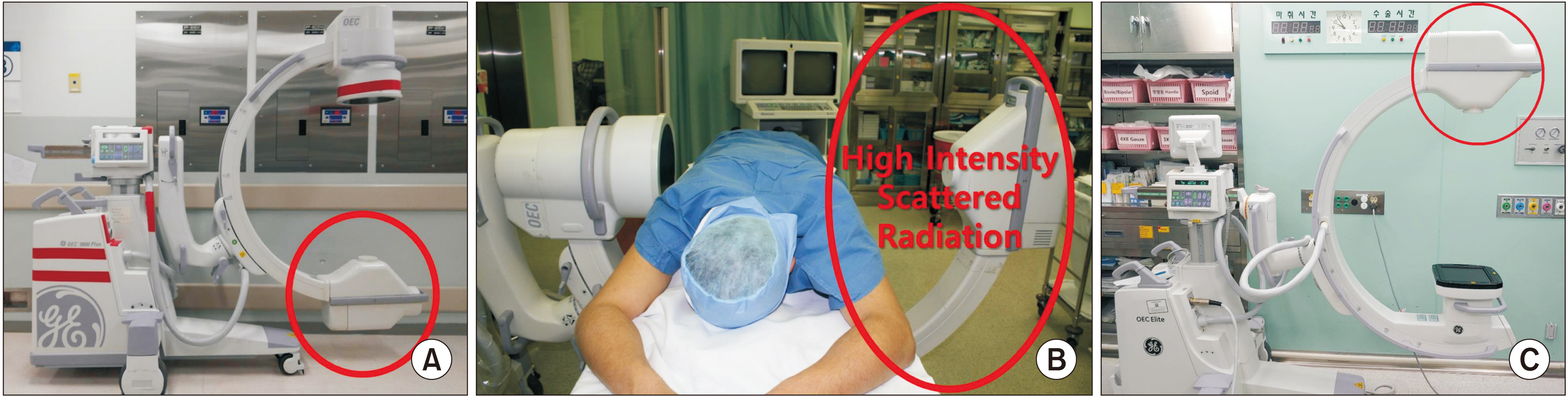
Fig. 2 Depending on the location of the X-ray generator, the parts of the body exposed to scattered X-rays vary. When an image is taken with the X-ray generator (red circle) positioned below (A), more scattered radiation is generated in the lower extremities of the physician and medical staff [9]. When a lateral view is taken (B), more scattered radiation is generated on the side where the X-ray generator is located [14]. If the flat panel detector is positioned downwards to obtain an image (C), a large amount of scattered radiation is generated to the upper body, neck, and head. The red circle is X-ray generator.

Fig. 3 Personal protective devices. Three types of commercially available lead aprons: (A) front type, (B) wraparound type, and (C) skirt and vest type. Various types of eye shields: (D) wraparound type, (E) side shield type, and (F) face shield.
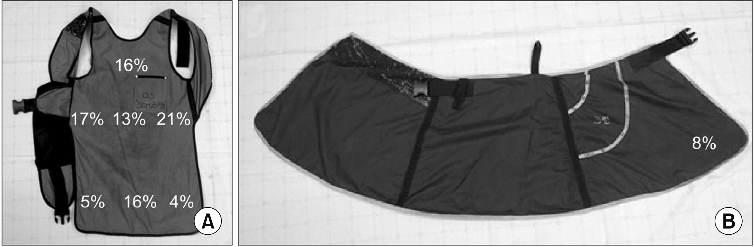
Fig. 4 Defects in the shields are found using fluoroscopic images. The most common site of damage to the radiation-protective shields was at the waist of the aprons (51%) [51]. (A) One-piece-type apron and (B) skirt-type apron. Adapted from the article of Ryu et al. (Korean J Pain 2013; 26: 142-7) [51].

Fig. 5 Reduce radiation exposure by changing the C-arm fluoroscopy mode. (A) Comparison of the radiation absorbed dose (RAD) in the C-arm fluoroscopic mode [54]. The graph shows the time, RADs, mean RADs/mean time, and current (mA) according to the C-arm modes. The pulsed fluoroscopic mode of 15 frames per second is used. *P < 0.050. aUnit is expressed as second for time, mRADs/cm2 for RADs, mRADs/cm2 • second for RADs/Time, and mA for current. (B) The differences in radiation exposure in relation to collimation in the medial branch block [56]. Comparison between the effective dose on left chest of the operator and the side of the table among groups. Chest, *P = 0.042; table, *P = 0.025. (C) Comparison of RAD between the control and collimation groups, *P = 0.001. Adapted from the article of Cho et al. (Korean J Pain 2011; 24: 199-204) [54]; Baek et al. (Korean J Pain 2013; 26: 148-53) [56].
Reference
-
1. Tapio S, Little MP, Kaiser JC, Impens N, Hamada N, Georgakilas AG, et al. 2021; Ionizing radiation-induced circulatory and metabolic diseases. Environ Int. 146:106235. DOI: 10.1016/j.envint.2020.106235. PMID: 33157375.
Article2. Paulo G, Bartal G, Vano E. 2021; Radiation dose of patients in fluoroscopically guided interventions: an update. Cardiovasc Intervent Radiol. 44:842–8. DOI: 10.1007/s00270-020-02667-3. PMID: 33034703.
Article3. Vera GV, Aleksandra F, Dragan K, Andrija H. 1997; Assessment of genome damage in occupational exposure to ionising radiation and ultrasound. Mutat Res. 395:101–5. DOI: 10.1016/S1383-5718(97)00149-6. PMID: 9465919.
Article4. Maffei F, Angelini S, Forti GC, Violante FS, Lodi V, Mattioli S, et al. 2004; Spectrum of chromosomal aberrations in peripheral lymphocytes of hospital workers occupationally exposed to low doses of ionizing radiation. Mutat Res. 547:91–9. DOI: 10.1016/j.mrfmmm.2003.12.003. PMID: 15013703.
Article5. Milacic S. 2009; Risk of occupational radiation-induced cataract in medical workers. Med Lav. 100:178–86. PMID: 19601402.6. Mastrangelo G, Fedeli U, Fadda E, Giovanazzi A, Scoizzato L, Saia B. 2005; Increased cancer risk among surgeons in an orthopaedic hospital. Occup Med (Lond). 55:498–500. DOI: 10.1093/occmed/kqi048. PMID: 16140840.
Article7. Roguin A, Goldstein J, Bar O, Goldstein JA. 2013; Brain and neck tumors among physicians performing interventional procedures. Am J Cardiol. 111:1368–72. DOI: 10.1016/j.amjcard.2012.12.060. PMID: 23419190.
Article8. Chartier H, Fassier P, Leuraud K, Jacob S, Baudin C, Laurier D, et al. 2020; Occupational low-dose irradiation and cancer risk among medical radiation workers. Occup Med (Lond). 70:476–84. DOI: 10.1093/occmed/kqaa130. PMID: 32756890.
Article9. Schueler BA. 2010; Operator shielding: how and why. Tech Vasc Interv Radiol. 13:167–71. DOI: 10.1053/j.tvir.2010.03.005. PMID: 20723831.
Article10. Dagal A. 2011; Radiation safety for anesthesiologists. Curr Opin Anaesthesiol. 24:445–50. DOI: 10.1097/ACO.0b013e328347f984. PMID: 21577097.
Article11. Cheon BK, Kim CL, Kim KR, Kang MH, Lim JA, Woo NS, et al. 2018; Radiation safety: a focus on lead aprons and thyroid shields in interventional pain management. Korean J Pain. 31:244–52. DOI: 10.3344/kjp.2018.31.4.244. PMID: 30310549. PMCID: PMC6177538.
Article12. Valentin J. 2000; Avoidance of radiation injuries from medical interventional procedures. Ann ICRP. 30:7–67. DOI: 10.1016/S0146-6453(00)00026-9. PMID: 11459599.
Article13. Vano E, Gonzalez L, Fernandez JM, Prieto C, Guibelalde E. 2006; Influence of patient thickness and operation modes on occupational and patient radiation doses in interventional cardiology. Radiat Prot Dosimetry. 118:325–30. DOI: 10.1093/rpd/nci369. PMID: 16439516.
Article14. Manchikanti L, Cash KA, Moss TL, Pampati V. 2002; Radiation exposure to the physician in interventional pain management. Pain Physician. 5:385–93. Erratum in: Pain Physician 2003; 6: 141. DOI: 10.36076/ppj.2002/5/385. PMID: 16886017.
Article15. Madder RD, VanOosterhout S, Mulder A, Ten Brock T, Clarey AT, Parker JL, et al. 2019; Patient body mass index and physician radiation dose during coronary angiography. Circ Cardiovasc Interv. 12:e006823. DOI: 10.1161/CIRCINTERVENTIONS.118.006823. PMID: 30599769.
Article16. Chang YJ, Kim AN, Oh IS, Woo NS, Kim HK, Kim JH. 2014; The radiation exposure of radiographer related to the location in C-arm fluoroscopy-guided pain interventions. Korean J Pain. 27:162–7. DOI: 10.3344/kjp.2014.27.2.162. PMID: 24748945. PMCID: PMC3990825.
Article17. Fink GE. 2009; Radiation safety in fluoroscopy for neuraxial injections. AANA J. 77:265–9. PMID: 19731844.18. Peled A, Moshe S, Chodick G. 2018; [Ionizing radiation and the risk for cataract and lens opacities]. Harefuah. 157:650–4. Hebrew. PMID: 30343544.19. Albi E, Cataldi S, Lazzarini A, Codini M, Beccari T, Ambesi-Impiombato FS, et al. 2017; Radiation and thyroid cancer. Int J Mol Sci. 18:911. DOI: 10.3390/ijms18050911. PMID: 28445397. PMCID: PMC5454824.
Article20. National Council on Radiation Protection and Measurements. 1993. Limitation of exposure to ionizing radiation: recommendations of the National Council on Radiation Protection and Measurements. National Council on Radiation Protection and Measurements;Bethesda: p. 1–86. https://books.google.co.kr/books?hl=ko&lr=&id=uFzLJu-9x8wC&oi=fnd&pg=PR3&dq=C.B,+Abrahamson+M,+Meinhold+CB,+Abrahamson+S,+Adelstein+SJ,+Bair+WJ,+et+al.+Limitation+of+exposure+to+ionizing+radiation.+1993,&ots=Olt2SDiwGf&sig=lp6ykvzJqgDjPOLRLS9ASw6VsBE#v=onepage&q&f=false.21. Hamada N, Fujimichi Y. 2015; Role of carcinogenesis related mechanisms in cataractogenesis and its implications for ionizing radiation cataractogenesis. Cancer Lett. 368:262–74. DOI: 10.1016/j.canlet.2015.02.017. PMID: 25687882.
Article22. Stewart FA, Akleyev AV, Hauer-Jensen M, Hendry JH, Kleiman NJ, et al. Authors on behalf of ICRP. 2012; ICRP publication 118: ICRP statement on tissue reactions and early and late effects of radiation in normal tissues and organs--threshold doses for tissue reactions in a radiation protection context. Ann ICRP. 41:1–322. DOI: 10.1016/j.icrp.2012.02.001. PMID: 22925378.
Article23. Dauer LT, Ainsbury EA, Dynlacht J, Hoel D, Klein BEK, Mayer D, et al. 2017; Guidance on radiation dose limits for the lens of the eye: overview of the recommendations in NCRP Commentary No. 26. Int J Radiat Biol. 93:1015–23. DOI: 10.1080/09553002.2017.1304669. PMID: 28346025.
Article24. Kim MJ, Kim JH. 2017; Radiation exposure and protection for eyes in pain management. Anesth Pain Med. 12:297–305. DOI: 10.17085/apm.2017.12.4.297.
Article25. Choi EJ, Go G, Han WK, Lee PB. 2020; Radiation exposure to the eyes and thyroid during C-arm fluoroscopy-guided cervical epidural injections is far below the safety limit. Korean J Pain. 33:73–80. DOI: 10.3344/kjp.2020.33.1.73. PMID: 31888321. PMCID: PMC6944368.
Article26. National Council on Radiation Protection and Measurements. 2018. Management of exposure to ionizing radiation: radiation protection guidance for the United States. National Council on Radiation Protection and Measurements;Bethesda (MD): Report No.: 180. https://www.worldcat.org/title/management-of-exposure-to-ionizing-radiation-radiation-protection-guidance-for-the-united-states/oclc/1121052530&referer=brief_results.27. Rivett C, Dixon M, Matthews L, Rowles N. 2016; An assessment of the dose reduction of commercially available lead protective glasses for interventional radiology staff. Radiat Prot Dosimetry. 172:443–52. DOI: 10.1093/rpd/ncv540. PMID: 26769907.
Article28. Magee JS, Martin CJ, Sandblom V, Carter MJ, Almén A, Cederblad Å, et al. 2014; Derivation and application of dose reduction factors for protective eyewear worn in interventional radiology and cardiology. J Radiol Prot. 34:811–23. DOI: 10.1088/0952-4746/34/4/811. PMID: 25332300.
Article29. Miller DL, Vañó E, Bartal G, Balter S, Dixon R, Padovani R, et al. 2010; Occupational radiation protection in interventional radiology: a joint guideline of the Cardiovascular and Interventional Radiology Society of Europe and the Society of Interventional Radiology. Cardiovasc Intervent Radiol. 33:230–9. DOI: 10.1007/s00270-009-9756-7. PMID: 20020300. PMCID: PMC2841268.
Article30. Kuipers G, Velders XL, de Winter RJ, Reekers JA, Piek JJ. 2008; Evaluation of the occupational doses of interventional radiologists. Cardiovasc Intervent Radiol. 31:483–9. DOI: 10.1007/s00270-008-9307-7. PMID: 18266030. PMCID: PMC2367387.
Article31. Wang RR, Kumar AH, Tanaka P, Macario A. 2017; Occupational Radiation exposure of anesthesia providers: a summary of key learning points and resident-led radiation safety projects. Semin Cardiothorac Vasc Anesth. 21:165–71. DOI: 10.1177/1089253217692110. PMID: 28190371.
Article32. Bryant PA, Croft J, Cole P. 2018; Integration of risks from multiple hazards into a holistic ALARA/ALARP demonstration. J Radiol Prot. 38:81–91. DOI: 10.1088/1361-6498/aa8e53. PMID: 29211686.
Article33. Kim JH. 2018; Three principles for radiation safety: time, distance, and shielding. Korean J Pain. 31:145–6. DOI: 10.3344/kjp.2018.31.3.145. PMID: 30013728. PMCID: PMC6037814.
Article34. Shuler FD, Daigre JL, Pham D, Kish VL. 2013; Laser targeting with C-arm fluoroscopy: effect on image acquisition and radiation exposure. J Orthop Trauma. 27:e97–102. DOI: 10.1097/BOT.0b013e31826625df. PMID: 22773019.35. Kim AN, Chang YJ, Cheon BK, Kim JH. 2014; How effective are radiation reducing gloves in C-arm fluoroscopy-guided pain interventions? Korean J Pain. 27:145–51. DOI: 10.3344/kjp.2014.27.2.145. PMID: 24748943. PMCID: PMC3990823.
Article36. Jung CH, Ryu JS, Baek SW, Oh JH, Woo NS, Kim HK, et al. 2013; Radiation exposure of the hand and chest during C-arm fluoroscopy-guided procedures. Korean J Pain. 26:51–6. DOI: 10.3344/kjp.2013.26.1.51. PMID: 23342208. PMCID: PMC3546211.
Article37. Livingstone RS, Varghese A. 2018; A simple quality control tool for assessing integrity of lead equivalent aprons. Indian J Radiol Imaging. 28:258–62. DOI: 10.4103/ijri.IJRI_374_17. PMID: 30050253. PMCID: PMC6038217.
Article38. Fakhoury E, Provencher JA, Subramaniam R, Finlay DJ. 2019; Not all lightweight lead aprons and thyroid shields are alike. J Vasc Surg. 70:246–50. DOI: 10.1016/j.jvs.2018.07.055. PMID: 30292602.
Article39. Singer G. 2005; Occupational radiation exposure to the surgeon. J Am Acad Orthop Surg. 13:69–76. DOI: 10.5435/00124635-200501000-00009. PMID: 15712984.
Article40. Hong SW, Kim TW, Kim JH. 2021; Radiation exposure to the back with different types of aprons. Radiat Prot Dosimetry. 193:185–9. DOI: 10.1093/rpd/ncab044. PMID: 33839791.
Article41. Livingstone RS, Varghese A, Keshava SN. 2018; A study on the use of radiation-protective apron among interventionists in radiology. J Clin Imaging Sci. 8:34. DOI: 10.4103/jcis.JCIS_34_18. PMID: 30197825. PMCID: PMC6118106.
Article42. Chou LB, Cox CA, Tung JJ, Harris AH, Brooks-Terrell D, Sieh W. 2010; Prevalence of cancer in female orthopaedic surgeons in the United States. J Bone Joint Surg Am. 92:240–4. DOI: 10.2106/JBJS.H.01691. PMID: 20048119.
Article43. Müller LP, Suffner J, Wenda K, Mohr W, Rommens PM. 1998; Radiation exposure to the hands and the thyroid of the surgeon during intramedullary nailing. Injury. 29:461–8. DOI: 10.1016/S0020-1383(98)00088-6. PMID: 9813705.
Article44. Lee SY, Min E, Bae J, Chung CY, Lee KM, Kwon SS, et al. 2013; Types and arrangement of thyroid shields to reduce exposure of surgeons to ionizing radiation during intraoperative use of C-arm fluoroscopy. Spine (Phila Pa 1976). 38:2108–12. DOI: 10.1097/BRS.0b013e3182a8270d. PMID: 23963017.
Article45. Wagner LK, Mulhern OR. 1996; Radiation-attenuating surgical gloves: effects of scatter and secondary electron production. Radiology. 200:45–8. DOI: 10.1148/radiology.200.1.8657942. PMID: 8657942.
Article46. Cousin AJ, Lawdahl RB, Chakraborty DP, Koehler RE. 1987; The case for radioprotective eyewear/facewear. Practical implications and suggestions. Invest Radiol. 22:688–92. DOI: 10.1097/00004424-198708000-00012. PMID: 3667176.47. Kirkwood ML, Klein A, Guild J, Arbique G, Xi Y, Tsai S, et al. 2020; Novel modification to leaded eyewear results in significant operator eye radiation dose reduction. J Vasc Surg. 72:2139–44. DOI: 10.1016/j.jvs.2020.02.049. PMID: 32276011.
Article48. Koukorava C, Farah J, Struelens L, Clairand I, Donadille L, Vanhavere F, et al. 2014; Efficiency of radiation protection equipment in interventional radiology: a systematic Monte Carlo study of eye lens and whole body doses. J Radiol Prot. 34:509–28. DOI: 10.1088/0952-4746/34/3/509. PMID: 24938591.
Article49. Lian Y, Xiao J, Ji X, Guan S, Ge H, Li F, et al. 2015; Protracted low-dose radiation exposure and cataract in a cohort of Chinese industry radiographers. Occup Environ Med. 72:640–7. DOI: 10.1136/oemed-2014-102772. PMID: 26163545.
Article50. Maina PM, Motto JA, Hazell LJ. 2020; Investigation of radiation protection and safety measures in Rwandan public hospitals: readiness for the implementation of the new regulations. J Med Imaging Radiat Sci. 51:629–38. DOI: 10.1016/j.jmir.2020.07.056. PMID: 32839139.
Article51. Ryu JS, Baek SW, Jung CH, Cho SJ, Jung EG, Kim HK, et al. 2013; The survey about the degree of damage of radiation-protective shields in operation room. Korean J Pain. 26:142–7. DOI: 10.3344/kjp.2013.26.2.142. PMID: 23614075. PMCID: PMC3629340.
Article52. Christodoulou EG, Goodsitt MM, Larson SC, Darner KL, Satti J, Chan HP. 2003; Evaluation of the transmitted exposure through lead equivalent aprons used in a radiology department, including the contribution from backscatter. Med Phys. 30:1033–8. DOI: 10.1118/1.1573207. PMID: 12852526.
Article53. Brown PH, Thomas RD, Silberberg PJ, Johnson LM. 2000; Optimization of a fluoroscope to reduce radiation exposure in pediatric imaging. Pediatr Radiol. 30:229–35. DOI: 10.1007/s002470050728. PMID: 10789900.
Article54. Cho JH, Kim JY, Kang JE, Park PE, Kim JH, Lim JA, et al. 2011; A study to compare the radiation absorbed dose of the C-arm fluoroscopic modes. Korean J Pain. 24:199–204. DOI: 10.3344/kjp.2011.24.4.199. PMID: 22220241. PMCID: PMC3248583.
Article55. Aufrichtig R, Xue P, Thomas CW, Gilmore GC, Wilson DL. 1994; Perceptual comparison of pulsed and continuous fluoroscopy. Med Phys. 21:245–56. DOI: 10.1118/1.597285. PMID: 8177157.
Article56. Baek SW, Ryu JS, Jung CH, Lee JH, Kwon WK, Woo NS, et al. 2013; A randomized controlled trial about the levels of radiation exposure depends on the use of collimation C-arm fluoroscopic-guided medial branch block. Korean J Pain. 26:148–53. DOI: 10.3344/kjp.2013.26.2.148. PMID: 23614076. PMCID: PMC3629341.
Article57. Mahesh M. 2001; Fluoroscopy: patient radiation exposure issues. Radiographics. 21:1033–45. DOI: 10.1148/radiographics.21.4.g01jl271033. PMID: 11452079.
Article58. Kim TH, Hong SW, Woo NS, Kim HK, Kim JH. 2017; The radiation safety education and the pain physicians' efforts to reduce radiation exposure. Korean J Pain. 30:104–15. DOI: 10.3344/kjp.2017.30.2.104. PMID: 28416994. PMCID: PMC5392654.
Article
- Full Text Links
-
- Actions
-
Cited
- CITED
-
- Close
- Share
-
- Similar articles
-
- The radiation safety education and the pain physicians' efforts to reduce radiation exposure
- Radiation safety: a focus on lead aprons and thyroid shields in interventional pain management
- Radiation Safety in Nuclear Medicine Procedures
- Three principles for radiation safety: time, distance, and shielding
- Radiation exposure and protection for eyes in pain management
