Full mouth rehabilitation for a Parkinson's diseases patient with chronic periodontitis: a case report
- Affiliations
-
- 1Divison of Prosthodontics, Department of Dentistry, Asan Medical Center, Seoul, Republic of Korea.
- 2Divison of Prosthodontics, Department of Dentistry, Asan Medical Center, College of Medicine, University of Ulsan, Seoul, Republic of Korea. ljhl11911@hanmail.net
- KMID: 2470628
- DOI: http://doi.org/10.14368/jdras.2019.35.4.228
Abstract
- Parkinson's disease is a neurological disorder characterized by tremor, bradykinesia, akinesia, postural instability, and muscular rigidity, which is caused by the depletion of neurotransmitters such as dopamine. Cooperative dental treatment is more challenging because of tremor of Parkinson's disease. In this case, a 47-year-old Parkinson's disease patient with chronic periodontitis was treated with full-mouth rehabilitation using conventional fixed prostheses and implant fixed partial denture, which attained satisfactory outcomes functionally and esthetically. Short term periodic follow-ups will be needed with consideration for the characteristics of Parkinson's disease such as decreased manual dexterity.
MeSH Terms
Figure
-
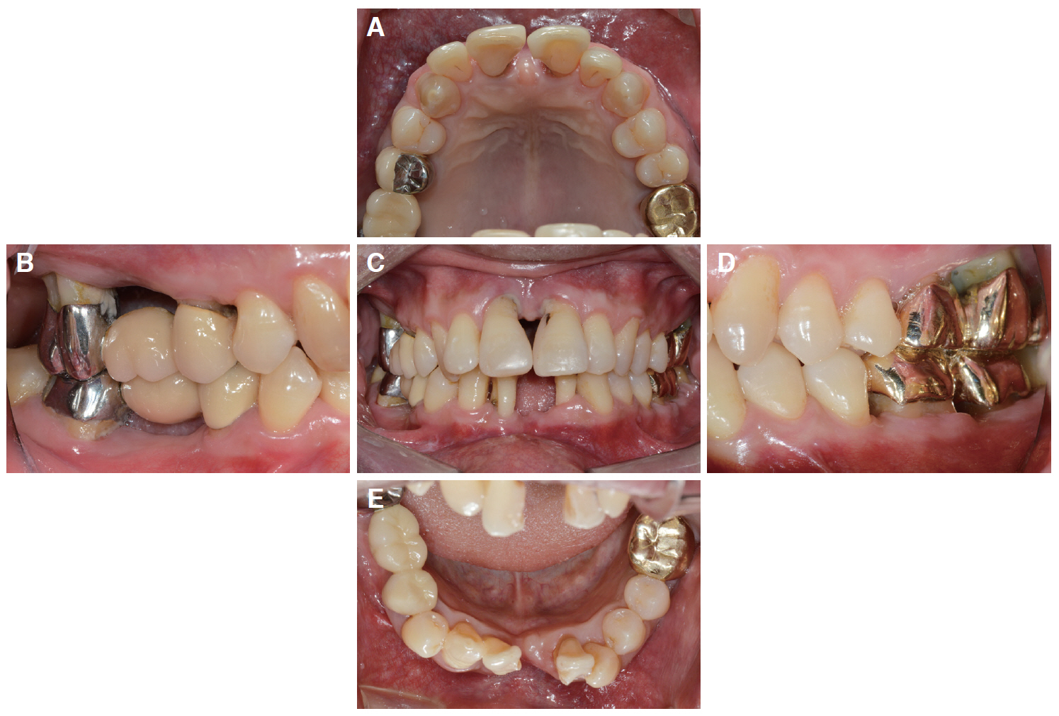
Fig. 1 Preoperative intraoral photographs. (A) Maxillary occlusal view, (B) Right lateral view, (C) Frontal view, (D) Left lateral view, (E) Mandibular occlusal view.

Fig. 2 Radiographic evaluation before treatment (Panoramic view).
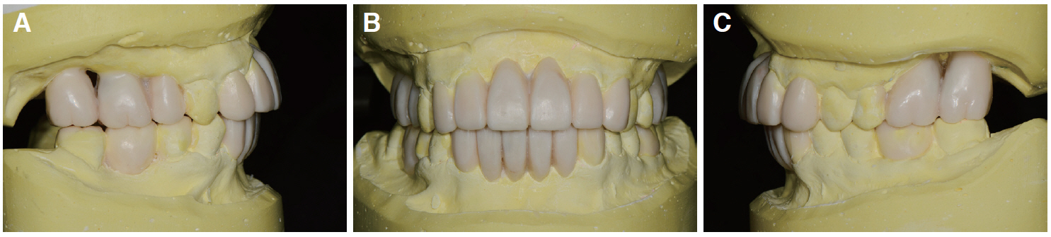
Fig. 3 Diagnostic wax-up. (A) Right lateral view, (B) Frontal view, (C) Left lateral view.
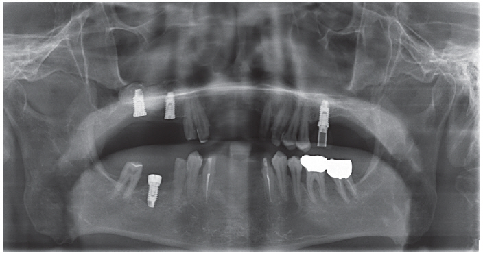
Fig. 4 Panoramic radiograph after implant installation.
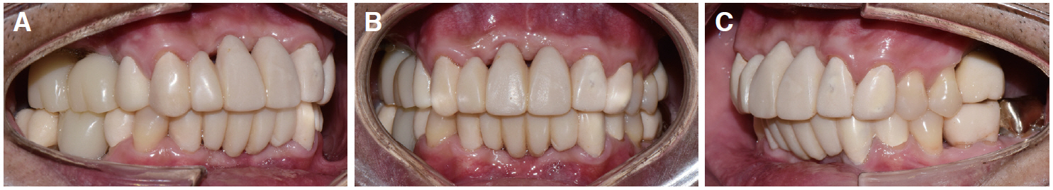
Fig. 5 Provisional restoration after implant installation. (A) Right lateral view, (B) Frontal view, (C) Left lateral view.
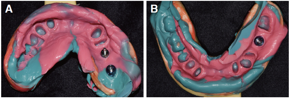
Fig. 6 Final impression taking. (A) Maxilla, (B) Mandible.
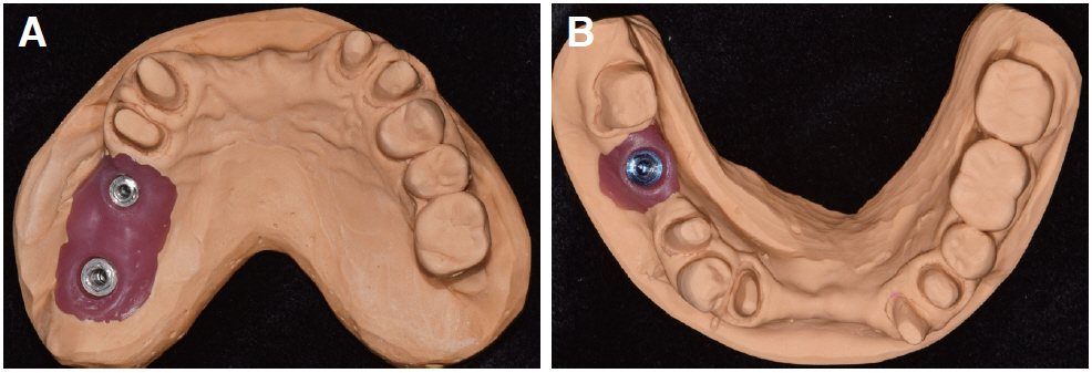
Fig. 7 Master cast fabrication. (A) Maxilla, (B) Mandible.
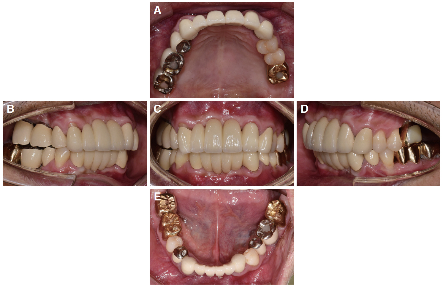
Fig. 8 Placement of the definitive prostheses. (A) Maxillary occlusal view, (B) Right lateral view, (C) Frontal view, (D) Left lateral view, (E) Mandibular occlusal view.

Fig. 9 Panoramic radiograph after placement of the definitive prostheses.
Reference
-
References
1. Rajeswari CL. 2010; Prosthodontic considerations in Parkinson's disease. PJSR. 3:45–7.2. Packer M, Nikitin V, Coward T, Davis DM, Fiske J. 2009; The potential benefits of dental implants on the oral health quality of life of people with Parkinson's disease. Gerodontology. 26:11–8. DOI: 10.1111/j.1741-2358.2008.00233.x. PMID: 19278520.3. Haralur SB. 2015; Clinical strategies for complete denture rehabilitation in a patient with Parkinson disease and reduced neuromuscular control. Case Rep Dent. 2015:352878. DOI: 10.1155/2015/352878. PMID: 25737785. PMCID: PMC4337048.4. Al-Omari FA, Al Moaleem MM, Al-Qahtani SS, Al Garni AS, Sadatullah S, Luqman M. 2014; Oral rehabilitation of Parkinson's disease patient: a review and case report. Case Rep Dent. 2014:432475. DOI: 10.1155/2014/432475. PMID: 24551462. PMCID: PMC3914341.5. Singh Y, Saini M, Garg N. 2013; Oral rehabilitation of a Parkinson's patient: A case report. World J Clin Cases. 1:67–70. DOI: 10.12998/wjcc.v1.i1.67. PMID: 24303468. PMCID: PMC3845922.
- Full Text Links
-
- Actions
-
Cited
- CITED
-
- Close
- Share
-
- Similar articles
-
- Full mouth fixed implant rehabilitation in a patient with generalized aggressive periodontitis
- Full mouth rehabilitation with a few remaining teeth and implants for a patient with chronic periodontitis: a case report
- Full mouth rehabilitation in a patient with peri-implantitis: A case report
- Full mouth rehabilitation of a patient with severe periodontitis using immediate loading after computer aided flapless implant surgery
- Ten-year follow-up of full mouth rehabilitation with fixed prostheses using implants and natural tooth
