Clinical Outcomes of Small Incision Lenticule Extraction in Myopia: Study of Vector Parameters and Corneal Aberrations
- Affiliations
-
- 1Institute of Vision Research, Department of Ophthalmology, Yonsei University College of Medicine, Seoul, Korea. tikim@yuhs.ac
- 2Corneal Dystrophy Research Institute, Severance Biomedical Science Institute, and Brain Korea 21 Project for Medical Science, Department of Ophthalmology, Yonsei University College of Medicine, Seoul, Korea.
- KMID: 2469289
- DOI: http://doi.org/10.3341/kjo.2019.0109
Abstract
- PURPOSE
To investigate clinical outcomes of small incision lenticule extraction (SMILE) including vector parameters and corneal aberrations in myopic patients.
METHODS
This retrospective, observational case series included 57 eyes (29 patients) that received treatment for myopia using SMILE. Visual acuity measurement, manifest refraction, slit-lamp examination, autokeratometry, corneal topography, and evaluation of corneal wavefront aberration were performed preoperatively and at 1 and 3 months after surgery. We analyzed the safety, efficacy, vector parameters, and corneal aberrations at 3 months after surgery.
RESULTS
Preoperatively, mean manifest refraction spherical equivalent refraction was −4.94 ± 1.94 D (range, −8.25 to 0 diopters [D]), and the cylinder was −1.14 ± 0.82 D (range, −3 to 0 D). Mean manifest refraction spherical equivalent improved to −0.10 ± 0.23 D at 3 months postoperatively, when uncorrected distance visual acuity was 20 / 20 or better in 55 (96%) eyes. The linear regression model of target induced astigmatism vector versus surgically induced astigmatism vector exhibited slopes and coefficients (R²) of 0.9618 and 0.9748, respectively (y = 0.9618x + 0.0006, R² = 0.9748). While total corneal root mean square higher order aberrations, coma and trefoil showed statistically significant increase, spherical aberration did not show statistically significant change after SMILE.
CONCLUSIONS
SMILE has proven to be effective and safe for correcting myopia and astigmatism. We showed that SMILE did not induce spherical aberrations. A small increase in postoperative corneal higher order aberration may be associated with increase in coma and trefoil.
Keyword
MeSH Terms
Figure
-
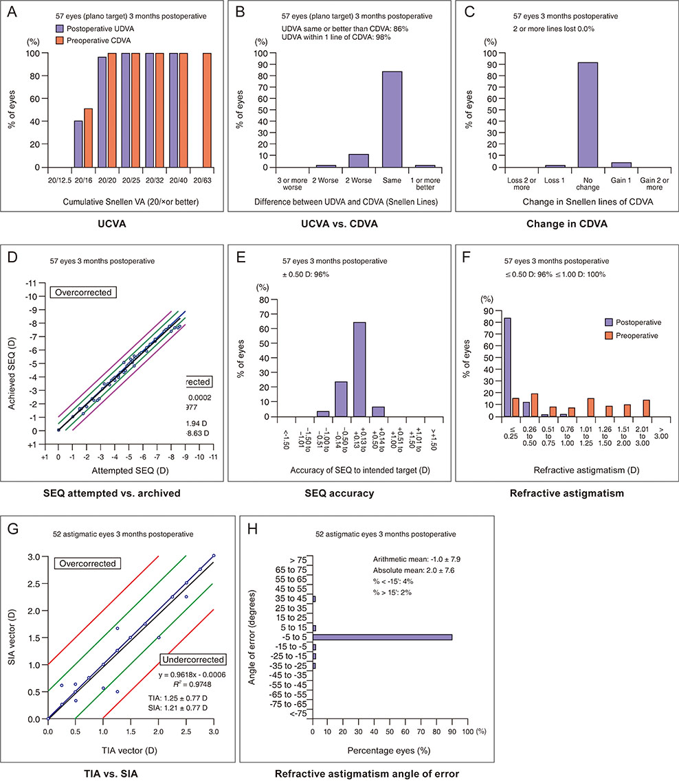
Fig. 1 Visual outcomes after small incision lenticule extraction. (A) Cumulative 3-month postoperative uncorrected distance visual acuity (UDVA) and preoperative corrected distance visual acuity (CDVA). Changes in Snellen lines of (B) postoperative UDVA and (C) CDVA relative to preoperative CDVA. (D) Attempted versus achieved changes in spherical equivalent refraction (SEQ) at 3 months after surgery. (E) The accuracy of SEQ to the intended target. (F) Comparative distribution of preoperative and 3-month postoperative cylinder and (G) target induced astigmatism (TIA) versus surgically induced astigmatism (SIA) vectors at 3 months. (H) Refractive astigmatism angle of error distribution at 3 months after surgery. D = diopters.

Fig. 2 Single-angle polar plots of (A) target induced astigmatism vector, (B) surgically induced astigmatism vector, (C) difference vector, and (D) correction index at 3 months after small incision lenticule extraction (SMILE). D = diopters.
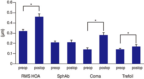
Fig. 3 Changes in higher order aberrations (HOA) at 3 months after small incision lenticule extraction. Data are presented as mean values ± standard error of the mean. RMS = root mean square; SphAb = spherical aberration; preop = preoperative; postop = postoperative. *Significant difference.
Reference
-
1. Reinstein DZ, Archer TJ, Gobbe M. The history of LASIK. J Refract Surg. 2012; 28:291–298.
Article2. Kim TI, Alio Del, Wilkins M, et al. Refractive surgery. Lancet. 2019; 393:2085–2098.
Article3. Sekundo W, Kunert K, Russmann C, et al. First efficacy and safety study of femtosecond lenticule extraction for the correction of myopia: six-month results. J Cataract Refract Surg. 2008; 34:1513–1520.4. Tan DK, Tay WT, Chan C, et al. Postoperative ocular higher-order aberrations and contrast sensitivity: femtosecond lenticule extraction versus pseudo small-incision lenticule extraction. J Cataract Refract Surg. 2015; 41:623–634.
Article5. Sekundo W, Kunert KS, Blum M. Small incision corneal refractive surgery using the small incision lenticule extraction (SMILE) procedure for the correction of myopia and myopic astigmatism: results of a 6 month prospective study. Br J Ophthalmol. 2011; 95:335–339.
Article6. Shah R, Shah S, Sengupta S. Results of small incision lenticule extraction: all-in-one femtosecond laser refractive surgery. J Cataract Refract Surg. 2011; 37:127–137.
Article7. Jun I, Kang DS, Reinstein DZ, et al. Clinical outcomes of SMILE with a triple centration technique and corneal wavefront-guided transepithelial PRK in high astigmatism. J Refract Surg. 2018; 34:156–163.
Article8. Alpins N. Astigmatism analysis by the Alpins method. J Cataract Refract Surg. 2001; 27:31–49.
Article9. Read SA, Collins MJ, Carney LG. A review of astigmatism and its possible genesis. Clin Exp Optom. 2007; 90:5–19.
Article10. Blum M, Taubig K, Gruhn C, et al. Five-year results of Small Incision Lenticule Extraction (ReLEx SMILE). Br J Ophthalmol. 2016; 100:1192–1195.
Article11. Xia LK, Ma J, Liu HN, et al. Three-year results of small incision lenticule extraction and wavefront-guided femtosecond laser-assisted laser in situ keratomileusis for correction of high myopia and myopic astigmatism. Int J Ophthalmol. 2018; 11:470–477.
Article12. Sanchez-Gonzalez JM, Alonso-Aliste F. Visual and refractive outcomes of 100 small incision lenticule extractions (SMILE) in moderate and high myopia: a 24-month follow-up study. Graefes Arch Clin Exp Ophthalmol. 2019; 257:1561–1567.
Article13. Sekundo W, Gertnere J, Bertelmann T, Solomatin I. Oneyear refractive results, contrast sensitivity, high-order aberrations and complications after myopic small-incision lenticule extraction (ReLEx SMILE). Graefes Arch Clin Exp Ophthalmol. 2014; 252:837–843.
Article14. Han T, Xu Y, Han X, et al. Three-year outcomes of small incision lenticule extraction (SMILE) and femtosecond laser-assisted laser in situ keratomileusis (FS-LASIK) for myopia and myopic astigmatism. Br J Ophthalmol. 2019; 103:565–568.
Article15. Pinero DP, Teus MA. Clinical outcomes of small-incision lenticule extraction and femtosecond laser-assisted wavefront-guided laser in situ keratomileusis. J Cataract Refract Surg. 2016; 42:1078–1093.16. Pedersen IB, Ivarsen A, Hjortdal J. Changes in astigmatism, densitometry, and aberrations After SMILE for low to high myopic astigmatism: a 12-month prospective study. J Refract Surg. 2017; 33:11–17.
Article17. Chan TC, Yu MC, Ng A, et al. Early outcomes after small incision lenticule extraction and photorefractive keratectomy for correction of high myopia. Sci Rep. 2016; 6:32820.
Article18. Qin B, Zhao J, Li M, et al. The comparison of visual outcomes, aberrations, and Bowman's layer micro-distortions after femtosecond laser small-incision lenticule extraction (SMILE) for the correction of high and moderate myopia and myopic astigmatism. BMC Ophthalmol. 2019; 19:138.
Article19. Gyldenkerne A, Ivarsen A, Hjortdal J. Optical and visual quality after small-incision lenticule extraction. J Cataract Refract Surg. 2019; 45:54–61.
Article20. Jin HY, Wan T, Yu XN, et al. Corneal higher-order aberrations of the anterior surface, posterior surface, and total cornea after small incision lenticule extraction (SMILE): high myopia versus mild to moderate myopia. BMC Ophthalmol. 2018; 18:295.
Article21. Mrochen M, Kaemmerer M, Mierdel P, Seiler T. Increased higher-order optical aberrations after laser refractive surgery: a problem of subclinical decentration. J Cataract Refract Surg. 2001; 27:362–369.22. Yu M, Chen M, Liu W, Dai J. Comparative study of wave-front aberration and corneal Asphericity after SMILE and LASEK for myopia: a short and long term study. BMC Ophthalmol. 2019; 19:80.
Article23. Wu W, Wang Y. Corneal higher-order aberrations of the anterior surface, posterior surface, and total cornea after SMILE, FS-LASIK, and FLEx surgeries. Eye Contact Lens. 2016; 42:358–365.
Article24. Zhang H, Wang Y, Li H. Corneal spherical aberration and corneal asphericity after small incision lenticule extraction and femtosecond laser-assisted LASIK. J Ophthalmol. 2017; 2017:4921090.
Article25. Dong Z, Zhou X, Wu J, et al. Small incision lenticule extraction (SMILE) and femtosecond laser LASIK: comparison of corneal wound healing and inflammation. Br J Ophthalmol. 2014; 98:263–269.
Article26. Shetty R, Matalia H, Nandini C, et al. Wavefront-guided LASIK has comparable ocular and corneal aberrometric outcomes but better visual acuity outcomes than SMILE in myopic eyes. J Refract Surg. 2018; 34:527–532.
Article27. Jin HY, Wan T, Wu F, Yao K. Comparison of visual results and higher-order aberrations after small incision lenticule extraction (SMILE): high myopia vs. mild to moderate myopia. BMC Ophthalmol. 2017; 17:118.
Article28. Damgaard IB, Ang M, Mahmoud AM, et al. Functional optical zone and centration following SMILE and LASIK: a prospective, randomized, contralateral eye study. J Refract Surg. 2019; 35:230–237.
Article29. Lin F, Xu Y, Yang Y. Comparison of the visual results after SMILE and femtosecond laser-assisted LASIK for myopia. J Refract Surg. 2014; 30:248–254.
Article30. Yao P, Zhao J, Li M, et al. Microdistortions in Bowman's layer following femtosecond laser small incision lenticule extraction observed by Fourier-Domain OCT. J Refract Surg. 2013; 29:668–674.
Article31. Vestergaard AH, Grauslund J, Ivarsen AR, Hjortdal JO. Efficacy, safety, predictability, contrast sensitivity, and aberrations after femtosecond laser lenticule extraction. J Cataract Refract Surg. 2014; 40:403–411.
Article32. Al-Zeraid FM, Osuagwu UL. Induced higher-order aberrations after laser in situ keratomileusis (LASIK) performed with wavefront-guided IntraLase femtosecond laser in moderate to high astigmatism. BMC Ophthalmol. 2016; 16:29.
Article33. Yu M, Chen M, Wang B, et al. Comparison of visual quality after SMILE and LASEK for mild to moderate myopia. J Refract Surg. 2015; 31:795–800.
Article
- Full Text Links
-
- Actions
-
Cited
- CITED
-
- Close
- Share
-
- Similar articles
-
- Short-term Clinical Outcomes of Small Incision Lenticule Extraction for Correction of Myopia Patients with Corneal Opacity
- Comparison of the Early Clinical Outcomes between Combined SMILE and Collagen Cross-linking versus SMILE
- Clear lens Extraction and Epikeratophakic Lenticule Removal in Complicated Epikeratophakic Patients
- Outcomes of Small Incision Lenticule Extraction: Mild to Moderate Myopia versus High Myopia
- Clinical Outcome of Small Incision Lenticule Extraction including Visual Quality Analysis
