Nonarteritic Anterior Ischemic Optic Neuropathy with an Atypical Visual Field Defect
- Affiliations
-
- 1Department of Ophthalmology, Maryknoll Medical Center, Busan, Korea. ddalkieco@gmail.com
- KMID: 2466199
- DOI: http://doi.org/10.3341/jkos.2019.60.12.1374
Abstract
- PURPOSE
To report a case of nonarteritic anterior ischemic optic neuropathy (NA-AION) with an atypical visual field defect after intraocular surgery.
CASE SUMMARY
A 61-year-old male presented with a visual field defect in his right eye 1 day after uneventful cataract surgery with pars plana vitrectomy for epiretinal membrane. His best-corrected visual acuity (BCVA) in the right eye was 20/400 with a relative afferent pupillary defect. A color vision test revealed failure only in the right eye. A slit-lamp examination revealed no abnormality in the anterior part of the eyes. A fundoscopic examination also revealed no abnormality in the posterior part of the eyes, including the optic disc. The Humphrey visual field test revealed a nasal vertical defect in the right eye. Orbital and brain magnetic resonance imaging were normal. After 14 days from the initial symptom, fundus photography and optical coherence tomography revealed an optic disc swelling and splinter hemorrhage. Fluorescein angiography revealed a delayed filling on the temporal half of the optic disc and inferotemporal peripapillary choroid. A diagnosis of NA-AION was made. The patient was treated with oral steroids and aspirin. After 3 months, the BCVA was 20/125. The visual field defect was maintained and segmental atrophy developed on the superior and inferior sides of the right optic disc.
CONCLUSIONS
AION may present as vertical hemianopsia. With the risk factors of ischemic optic neuropathy, the possibility of AION should be considered in the differential diagnoses of postoperative visual impairments or field defects after intraocular surgery.
MeSH Terms
-
Aspirin
Atrophy
Brain
Cataract
Choroid
Color Vision
Diagnosis
Diagnosis, Differential
Epiretinal Membrane
Fluorescein Angiography
Hemianopsia
Hemorrhage
Humans
Magnetic Resonance Imaging
Male
Middle Aged
Optic Neuropathy, Ischemic*
Orbit
Photography
Pupil Disorders
Risk Factors
Steroids
Tomography, Optical Coherence
Vision Disorders
Visual Acuity
Visual Field Tests
Visual Fields*
Vitrectomy
Aspirin
Steroids
Figure
-

Figure 1 Postoperative ophthalmic examinations performed 1 week after onset of visual field loss. (A) Ultrawide fundus photographshows that the epiretinal membrane was removed and there was no apparent edema or hyperemia of right optic disc. (B) Humphreyvisual field test demonstrates a nasal visual field defect respecting the vertical meridian in his right eye.
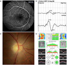
Figure 2 Postoperative ophthalmic examinations performed 1 week after onset of visual field loss. (A) Ultrawide fundus photographshows that the epiretinal membrane was removed and there was no apparent edema or hyperemia of right optic disc. (B) Humphreyvisual field test demonstrates a nasal visual field defect respecting the vertical meridian in his right eye.

Figure 3 Postoperative ophthalmic examinations performed 1 week after onset of visual field loss. (A) Ultrawide fundus photographshows that the epiretinal membrane was removed and there was no apparent edema or hyperemia of right optic disc. (B) Humphreyvisual field test demonstrates a nasal visual field defect respecting the vertical meridian in his right eye.
Reference
-
1. Hayreh SS. Ischemic optic neuropathy. Prog Retin Eye Res. 2009; 28:34–62.2. McCulley TJ, Lam BL, Feuer WJ. Incidence of nonarteritic anterior ischemic optic neuropathy associated with cataract extraction. Ophthalmology. 2001; 108:1275–1278.3. Taban M, Lewis H, Lee MS. Nonarteritic anterior ischemic optic neuropathy and ‘visual field defects’ following vitrectomy: could they be related? Graefes Arch Clin Exp Ophthalmol. 2007; 245:600–605.4. Uchida A, Shinoda K, Matsumoto CS, et al. Acute visual field defect following vitrectomy determined to originate from optic nerve by electrophysiological tests. Case Rep Ophthalmol. 2012; 3:396–405.5. Cunha LP, Cunha LV, Costa CF, Monteiro ML. Nonarteritic anterior ischemic optic neuropathy following pars plana vitrectomy for macular hole treatment: case report. Arq Bras Oftalmol. 2016; 79:342–345.6. Taban M, Sharma MC, Lee MS. Anterior ischemic optic neuropathy after uncomplicated scleral buckling surgery. Graefes Arch Clin Exp Ophthalmol. 2006; 244:1370–1372.7. Lee AG, Kohnen T, Ebner R, et al. Optic neuropathy associated with laser in situ keratomileusis. J Cataract Refract Surg. 2000; 26:1581–1584.8. Mahroo OA, Hammond CJ. Anterior ischemic optic neuropathy after strabismus surgery. J Neuroophthalmol. 2009; 29:157–158.9. Ganssauge M, Wilhelm H, Bartz-Schmidt KU, Aisenbrey S. Non-arteritic anterior ischemic optic neuropathy (NA-AION) after intravitreal injection of bevacizumab (Avastin) for treatment of angoid streaks in pseudoxanthoma elasticum. Graefes Arch Clin Exp Ophthalmol. 2009; 247:1707–1710.10. Hosseini H, Razeghinejad MR. Anterior ischemic optic neuropathy after intravitreal injection of bevacizumab. J Neuroophthalmol. 2009; 29:160–161.11. Bodla AA, Rao P. Non-arteritic ischemic optic neuropathy followed by intravitreal bevacizumab injection: is there an association? Indian J Ophthalmol. 2010; 58:349–350.12. Huang JY, Ozaki H, Hayashi H, Uchio E. Anterior ischemic optic neuropathy following intravitreal bevacizumab. Jpn J Ophthalmol. 2010; 54:252–254.13. McCulley TJ, Lam BL, Feuer WJ. Nonarteritic anterior ischemicoptic neuropathy and surgery of the anterior segment: temporal relationship analysis. Am J Ophthalmol. 2003; 136:1171–1172.14. Kawashima H, Nagai N, Shinoda H, et al. Optic neuropathy causing vertical unilateral hemianopsia after pars plana vitrectomy for a macular hole: a case report. Medicine (Baltimore). 2018; 97:e0321.15. Mandelcorn E, Khan Y, Javorska L, et al. Idiopathic epiretinal membranes: cell type, growth factor expression, and fluorescein angiographic and retinal photographic correlations. Can J Ophthalmol. 2003; 38:457–463.16. Harada C, Mitamura Y, Harada T. The role of cytokines and trophic factors in epiretinal membranes: involvement of signal transduction in glial cells. Prog Retin Eye Res. 2006; 25:149–164.17. Creuzot-Garcher C, Wolf S. Macular edema. Miscellaneous. Dev Ophthalmol. 2010; 47:183–198.18. Chang YC, Lin CC, Wu WC. Long-term anatomical and functional outcome of three intravitreal bevacizumab injections for persistent macular edema after idiopathic macular epiretinal membrane peeling. Ophthalmologica Extra. 2014; 1:1–8.19. Bennett JL, Thomas S, Olson JL, Mandava N. Treatment of nonarteritic anterior ischemic optic neuropathy with intravitreal bevacizumab. J Neuroophthalmol. 2007; 27:238–240.20. Dave VP, Pappuru RR. An unusual presentation of nonarteritic ischemic optic neuropathy with subretinal fluid treated with intravitreal bevacizumab. Indian J Ophthalmol. 2016; 64:87–88.21. Prescott CR, Sklar CA, Lesser RL, Adelman RA. Is intravitreal bevacizumab an effective treatment option for nonarteritic anterior ischemic optic neuropathy? J Neuroophthalmol. 2012; 32:51–53.22. Rootman DB, Gill HS, Margolin EA. Intravitreal bevacizumab for the treatment of nonarteritic anterior ischemic optic neuropathy: a prospective trial. Eye (Lond). 2013; 27:538–544.23. Hayreh SS. Anterior ischaemic optic neuropathy. II. Fundus on ophthalmoscopy and fluorescein angiography. Br J Ophthalmol. 1974; 58:964–980.24. Hayreh SS. Anterior ischaemic optic neuropathy. Differentiation of arteritic from non-arteritic type and its management. Eye (Lond). 1990; 4:25–41.25. Arnold AC, Hepler RS. Fluorescein angiography in acute nonarteritic anterior ischemic optic neuropathy. Am J Ophthalmol. 1994; 117:222–230.26. Shin SY, Kim DS, Ko MK. Fluorescein angiographic features of choroidal insufficiency in anterior ischemic optic neuropathy. Korean J Ophthalmol. 1999; 13:100–104.27. Kim SB, Kyung SE. Fluorescein angiographic findings of nonarteritic anterior ischemic optic neuropathy and optic neuritis. J Korean Ophthalmol Soc. 2012; 53:1143–1149.28. Hayreh SS. Posterior ciliary artery occlusion. Ophthalmol Retina. 2018; 2:106–111.29. Hayreh SS, Zimmerman B. Visual field abnormalities in nonarteritic anterior ischemic optic neuropathy their pattern and prevalence at initial examination. Arch Ophthalmol. 2005; 123:1554–1562.
- Full Text Links
-
- Actions
-
Cited
- CITED
-
- Close
- Share
-
- Similar articles
-
- The Function of the Fellow Eye in Patients with Unilateral Nonarteritic Anterior Ischemic Optic Neuropathy
- Nonarteritic Anterior Ischemic Optic Neuropathy Simultaneously Associated with Hashimoto’s Thyroiditis
- A Case of Decreased Visual Field after Uneventful Cataract Surgery: Nonarteritic Anterior Ischemic Optic Neuropathy
- A Case of Anterior Ischemic Optic Neuropethy
- A Case of Non-Arteritic Anterior Ischemic Optic Neuropathy after Bilateral Selective Neck Dissection
