High-Altitude Cerebral Edema Evaluated with MRI: A Case Report
- Affiliations
-
- 1Department of Radiology, Ilsan Paik Hospital, Inje University College of Medicine, Goyang, Korea. bhlee@paik.ac.kr
- KMID: 2464922
- DOI: http://doi.org/10.3348/jksr.2019.80.6.1247
Abstract
- High-altitude cerebral edema (HACE) is a rare life-threatening condition observed in individuals who climb high altitudes. This report describes the case of a 38-year-old man who recently climbed a 5000-m-high mountain, showing the following radiologic findings at 3 different anatomical locations: 1) increased T2 signal intensity (SI) without restricted diffusion, with full recovery in the posterior limb of the left internal capsule; 2) increased T2 SI with restricted diffusion, with full recovery in the splenium of the corpus callosum; and 3) increased T2 SI with restricted diffusion and microbleeds, resulting in bilateral encephalomalacia in the globus pallidus. Herein, we report the concurrent typical and atypical radiologic findings of this rare condition caused by vasogenic and cytotoxic edema.
MeSH Terms
Figure
-
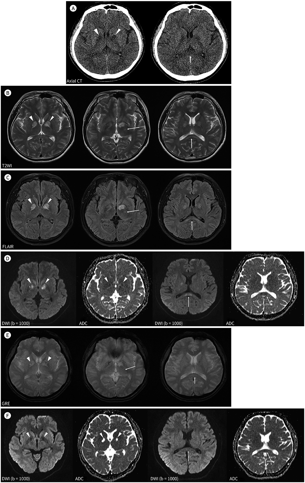
Fig. 1 CT and MRI findings of a 38-year-old man with general weakness and urinary incontinence after climbing a 5000-m-high mountain. A. Axial CT scan image shows low attenuation in bilateral globus pallidus (arrowheads) and the splenium of the corpus callosum (arrow). B. Axial T2WI shows a high SI in bilateral globus pallidus (arrowheads), the posterior limb of the left internal capsule (arrow in the middle image), and the splenium of the corpus callosum (arrow in the right image). C. Axial FLAIR image shows high SI in bilateral globus pallidus (arrowheads), the posterior limb of the left internal capsule (arrow in the middle image), and the splenium of the corpus callosum (arrow in the right image). D. There are high SIs on the axial DWI image of high b-value (1000 s/mm2) and dark SI on the axial ADC map, revealing diffusion restriction in bilateral globus pallidus (arrowheads) and the splenium of the corpus callosum (arrows). E. Axial GRE image shows dark SI with microbleeds in bilateral globus pallidus (arrowheads). No abnormal SI is noted in the posterior limb of the left internal capsule (arrow in the middle image) and splenium of the corpus callosum (arrow in the right image). F. Follow-up axial DWI of high b-value (1000 s/mm2) and ADC map images show focal encephalomalacia in bilateral globus pallidus (arrowheads), contrary to the fully recovered previously restricted diffusion in the splenium of the corpus callosum (arrows). FLAIR = fluid-attenuated inversion recovery, SI = signal intensity, T2WI = T2-weighted image, ADC = apparent diffusion coefficient, DWI = diffusion-weighted imaging, GRE = gradient echo sequences, SI = signal intensity
Reference
-
1. Hackett PH, Roach RC. High altitude cerebral edema. High Alt Med Biol. 2004; 5:136–146.2. Houston CS, Dickinson J. Cerebral form of high-altitude illness. Lancet. 1975; 2:758–761.3. Hackett PH, Roach RC. High-altitude illness. N Engl J Med. 2001; 345:107–114.4. Bailey DM, Roukens R, Knauth M, Kallenberg K, Christ S, Mohr A, et al. Free radical-mediated damage to barrier function is not associated with altered brain morphology in high-altitude headache. J Cereb Blood Flow Metab. 2006; 26:99–111.5. Klatzo I. Pathophysiological aspects of brain edema. Acta Neuropathol. 1987; 72:236–239.6. Hassel B, Boldingh KA, Narvesen C, Iversen EG, Skrede KK. Glutamate transport, glutamine synthetase and phosphate-activated glutaminase in rat CNS white matter. A quantitative study. J Neurochem. 2003; 87:230–237.7. Kang EG, Jeon SJ, Choi SS, Song CJ, Yu IK. Diffusion MR imaging of hypoglycemic encephalopathy. AJNR Am J Neuroradiol. 2010; 31:559–564.8. Gutierrez LG, Rovira A, Portela LA, Leite Cda C, Lucato LT. CT and MR in non-neonatal hypoxic-ischemic encephalopathy: radiological findings with pathophysiological correlations. Neuroradiology. 2010; 52:949–976.9. Kim YH, Foo M, Terry RD. Cyanide encephalopathy following therapy with sodium nitroprusside: report of a case. Arch Pathol Lab Med. 1982; 106:392–393.10. Hackett PH, Yarnell PR, Weiland DA, Reynard KB. Acute and evolving MRI of high-altitude cerebral edema: microbleeds, edema, and pathophysiology. AJNR Am J Neuroradiol. 2019; 40:464–469.
- Full Text Links
-
- Actions
-
Cited
- CITED
-
- Close
- Share
-
- Similar articles
-
- Treatment and Prevention of High Altitude Illness and Mountain Sickness
- Acute High-Altitude Cerebral Edema Presenting as Extensive Microbleeds along the Corpus Callosum without T2 Hyperintensity: A Case Report and Literature Review
- Altitude, Immigration and Suicide Rates: A Study from Turkey
- High Altitude Remains Associated with Elevated Suicide Rates after Adjusting for Socioeconomic Status: A Study from South Korea
- Altitude Stress During Participation of Medical Congress
