Preclinical Efficacy of [Vâ´Qâµ]dDAVP, a Second Generation Vasopressin Analog, on Metastatic Spread and Tumor-Associated Angiogenesis in Colorectal Cancer
- Affiliations
-
- 1Laboratory of Molecular Oncology, Science and Technology Department, National University of Quilmes, Buenos Aires, Argentina. juan.garona@unq.edu.ar
- KMID: 2464391
- DOI: http://doi.org/10.4143/crt.2018.040
Abstract
- PURPOSE
Control of metastatic spread of colorectal cancer (CRC) remains as a major therapeutic challenge. [V4 Q5 ]dDAVP is a vasopressin peptide analog with previously reported anticancer activity against carcinoma tumors. By acting as a selective agonist of arginine vasopressin type 2 membrane receptor (AVPR2) present in endothelial and tumor cells, [Vâ´Qâµ]dDAVP is able to impair tumor aggressiveness and distant spread. Our aim was to evaluate the potential therapeutic benefits of [Vâ´Qâµ]dDAVP on highly aggressive CRC disease using experimental models with translational relevance.
MATERIALS AND METHODS
Murine CT-26 and human Colo-205 AVPR2-expressing CRC cell lines were used to test the preclinical efficacy of [Vâ´Qâµ]dDAVP, both in vitro and in vivo.
RESULTS
In syngeneic mice surgically implanted with CT-26 cells in the spleen, sustained intravenous treatment with [Vâ´Qâµ]dDAVP (0.3 µg/kg) dramatically impaired metastatic progression to liver without overt signs of toxicity, and also reduced experimental lung colonization. The compound inhibited in vivo angiogenesis driven by Colo-205 cells in athymic mice, as well as in vitro endothelial cell migration and capillary tube formation. [Vâ´Qâµ]dDAVP exerted AVPR2-dependent cytostatic activity in vitro (ICâ‚…â‚€ 1.08 µM) and addition to 5-fluorouracil resulted in synergistic antiproliferative effects both in CT-26 and Colo-205 cells.
CONCLUSION
The present preclinical study establishes for the first time the efficacy of [Vâ´Qâµ]dDAVP on CRC. These encouraging results suggest that the novel second generation vasopressin analog could be used for the management of aggressive CRC as an adjuvant agent during surgery or to complement standard chemotherapy, limiting tumor angiogenesis and metastasis and thus protecting the patient from CRC recurrence.
MeSH Terms
-
Animals
Arginine Vasopressin
Capillaries
Cell Line
Colon
Colorectal Neoplasms*
Complement System Proteins
Drug Therapy
Endothelial Cells
Fluorouracil
Humans
In Vitro Techniques
Liver
Lung
Membranes
Mice
Mice, Nude
Models, Theoretical
Neoplasm Metastasis
Recurrence
Robenidine
Spleen
Vasopressins*
Arginine Vasopressin
Complement System Proteins
Fluorouracil
Robenidine
Vasopressins
Figure
-

Fig. 1. Intravenous administration of [V4Q5]dDAVP inhibits metastatic progression of colorectal cancer cells to liver. Mice were inoculated intrasplenically with 1.5×10⁴ CT-26 colorectal carcinoma cells, and after 21-day hepatic metastases were evaluated. [V4Q5]dDAVP peptide was injected at a dose of 0.3 μg/kg intravenous 30 minutes before tumor cell injection, and continued on a three times a week basis until the end of the protocol. (A) Spleens with CT-26 primary tumors from control or [V4Q5]dDAVP-treated mice after 21 days of tumor cell inoculation. (B) Superficial macroscopic colorectal metastasis in liver from control (top) or [V4Q5]dDAVP group (bottom) are depicted. (C) Quantification of total (left) or macrometastatic (> 1 mm of diameter) (right) liver nodules per mice. Metastatic nodules per mouse are expressed as median with range (box and whiskers). Mice weight is expressed as mean and SD. *p < 0.05 and **p < 0.01, unpaired t test.

Fig. 2. Histopathological assessment of colorectal cancer liver metastatic spread inhibition by [V4Q5]dDAVP. Representative microphotographs of livers from control (A, B) mice receiving saline solution, and mice treated with [V4Q5]dDAVP (C, D). Superficial (A, C) and intrahepatic (B, D) metastatic lesions are indicated with “M” in bold (H&E staining, ×40; insets ×200 [magnificated dashed square]). Scale bars=500 μm and 200 μm, respectively.

Fig. 3. Reduction of pulmonary experimental metastatic spread of colorectal cancer cells by intravenous administration of [V4Q5]dDAVP. Mice were inoculated with 2×10⁵ CT-26 colorectal carcinoma cells in the lateral tail vein, and after 21-day pulmonary metastases were evaluated. [V4Q5]dDAVP peptide was injected at a dose of 0.3 μg/kg intravenous 30 minutes before tumor cell injection and 24 hours after. (A) Representative left lung lobes (five most colonized lobes from each group) are depicted. Bouin dye. (B) Weight of colonized lungs from different experimental groups. Lung weights are expressed as median with range (box and whiskers). *p < 0.05, unpaired t test. n=12 animals per experimental group. (C) Representative sections of lungs from control (top) and treated (bottom) animals are depicted (H&E staining, ×40). Scale bars=500 μm.
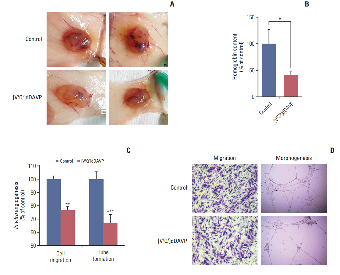
Fig. 4. Effect of [V4Q5]dDAVP on colorectal tumor-induced angiogenesis and microvascular cell migration and morphogenesis. (A) Colo-205 cell-induced angiogenesis evaluated by a modified-Matrigel plug assay. Representative images of plugs recovered from vehicle or [V4Q5]dDAVP (0.3 μg/kg intravenously, thrice-weekly for 2 weeks) treated mice are depicted. (B) Quantitative assessment of angiogenesis was achieved by the determination of relative hemoglobin content in Matrigel plugs using the Drabkin's method. n=10 animals per experimental group. (C) Quantification of in vitro migration and tube formation by arginine vasopressin type 2 membrane receptor‒expressing microvascular HMVEC-L cells incubated overnight with [V4Q5]dDAVP (1 μM) in comparison to saline vehicle. (D) Crystal violet staining of microvascular endothelial cells that crossed the polycarbonate membrane in the Transwell migration assay (left) and representative images of vasculature-like structures formed by HMVEC-L cells in the morphogenesis assay (right, ×100). Data are presented as mean±standard error of mean and are representative of at least two independent experiments. *p < 0.05, **p < 0.01, ***p < 0.001 vs. control, unpaired t test.

Fig. 5. In vitro direct cytostatic effects of [V4Q5]dDAVP on colorectal cancer (CRC) cells alone or in combination with standard-of-care chemotherapy. (A) IC50 calculation and reduction of in vitro CT-26 cell colony formation by [V4Q5]dDAVP (right). Dotted line indicates 50% inhibition. Statistical significance (*p < 0.05) in IC50 calculation studies for [V4Q5]dDAVP was achieved with drug concentrations ≥ 1 μM. Representative photographs of vehicle- or [V4Q5]dDAVP-treated low density CT-26 cultures are depicted (left). (B) Reversal of [V4Q5]dDAVP cytostatic effect by addition of arginine vasopressin type 2 membrane receptor selective chemical antagonist tolvaptan. ***p < 0.001, [V4Q5]dDAVP vs. control or [V4Q5]dDAVP plus tolvaptan. (C) IC50 calculation of cytotoxic agent 5-fluorouracil (5-FU) on exponentially growing CT-26 and Colo-205 CRC cells. Statistical significance (*p < 0.05) in IC50 calculation studies for 5-FU in CT-26 and Colo-205 was achieved with drug concentrations ≥ 0.5 or 5 μM, respectively. PBS, phosphate buffered saline. (D) Addition of [V4Q5]dDAVP to sub-IC50 concentrations of 5-FU enhances in vitro cytostatic effect on high density AVPR2-expressing CRC cell cultures. *p < 0.05, **p < 0.01, ***p < 0.001 vs. control. ##p < 0.01 and ###p < 0.001, [V4Q5]dDAVP plus 5-FU vs. [V4Q5]dDAVP or 5-FU individual therapies. Results are representative of at least three independent experiments. Data are presented as mean±standard error of mean. ANOVA plus Dunnet´s test.
Reference
-
References
1. Ferlay J, Soerjomataram I, Dikshit R, Eser S, Mathers C, Rebelo M, et al. Cancer incidence and mortality worldwide: sources, methods and major patterns in GLOBOCAN 2012. Int J Cancer. 2015; 136:E359–86.
Article2. Alonso DF, Skilton G, Farias EF, Bal de Kier Joffe E, Gomez DE. Antimetastatic effect of desmopressin in a mouse mammary tumor model. Breast Cancer Res Treat. 1999; 57:271–5.
Article3. Ripoll GV, Garona J, Pifano M, Farina HG, Gomez DE, Alonso DF. Reduction of tumor angiogenesis induced by desmopressin in a breast cancer model. Breast Cancer Res Treat. 2013; 142:9–18.
Article4. Kaufmann JE, Oksche A, Wollheim CB, Gunther G, Rosenthal W, Vischer UM. Vasopressin-induced von Willebrand factor secretion from endothelial cells involves V2 receptors and cAMP. J Clin Invest. 2000; 106:107–16.
Article5. Pifano M, Garona J, Capobianco CS, Gonzalez N, Alonso DF, Ripoll GV. Peptide agonists of vasopressin V2 receptor reduce expression of neuroendocrine markers and tumor growth in human lung and prostate tumor cells. Front Oncol. 2017; 7:11.
Article6. Ripoll GV, Garona J, Hermo GA, Gomez DE, Alonso DF. Effects of the synthetic vasopressin analog desmopressin in a mouse model of colon cancer. Anticancer Res. 2010; 30:5049–54.7. Mochizuki S, Soejima K, Shimoda M, Abe H, Sasaki A, Okano HJ, et al. Effect of ADAM28 on carcinoma cell metastasis by cleavage of von Willebrand factor. J Natl Cancer Inst. 2012; 104:906–22.
Article8. Terraube V, Marx I, Denis CV. Role of von Willebrand factor in tumor metastasis. Thromb Res. 2007; 120 Suppl 2:S64–70.
Article9. Kawecki C, Lenting PJ, Denis CV. von Willebrand factor and inflammation. J Thromb Haemost. 2017; 15:1285–94.
Article10. Starke RD, Ferraro F, Paschalaki KE, Dryden NH, McKinnon TA, Sutton RE, et al. Endothelial von Willebrand factor regulates angiogenesis. Blood. 2011; 117:1071–80.
Article11. Weinberg RS, Grecco MO, Ferro GS, Seigelshifer DJ, Perroni NV, Terrier FJ, et al. A phase II dose-escalation trial of perioperative desmopressin (1-desamino-8-d-arginine vasopressin) in breast cancer patients. Springerplus. 2015; 4:428.
Article12. Czaplewski C, Kazmierkiewicz R, Ciarkowski J. Molecular modeling of the human vasopressin V2 receptor/agonist complex. J Comput Aided Mol Des. 1998; 12:275–87.13. Kowalczyk W, Prahl A, Derdowska I, Sobolewski D, Olejnik J, Zabrocki J, et al. Analogues of neurohypophyseal hormones, oxytocin and arginine vasopressin, conformationally restricted in the N-terminal part of the molecule. J Med Chem. 2006; 49:2016–21.
Article14. Pastrian MB, Guzman F, Garona J, Pifano M, Ripoll GV, Cascone O, et al. Structure-activity relationship of 1-desamino-8-D-arginine vasopressin as an antiproliferative agent on human vasopressin V2 receptor-expressing cancer cells. Mol Med Rep. 2014; 9:2568–72.
Article15. Garona J, Pifano M, Orlando UD, Pastrian MB, Iannucci NB, Ortega HH, et al. The novel desmopressin analogue [V4Q5]dDAVP inhibits angiogenesis, tumour growth and metastases in vasopressin type 2 receptor-expressing breast cancer models. Int J Oncol. 2015; 46:2335–45.16. Iannucci NB, Ripoll GV, Garona J, Cascone O, Ciccia GN, Gomez DE, et al. Antiproliferative effect of 1-deamino-8-D-arginine vasopressin analogs on human breast cancer cells. Future Med Chem. 2011; 3:1987–93.
Article17. Monstein HJ, Truedsson M, Ryberg A, Ohlsson B. Vasopressin receptor mRNA expression in the human gastrointestinal tract. Eur Surg Res. 2008; 40:34–40.
Article18. Marques I, Araujo A, de Mello RA. Anti-angiogenic therapies for metastatic colorectal cancer: current and future perspectives. World J Gastroenterol. 2013; 19:7955–71.
Article19. Castle JC, Loewer M, Boegel S, de Graaf J, Bender C, Tadmor AD, et al. Immunomic, genomic and transcriptomic characterization of CT26 colorectal carcinoma. BMC Genomics. 2014; 15:190.
Article20. Khan S, Cameron S, Blaschke M, Moriconi F, Naz N, Amanzada A, et al. Differential gene expression of chemokines in KRAS and BRAF mutated colorectal cell lines: role of cytokines. World J Gastroenterol. 2014; 20:2979–94.21. Riihimaki M, Hemminki A, Sundquist J, Hemminki K. Patterns of metastasis in colon and rectal cancer. Sci Rep. 2016; 6:29765.
Article22. Kim KY, Kim NK, Cha IH, Ahn JB, Choi JS, Choi GH, et al. Novel methods for clinical risk stratification in patients with colorectal liver metastases. Cancer Res Treat. 2015; 47:242–50.
Article23. Rmali KA, Puntis MC, Jiang WG. Tumour-associated angiogenesis in human colorectal cancer. Colorectal Dis. 2007; 9:3–14.
Article24. Ripoll G, Iannucci N, Giron S, Cascone O, Gomez D, Alonso D. Angiostatic activity of 1-deamino-8-D-arginine vasopressin and novel peptide analogues in breast cancer cells. Cancer Res. 2008; 68(9 Suppl):295.25. Horowitz M, Neeman E, Sharon E, Ben-Eliyahu S. Exploiting the critical perioperative period to improve long-term cancer outcomes. Nat Rev Clin Oncol. 2015; 12:213–26.
Article26. Hirai T, Matsumoto H, Kubota H, Yamaguchi Y. Regulating surgical oncotaxis to improve the outcomes in cancer patients. Surg Today. 2014; 44:804–11.
Article27. Pifano M, Garona J, Sobol NT, Alberto M, Alonso DF, Ripoll GV. Search of vasopressin analogs with antiproliferative activity on small-cell lung cancer: drug design based on two different approaches. Future Med Chem. 2018; 10:879–94.
Article28. McEwan DG, Brunton VG, Baillie GS, Leslie NR, Houslay MD, Frame MC. Chemoresistant KM12C colon cancer cells are addicted to low cyclic AMP levels in a phosphodiesterase 4-regulated compartment via effects on phosphoinositide 3-kinase. Cancer Res. 2007; 67:5248–57.
Article29. Loffler I, Grun M, Bohmer FD, Rubio I. Role of cAMP in the promotion of colorectal cancer cell growth by prostaglandin E2. BMC Cancer. 2008; 8:380.
Article30. Hopfner M, Maaser K, Barthel B, von Lampe B, Hanski C, Riecken EO, et al. Growth inhibition and apoptosis induced by P2Y2 receptors in human colorectal carcinoma cells: involvement of intracellular calcium and cyclic adenosine monophosphate. Int J Colorectal Dis. 2001; 16:154–66.
Article
- Full Text Links
-
- Actions
-
Cited
- CITED
-
- Close
- Share
-
- Similar articles
-
- Sick Sinus Syndrome Following Severe Hyponatremia Associated with Desmopressin Therapy
- Correlation between expression of matrix metalloproteinase-2 (MMP-2), and matrix metalloproteinase-9 (MMP-9) and angiogenesis in colorectal adenocarcinoma
- Current Status of Chemotherapy in Colorectal Cancer: Updated Treatment Strategies
- Correlation between Tumor Angiogenesis (Microvessel Density), Metastasis and Tumor Cell Proliferation in Colorectal Carcinomas
- Expression of Vascular Endothelial Growth Factor (VEGF) and p53 in Colorectal Cancer
