Ann Hepatobiliary Pancreat Surg.
2019 Nov;23(4):334-338. 10.14701/ahbps.2019.23.4.334.
Differentiation of gallbladder adenomyomatosis from early-stage gallbladder cancer before surgery
- Affiliations
-
- 1Department of Surgery, Ilsan Paik Hospital, Inje University College of Medicine, Goyang, Korea. ycshindr@gmail.com
- KMID: 2464321
- DOI: http://doi.org/10.14701/ahbps.2019.23.4.334
Abstract
- BACKGROUNDS/AIMS
This study aimed to compare the perioperative and clinical outcomes in patients undergoing laparoscopic cholecystectomy for gallbladder adenomyomatosis (GBA) or early-stage gallbladder cancer (GBC).
METHODS
The perioperative and clinical outcomes of 194 patients diagnosed with GBA and 30 patients diagnosed with GBC who underwent laparoscopic cholecystectomy in our institution from January 2011 to December 2017 were retrospectively compared.
RESULTS
There were no significant differences between the GBA and GBC groups in sex (male:female ratio 1.0:0.8 vs. 1.0:0.7, p=0.734), BMI (23.9±3.4 vs. 24.0±3.8 kg/m², p=0.916), or preoperative liver function tests. Patients in the GBC group were significantly older (50.5±14.1 vs. 65.9±10.6 years, p<0.001) and had a higher ASA grade (40.3 vs. 63.4% grade II or III, p=0.043) than patients in the GBA group. Although there was no significant difference in preoperative diagnostic methods (p=0.442), the GBC group showed a significantly higher rate of misdiagnosis on preoperative imaging compared with postoperative histopathologic findings (30.9% vs. 53.3%, p=0.011). There were significantly more patients with gallstones in the GBA group than in the GBC group (68.6% vs. 40.0%, p=0.004).
CONCLUSIONS
In older patients hospitalized for biliary colic without gallstones but with a thickened gallbladder wall with inflammation on preoperative diagnostic exam, the possibility of early-stage GBC should be considered.
MeSH Terms
Figure
-
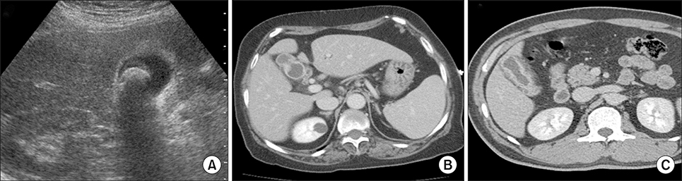
Fig. 1 Preoperative diagnostic images of gallbladder diseases. (A) Ultrasonographic finding of a patient with gallbladder cancer, considered as chronic calculous cholecystitis preoperatively. (B) Computed tomographic scan of a patient with gallbladder cancer, considered as gallbladder adenomyomatosis preoperatively. (C) Computed tomographic scan of a patient with gallbladder adenomyomatosis, considered as gallbladder cancer preoperatively.
Reference
-
1. Gerard PS, Berman D, Zafaranloo S. CT and ultrasound of gallbladder adenomyomatosis mimicking carcinoma. J Comput Assist Tomogr. 1990; 14:490–491.2. Ching BH, Yeh BM, Westphalen AC, Joe BN, Qayyum A, Coakley FV. CT differentiation of adenomyomatosis and gallbladder cancer. AJR Am J Roentgenol. 2007; 189:62–66.3. Stunell H, Buckley O, Geoghegan T, O'Brien J, Ward E, Torreggiani W. Imaging of adenomyomatosis of the gall bladder. J Med Imaging Radiat Oncol. 2008; 52:109–117.4. Yoshimitsu K, Honda H, Jimi M, Kuroiwa T, Hanada K, Irie H, et al. MR diagnosis of adenomyomatosis of the gallbladder and differentiation from gallbladder carcinoma: importance of showing Rokitansky-Aschoff sinuses. AJR Am J Roentgenol. 1999; 172:1535–1540.5. Haradome H, Ichikawa T, Sou H, Yoshikawa T, Nakamura A, Araki T, et al. The pearl necklace sign: an imaging sign of adenomyomatosis of the gallbladder at MR cholangiopancreatography. Radiology. 2003; 227:80–88.6. Joo I, Lee JY, Kim JH, Kim SJ, Kim MA, Han JK, et al. Differentiation of adenomyomatosis of the gallbladder from early-stage, wall-thickening-type gallbladder cancer using high-resolution ultrasound. Eur Radiol. 2013; 23:730–738.7. Oktar SO, Yücel C, Ozdemir H, Ulutürk A, Işik S. Comparison of conventional sonography, real-time compound sonography, tissue harmonic sonography, and tissue harmonic compound sonography of abdominal and pelvic lesions. AJR Am J Roentgenol. 2003; 181:1341–1347.8. Dahl JJ, Soo MS, Trahey GE. Clinical evaluation of combined spatial compounding and adaptive imaging in breast tissue. Ultrason Imaging. 2004; 26:203–216.9. Yen CL, Jeng CM, Yang SS. The benefits of comparing conventional sonography, real-time spatial compound sonography, tissue harmonic sonography, and tissue harmonic compound sonography of hepatic lesions. Clin Imaging. 2008; 32:11–15.10. Jung KW, Won YJ, Kong HJ, Lee ES. Cancer statistics in Korea: incidence, mortality, survival, and prevalence in 2016. Cancer Res Treat. 2019; 51:417–430.11. Kai K, Irie H, Ide T, Masuda M, Kitahara K, Miyoshi A, et al. Actual status of clinical diagnosis in patients with primary gallbladder cancer associated with adenomyomatosis. Indian J Gastroenterol. 2013; 32:386–391.12. Ozgonul A, Bitiren M, Guldur ME, Sogut O, Yilmaz LE. Fundal variant adenomyomatosis of the gallbladder: report of three cases and review of the literature. J Clin Med Res. 2010; 2:150–153.13. Pang L, Zhang Y, Wang Y, Kong J. Pathogenesis of gallbladder adenomyomatosis and its relationship with early-stage gallbladder carcinoma: an overview. Braz J Med Biol Res. 2018; 51:e7411.14. Kim BS, Oh JY, Nam KJ, Cho JH, Kwon HJ, Yoon SK, et al. Focal thickening at the fundus of the gallbladder: computed tomography differentiation of fundal type adenomyomatosis and localized chronic cholecystitis. Gut Liver. 2014; 8:219–223.15. Bang SH, Lee JY, Woo H, Joo I, Lee ES, Han JK, et al. Differentiating between adenomyomatosis and gallbladder cancer: revisiting a comparative study of high-resolution ultrasound, multidetector CT, and MR imaging. Korean J Radiol. 2014; 15:226–234.16. Ootani T, Shirai Y, Tsukada K, Muto T. Relationship between gallbladder carcinoma and the segmental type of adenomyomatosis of the gallbladder. Cancer. 1992; 69:2647–2652.17. Gore RM, Yaghmai V, Newmark GM, Berlin JW, Miller FH. Imaging benign and malignant disease of the gallbladder. Radiol Clin North Am. 2002; 40:1307–1323.18. Lowenfels AB, Maisonneuve P, Boyle P, Zatonski WA. Epidemiology of gallbladder cancer. Hepatogastroenterology. 1999; 46:1529–1532.
- Full Text Links
-
- Actions
-
Cited
- CITED
-
- Close
- Share
-
- Similar articles
-
- Adenocarcinoma Arising in Segmental Adenomyomatosis of the Gallbladder: A Case Report
- Localized Adenomyomatosis of Gallbladder Mimicking Advanced Hepatic Flexure Colon Cancer: A Case Report
- Surgical Management of Gallbladder Cancer How accurately can we assess the T-stage and resectability prior to GB cancer surgery?
- Heterotopic pancreas of the gallbladder associated with segmental adenomyomatosis of the gallbladder
- Cholelithiasis as a Risk Factor for Gallbladder Cancer
