Adenocarcinoma Arising in Segmental Adenomyomatosis of the Gallbladder: A Case Report
- Affiliations
-
- 1Department of Radiology, Chungnam University Hospital, Korea. jscho@cnu.ac.kr
- 2Department of Internal Medicine, Chungnam University Hospital, Korea.
- 3Department of Surgery, Chungnam University Hospital, Korea.
- 4Department of Pathology, Chungnam University Hospital, Korea.
- KMID: 2097917
- DOI: http://doi.org/10.3348/jksr.2010.63.5.453
Abstract
- We report here a rare case of adenocarcinoma arising from segmental adenomyomatosis of the gallbladder itself. CT images showed segmental annular wall thickening of the gallbladder body with regional lymph node enlargement, which could not exclude adenomyomatosis associated with malignancy. Therefore, we performed MRI and PET/CT for further evaluation. T2 weighted MRI revealed multiple tiny intramural cystic lesions in the focal wall thickening of the gallbladder, suggesting adenomyomatosis. However, PET/CT showed increased activity in the lesion of the gallbladder and regional lymph node, suggesting malignancy, which was pathologically confirmed as adenocarcinoma arising from adenomyomatosis.
Figure
-
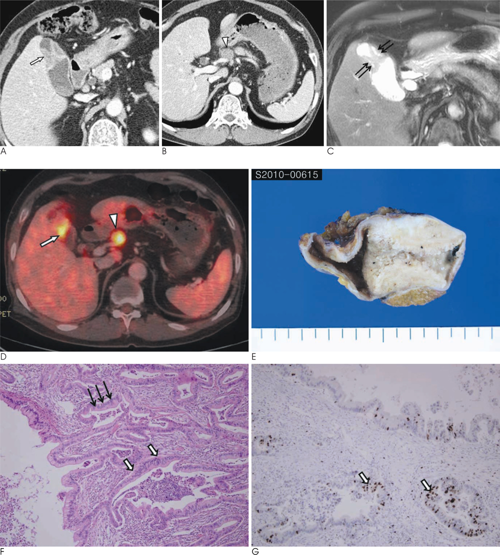
Fig. 1 Adenocarcinoma arising from segmental adenomyomatosis of the gallbladder in a 67-year-old man. A. Transverse contrast-enhanced MDCT image showing focal annular wall thickening (arrow) of the gallbladder. B. Transverse contranst-enhanced MDCT image showing an enlarged common hepatic lymph node (arrowhead), suggesting metastasis. C. Transverse T2-weighted MR image showing multiple tiny intramural hyperintense cystic lesions (thin arrows), which are not identified on the CT image. D. Transverse fused PET/CT image showing increased FDG activity in the annular wall thickening of the gallbladder (arrow) and common hepatic lymph nodes (arrowhead), which are consistent with gallbladder cancer and lymph node metastasis. E. Photograph of the cut surface of the gross specimen shows a yellowish-white colored wall thickening with multiple tiny intramural cysts. F. Photomicrograph showing normal epithelium of the Rokitansky-Aschoff sinus (thin arrows) and enlarged stratification of the nuclei and a high frequency of mitoses (arrows) representing adenocarcinoma (H & E, original magnification ×100). G. Photomicrograph of immunohistochemical stain with p53 showing positive staining (arrows) in adenocarcinoma (p53, original magnification ×100).
Reference
-
1. Aldridge MC, Gruffaz F, Castaing D, Bismuth H. Adenomyomatosis of the gallbladder. A premalignant lesion? Surgery. 1991; 109:107–110.2. Katoh T, Nakai T, Hayashi S, Satake T. Noninvasive carcinoma of the gallbladder arising in localized type adenomyomatosis. Am J Gastroenterol. 1988; 83:670–674.3. Kawarada Y, Sanda M, Mizumoto R, Yatani R. Early carcinoma of the gallbladder, noninvasive carcinoma originating in the Rokitansky-aschoff sinus: a case report. Am J Gastroenterol. 1986; 81:61–66.4. Nakafuji H, Koike Y, Wakabayashi M, Furihata R, Maruyama Y, Ogata H. Three cases of early stage carcinoma of the gallbladder. Gastroenterol Jpn. 1981; 16:134–140.5. Sakurai T, Saji Y, Kazui K, Yamaga S, Hirose K, Shinohara T, et al. A case of early carcinoma of the gall-bladder arising in adenomyomatosis detected by endoscopic ultrasonography. Nippon Shokakibyo Gakkai Zasshi. 1995; 92:1304–1308.6. Meguid MM, Aun F, Bradford ML. Adenomyomatosis of the gallbladder. Am J Surg. 1984; 147:260–262.7. Ootani T, Shirai Y, Tsukada K, Muto T. Relationship between gallbladder carcinoma and the segmental type of adenomyomatosis of the gallbladder. Cancer. 2006; 69:2647–2652.8. Yoshimitsu K, Honda H, Aibe H, Shinozaki K, Kuroiwa T, Irie H, et al. Radiologic diagnosis of adenomyomatosis of the gallbladder: comparative study among MRI, helical CT, and transabdominal US. J Comput Assist Tomogr. 2001; 25:843–850.9. Haradome H, Ichikawa T, Sou H, Yoshikawa T, Nakamura A, Araki T, et al. The pearl necklace sign: an imaging sign of adenomyomatosis of the gallbladder at MR cholangiopancreatography. Radiology. 2003; 227:80–88.
- Full Text Links
-
- Actions
-
Cited
- CITED
-
- Close
- Share
-
- Similar articles
-
- Heterotopic pancreas of the gallbladder associated with segmental adenomyomatosis of the gallbladder
- Localized Adenomyomatosis of Gallbladder Mimicking Advanced Hepatic Flexure Colon Cancer: A Case Report
- Segmental Adenomyomatosis of Gallbladder: CT Assessment of the Patterns of Cholecystolithiasis
- Intracystic Papillary Neoplasm of the Gallbladder Arising from a Localized Adenomyomatous Hyperplasia
- Report of a Case of Adenomyomatosis of Gallbladder
