Multidetector CT Findings and Differential Diagnoses of Malignant Pleural Mesothelioma and Metastatic Pleural Diseases in Korea
- Affiliations
-
- 1Department of Radiology, Gachon University Gil Medical Center, Incheon 21565, Korea.
- 2Department of Radiology, Dongguk University Ilsan Hospital, Goyang 10326, Korea. jeungkim@dumc.or.kr
- 3Department of Radiology, Seoul National University Bundang Hospital, Seongnam 13620, Korea.
- 4Department of Radiology, Samsung Medical Center, Sungkyunkwan University School of Medicine, Seoul 06351, Korea.
- 5Department of Radiology, Seoul National University College of Medicine, Seoul 03080, Korea.
- 6Department of Pathology, Yonsei University Wonju College of Medicine, Wonju 26426, Korea.
- KMID: 2455425
- DOI: http://doi.org/10.3348/kjr.2016.17.4.545
Abstract
OBJECTIVE
To compare the multidetector CT (MDCT) features of malignant pleural mesothelioma (MPM) and metastatic pleural disease (MPD).
MATERIALS AND METHODS
The authors reviewed the MDCT images of 167 patients, 103 patients with MPM and 64 patients with MPD. All 167 cases were pathologically confirmed by sonography-guided needle biopsy of pleura, thoracoscopic pleural biopsy, or open thoracotomy. CT features were evaluated with respect to pleural effusion, pleural thickening, invasion of other organs, lung abnormality, lymphadenopathy, mediastinal shifting, thoracic volume decrease, asbestosis, and the presence of pleural plaque.
RESULTS
Pleural thickening was the most common CT finding in MPM (96.1%) and MPD (93.8%). Circumferential pleural thickening (31.1% vs. 10.9%, odds ratio [OR] 3.670), thickening of fissural pleura (83.5% vs. 67.2%, OR 2.471), thickening of diaphragmatic pleura (90.3% vs. 73.4%, OR 3.364), pleural mass (38.8% vs. 23.4%, OR 2.074), pericardial involvement (56.3% vs. 20.3%, OR 5.056), and pleural plaque (66.0% vs. 21.9%, OR 6.939) were more frequently seen in MPM than in MPD. On the other hand, nodular pleural thickening (59.2% vs. 76.6%, OR 0.445), hilar lymph node metastasis (5.8% vs. 20.3%, OR 0.243), mediastinal lymph node metastasis (10.7% vs. 37.5%, OR 0.199), and hematogenous lung metastasis (9.7% vs. 29.2%, OR 0.261) were less frequent in MPM than in MPD. When we analyzed MPD from extrathoracic malignancy (EMPD) separately and compared them to MPM, circumferential pleural thickening, thickening of interlobar fissure, pericardial involvement and presence of pleural plaque were significant findings indicating MPM than EMPD. MPM had significantly lower occurrence of hematogenous lung metastasis, as compared with EMPD.
CONCLUSION
Awareness of frequent and infrequent CT findings could aid in distinguishing MPM from MPD.
Keyword
MeSH Terms
-
Adult
Aged
Diagnosis, Differential
Female
Humans
Image-Guided Biopsy
Lung Neoplasms/*diagnosis/diagnostic imaging/pathology/secondary
Lymphatic Metastasis
Male
Mesothelioma/*diagnosis/diagnostic imaging/pathology
Middle Aged
*Multidetector Computed Tomography
Odds Ratio
Pleural Neoplasms/*diagnosis/diagnostic imaging/pathology
Republic of Korea
Retrospective Studies
Figure
-
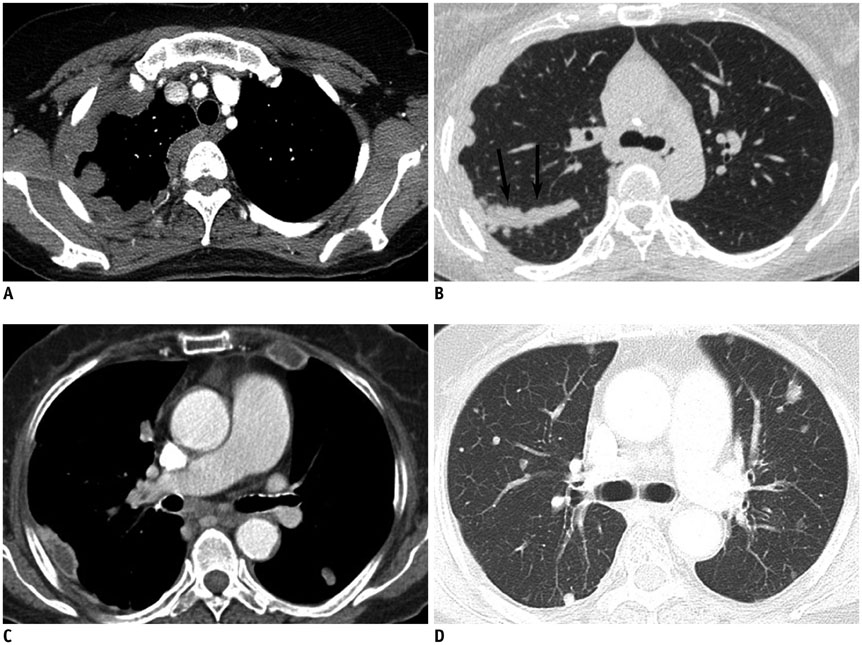
Fig. 1 CT findings of MPM and MPD. A, B. 48-year-old woman with epithelioid type MPM. Circumferential pleural thickening and fissural pleural thickening (arrows) involving right hemithorax. C, D. 82-year-old woman with MPD from renal cell carcinoma. Nodular pleural thickeing involving both hemithorax and hematogenous metastases in both lungs. CT = computed tomography, MPD = metastatic pleural disease, MPM = malignant pleural mesothelioma
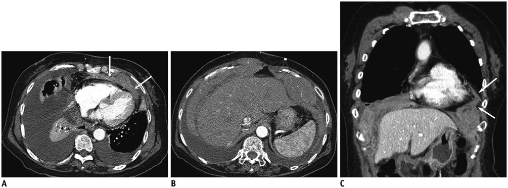
Fig. 2 63-year-old woman with bilateral MPM involving pericardium and both diaphragmatic pleura. Transverse CT images (A, B) and coronal reformatted image (C) show bilateral pleural effusion, pericardial thickening (arrows) and thickening of both diaphragmatic pleura. MPM = malignant pleural mesothelioma
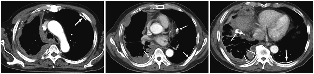
Fig. 3 71-year-old man with MPM. Transverse CT images show circumferential pleural thickening in right hemithorax. Note calcified pleural plaques in both hemithorax (arrows). CT = computed tomography, MPM = malignant pleural mesothelioma
Reference
-
1. Antman KH. Natural history and epidemiology of malignant mesothelioma. Chest. 1993; 103:4 Suppl. 373S–376S.2. Nickell LT Jr, Lichtenberger JP 3rd, Khorashadi L, Abbott GF, Carter BW. Multimodality imaging for characterization, classification, and staging of malignant pleural mesothelioma. Radiographics. 2014; 34:1692–1706.3. Robinson BW, Lake RA. Advances in malignant mesothelioma. N Engl J Med. 2005; 353:1591–1603.4. Leung AN, Müller NL, Miller RR. CT in differential diagnosis of diffuse pleural disease. AJR Am J Roentgenol. 1990; 154:487–492.5. Metintas M, Ucgun I, Elbek O, Erginel S, Metintas S, Kolsuz M, et al. Computed tomography features in malignant pleural mesothelioma and other commonly seen pleural diseases. Eur J Radiol. 2002; 41:1–9.6. Kawashima A, Libshitz HI. Malignant pleural mesothelioma: CT manifestations in 50 cases. AJR Am J Roentgenol. 1990; 155:965–969.7. Okten F, Köksal D, Onal M, Ozcan A, Sims¸ek C, Ertürk H. Computed tomography findings in 66 patients with malignant pleural mesothelioma due to environmental exposure to asbestos. Clin Imaging. 2006; 30:177–180.8. Seely JM, Nguyen ET, Churg AM, Müller NL. Malignant pleural mesothelioma: computed tomography and correlation with histology. Eur J Radiol. 2009; 70:485–491.9. Ng CS, Munden RF, Libshitz HI. Malignant pleural mesothelioma: the spectrum of manifestations on CT in 70 cases. Clin Radiol. 1999; 54:415–421.10. Tamer Dogan O, Salk I, Tas F, Epozturk K, Gumus C, Akkurt I, et al. Thoracic computed tomography findings in malignant mesothelioma. Iran J Radiol. 2012; 9:209–211.11. Jung SH, Kim HR, Koh SB, Yong SJ, Chung MJ, Lee CH, et al. A decade of malignant mesothelioma surveillance in Korea. Am J Ind Med. 2012; 55:869–875.12. Akira M, Yamamoto S, Yokoyama K, Kita N, Morinaga K, Higashihara T, et al. Asbestosis: high-resolution CT-pathologic correlation. Radiology. 1990; 176:389–394.13. Akira M, Yokoyama K, Yamamoto S, Higashihara T, Morinaga K, Kita N, et al. Early asbestosis: evaluation with high-resolution CT. Radiology. 1991; 178:409–416.14. Kim Y, Myong JP, Lee JK, Kim JS, Kim YK, Jung SH. CT characteristics of pleural plaques related to occupational or environmental asbestos exposure from South Korean asbestos mines. Korean J Radiol. 2015; 16:1142–1152.15. Kusaka Y, Hering KG, Parker JE. Coding CT-classification in occupational and environmental respiratory disease (OERD). In : Kusaka Y, Hering KG, Parker JE, editors. International classification of HRCT for occupational and environmental respiratory diseases. Tokyo: Springer;2005. p. 20–21.16. Wang ZJ, Reddy GP, Gotway MB, Higgins CB, Jablons DM, Ramaswamy M, et al. Malignant pleural mesothelioma: evaluation with CT, MR imaging, and PET. Radiographics. 2004; 24:105–119.17. Qureshi NR, Gleeson FV. Imaging of pleural disease. Clin Chest Med. 2006; 27:193–213.18. Sahin AA, Cöplü L, Selçuk ZT, Eryilmaz M, Emri S, Akhan O, et al. Malignant pleural mesothelioma caused by environmental exposure to asbestos or erionite in rural Turkey: CT findings in 84 patients. AJR Am J Roentgenol. 1993; 161:533–537.19. Feragalli B, Mantini C, Civitareale N, Polverosi R, Tartaro A, Cotroneo AR. Extrapleural and cardiophrenic lymph nodes: prevalence, clinical significance and diagnostic value. Radiol Med. 2014; 119:20–26.20. Abdel Rahman AR, Gaafar RM, Baki HA, El Hosieny HM, Aboulkasem F, Farahat EG, et al. Prevalence and pattern of lymph node metastasis in malignant pleural mesothelioma. Ann Thorac Surg. 2008; 86:391–395.21. Aberle DR, Gamsu G, Ray CS. High-resolution CT of benign asbestos-related diseases: clinical and radiographic correlation. AJR Am J Roentgenol. 1988; 151:883–891.22. Meirelles GS, Kavakama JI, Jasinowodolinski D, Nery LE, Terra-Filho M, Rodrigues RT, et al. Pleural plaques in asbestos-exposed workers: reproducibility of a new high-resolution CT visual semiquantitative measurement method. J Thorac Imaging. 2006; 21:8–13.
- Full Text Links
-
- Actions
-
Cited
- CITED
-
- Close
- Share
-
- Similar articles
-
- Differential CT Features between Malignant Mesothelioma and Pleural Metastasis from Lung Cancer or Extrathoracic Primary Tumor Mimicking Malignant Mesothelioma
- Malignant Mesothelioma Causing Bloody Pleural Effusion
- Differential Diagnosis of Tuberculous Pleural Effusion and Malignant Pleural Effusion: CT Accuracy and Findings
- Malignant Pleural Mesothelioma with Heterologous Osteoblastic Differentiation: Case Report of the Characteristic CT and Bone Scan Findings
- A Case of Malignant Mesothelioma with Pleural Effusion
