Cone-beam computed tomography-guided three-dimensional evaluation of treatment effectiveness of the Frog appliance
- Affiliations
-
- 1Clinical Research Center of Shaanxi Province for Dental and Maxillofacial Diseases, College of Stomatology, Xi'an Jiaotong University, Xi'an, China. sixinqin@mail.xjtu.edu.cn
- 2Department of Orthodontics, Stomatological Hospital, Xi'an Jiaotong University, Xi'an, China.
- 3Department of Orthodontics, Xi'an No.1 Hospital, Xi'an, China.
- KMID: 2450225
- DOI: http://doi.org/10.4041/kjod.2019.49.3.161
Abstract
OBJECTIVE
To evaluate the effectiveness of the Frog appliance in three dimensions by using cone-beam computed tomography (CBCT) images.
METHODS
Forty patients (21 boys and 19 girls), averaged 11.7 years old, with an Angle Class II division 1 malocclusion were included in our study. They had either late mixed dentition or early permanent dentition, and the maxillary second molars had not yet erupted. All patients underwent CBCT before and after the treatment for measuring changes in the maxillary first molars, second premolars, central incisors, and profile. Paired-samples t-test was used to compare the mean difference in each variable before treatment and after the first phase of treatment.
RESULTS
The maxillary first molars were effectively distalized by 4.25 mm (p < 0.001) and 3.53 mm (p < 0.05) in the dental crown and root apex, respectively. The tipping increased by 2.25°, but the difference was not significant. Moreover the teeth moved buccally by 0.84 mm (p < 0.05) and 2.87 mm (p < 0.01) in the mesiobuccal and distobuccal cusps, respectively, whereas no significant changes occurred in the root apex. Regarding the anchorage parts, the angle of the maxillary central incisor's long axis to the sella-nasion plane increased by 2.76° (p < 0.05) and the distance from the upper lip to the esthetic plane decreased by 0.52 mm (p = 0.01).
CONCLUSIONS
The Frog appliance effectively distalized the maxillary molars with an acceptable degree of tipping, distobuccal rotation, and buccal crown torque, with only slight anchorage loss. Furthermore, CBCT image demonstrated that it is a simple and reliable method for three-dimensional analysis.
Keyword
MeSH Terms
Figure
-
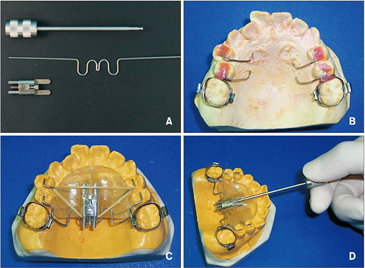
Figure 1 Structure of the Frog appliance (FORESTADENT Bernhard Förster GmbH, Pforzheim, Germany). A, Components of the Frog appliance, from top to bottom: screwdriver, preformed spring, and screw. B, Occlusal rest, one of the anchorage devices in the appliance. C, Fabrication of the complete appliance, each part of which is connected by an elastic band and Nance button. D, Activation of the appliance.

Figure 2 Intraoral view after treatment.
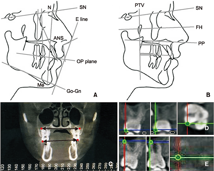
Figure 3 Cephalometric measurements performed in the study. A, B, Linear and angular measurements. C, Coronal measurements of the maxillary first molars. The upper two red dots indicate bilateral distobuccal root apices of the maxillary first molars; the lower two red dots indicate bilateral distobuccal cusps of the maxillary first molar. D, E, Position of the distobuccal cusp (D) and distobuccal root apex (E) of the right maxillary first molars in the coronal (left), sagittal (middle), and transverse (right) directions. N, Nasion; SN, sella-nasion; E line, esthetic plane; ANS, anterior nasal spine; OP, occlusion plane; Me, menton; Go-Gn, mandibular plane; PTV, pterygoid vertical; FH, Frankfort horizontal plane; PP, palatal plane.
Reference
-
1. Alexander SA. Diagnosis and treatment planning in orthodontics. Curr Opin Dent. 1992; 2:9–13.2. Alexander RG. The vari-simplex discipline. Part 2. Nonextraction treatment. J Clin Orthod. 1983; 17:474–482.3. Ludwig B, Glasl B, Kinzinger GS, Walde KC, Lisson JA. The skeletal frog appliance for maxillary molar distalization. J Clin Orthod. 2011; 45:77–84. quiz 91.4. Byloff FK, Darendeliler MA. Distal molar movement using the pendulum appliance. Part 1: clinical and radiological evaluation. Angle Orthod. 1997; 67:249–260.5. Bondemark L, Kurol J. Distalization of maxillary first and second molars simultaneously with repelling magnets. Eur J Orthod. 1992; 14:264–272.
Article6. Escobar SA, Tellez PA, Moncada CA, Villegas CA, Latorre CM, Oberti G. Distalization of maxillary molars with the bone-supported pendulum: a clinical study. Am J Orthod Dentofacial Orthop. 2007; 131:545–549.
Article7. Chiu PP, McNamara JA Jr, Franchi L. A comparison of two intraoral molar distalization appliances: distal jet versus pendulum. Am J Orthod Dentofacial Orthop. 2005; 128:353–365.
Article8. Mavropoulos A, Karamouzos A, Kiliaridis S, Papadopoulos MA. Efficiency of noncompliance simultaneous first and second upper molar distalization: a three-dimensional tooth movement analysis. Angle Orthod. 2005; 75:532–539.9. Nalcaci R, Kocoglu-Altan AB, Bicakci AA, Ozturk F, Babacan H. A reliable method for evaluating upper molar distalization: superimposition of three-dimensional digital models. Korean J Orthod. 2015; 45:82–88.
Article10. Duran GS, Görgülü S, Dindaroğlu F. Three-dimensional analysis of tooth movements after palatal miniscrew-supported molar distalization. Am J Orthod Dentofacial Orthop. 2016; 150:188–197.
Article11. Hourfar J, Ludwig B, Kanavakis G. An active, skeletally anchored transpalatal appliance for derotation, distalization and vertical control of maxillary first molars. J Orthod. 2014; 41:Suppl 1. S24–S32.
Article12. Uzuner FD, Kaygisiz E, Unver F, Tortop T. Comparison of transverse dental changes induced by the palatally applied Frog appliance and buccally applied Karad's integrated distalizing system. Korean J Orthod. 2016; 46:96–103.
Article13. Kapila SD, Nervina JM. CBCT in orthodontics: assessment of treatment outcomes and indications for its use. Dentomaxillofac Radiol. 2015; 44:20140282.
Article14. Özalp Ö, Tezerişener HA, Kocabalkan B, Büyükkaplan UŞ, Özarslan MM, Şimşek Kaya G, et al. Comparing the precision of panoramic radiography and cone-beam computed tomography in avoiding anatomical structures critical to dental implant surgery: a retrospective study. Imaging Sci Dent. 2018; 48:269–275.
Article15. Jacobs R, Quirynen M. Dental cone beam computed tomography: justification for use in planning oral implant placement. Periodontol 2000. 2014; 66:203–213.
Article16. Kinzinger GS, Fritz UB, Sander FG, Diedrich PR. Efficiency of a pendulum appliance for molar distalization related to second and third molar eruption stage. Am J Orthod Dentofacial Orthop. 2004; 125:8–23.
Article17. Flores-Mir C, McGrath L, Heo G, Major PW. Efficiency of molar distalization associated with second and third molar eruption stage. Angle Orthod. 2013; 83:735–742.
Article18. Joseph AA, Butchart CJ. An evaluation of the pendulum distalizing appliance. Semin Orthod. 2000; 6:129–135.19. Kang JM, Park JH, Bayome M, Oh M, Park CO, Kook YA, et al. A three-dimensional finite element analysis of molar distalization with a palatal plate, pendulum, and headgear according to molar eruption stage. Korean J Orthod. 2016; 46:290–300.
Article20. Burhan AS. Combined treatment with headgear and the Frog appliance for maxillary molar distalization: a randomized controlled trial. Korean J Orthod. 2013; 43:101–109.
Article21. Bayram M, Nur M, Kilkis D. The frog appliance for upper molar distalization: a case report. Korean J Orthod. 2010; 40:50–60.
Article22. Kinzinger G, Syrée C, Fritz U, Diedrich P. Molar distalization with different pendulum appliances: in vitro registration of orthodontic forces and moments in the initial phase. J Orofac Orthop. 2004; 65:389–409.
Article23. Kinzinger GS, Wehrbein H, Diedrich PR. Molar distalization with a modified pendulum appliance--in vitro analysis of the force systems and in vivo study in children and adolescents. Angle Orthod. 2005; 75:558–567.24. Karlsson I, Bondemark L. Intraoral maxillary molar distalization. Angle Orthod. 2006; 76:923–929.
Article25. Byloff FK, Darendeliler MA, Clar E, Darendeliler A. Distal molar movement using the pendulum appliance. Part 2: the effects of maxillary molar root uprighting bends. Angle Orthod. 1997; 67:261–270.26. Chaqués-Asensi J, Kalra V. Effects of the pendulum appliance on the dentofacial complex. J Clin Orthod. 2001; 35:254–257.27. Bussick TJ, McNamara JA Jr. Dentoalveolar and skeletal changes associated with the pendulum appliance. Am J Orthod Dentofacial Orthop. 2000; 117:333–343.
Article28. Nienkemper M, Wilmes B, Pauls A, Yamaguchi S, Ludwig B, Drescher D. Treatment efficiency of mini-implant-borne distalization depending on age and second-molar eruption. J Orofac Orthop. 2014; 75:118–132.
Article29. Fudalej P, Antoszewska J. Are orthodontic distalizers reinforced with the temporary skeletal anchorage devices effective? Am J Orthod Dentofacial Orthop. 2011; 139:722–729.
Article
- Full Text Links
-
- Actions
-
Cited
- CITED
-
- Close
- Share
-
- Similar articles
-
- Three-dimensional imaging modalities in endodontics
- A rare case of dilated invaginated odontome with talon cusp in a permanent maxillary central incisor diagnosed by cone beam computed tomography
- Management of root canal perforation by using cone-beam computed tomography
- A new minimally invasive guided endodontic microsurgery by cone beam computed tomography and 3-dimensional printing technology
- Detection of maxillary second molar with two palatal roots using cone beam computed tomography: a case report
